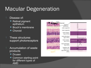
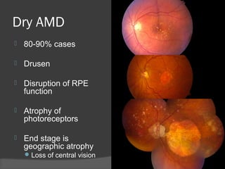
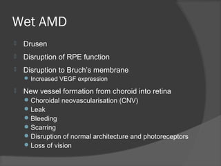
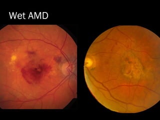
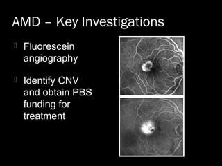
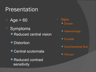
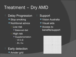
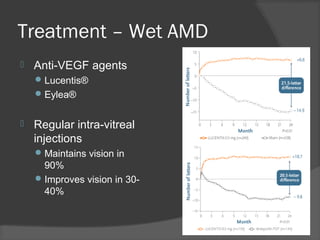
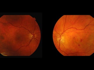
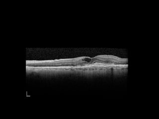
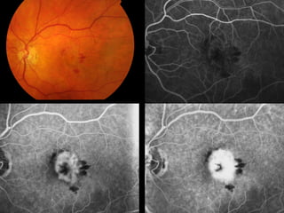
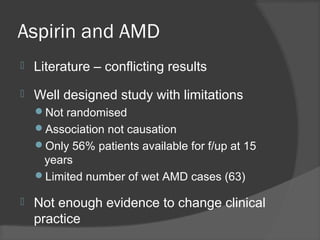
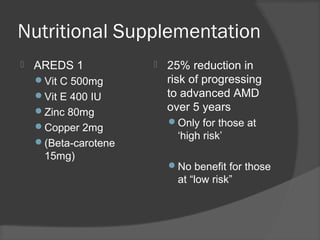
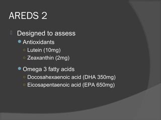
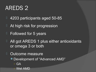
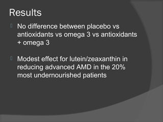
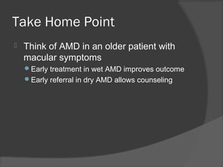
Raj Chalasani FRANZCO
Cataract Surgeon and Retinal Specialist
Hills Eye Surgery Pennant Hills
Staff Specialist Westmead Hospital
Macular Degeneration
 Disease of:
 Retinal pigment
epithelium
 Bruch’s membrane
 Choroid
 These structures
support photoreceptors
 Accumulation of waste
products
 Drusen
 Common starting point
for different types of
AMD
Dry AMD
 80-90% cases
 Drusen
 Disruption of RPE
function
 Atrophy of
photoreceptors
 End stage is
geographic atrophy
Loss of central vision
Wet AMD
 Drusen
 Disruption of RPE function
 Disruption to Bruch’s membrane
 Increased VEGF expression
 New vessel formation from choroid into retina
Choroidal neovascularisation (CNV)
Leak
Bleeding
Scarring
Disruption of normal architecture and photoreceptors
Loss of vision
Wet AMD
AMD – Key Investigations
 Optical coherence tomography (OCT)
 Cross sectional image of the macula
AMD – Key Investigations
 Fluorescein
angiography
 Identify CNV
and obtain PBS
funding for
treatment
Presentation
 Age > 60
 Symptoms
Reduced central vision
Distortion
Central scotomata
Reduced contrast
sensitivity
Presentation
 Age > 60
 Symptoms
Reduced central vision
Distortion
Central scotomata
Reduced contrast
sensitivity
 Signs
 Drusen
 Haemorrhage
 Exudate
 Sub/intraretinal fluid
 Fibrosis
Treatment – Dry AMD
 Delay Progression
Stop smoking
Nutritional advice
○ Low risk
Balanced diet
○ High risk
Supplementation
- Vit C,E
- Zn, Cu
 Early detection
Amsler grid
 Support
Vision Australia
Visual aids
Access to
benefits/support
Treatment – Wet AMD
 Anti-VEGF agents
Lucentis®
Eylea®
 Regular intra-vitreal
injections
Maintains vision in
90%
Improves vision in 30-
40%
Case
 Mr LS
 89 yo male
 4 day history of blurred vision LE and
‘edges looking crooked’
 PMhx
HT
Hypercholesterolaemia
 POHx
Bilateral cataract surgery 10 years ago
Examination
 Vision
RE 6/9
LE 6/18
 Normal IOP
 Anterior segment unremarkable
Examination
 Amsler
“I read in the
newspaper that aspirin
can cause AMD”
“Should I stop taking
my aspirin?”
Aspirin and AMD
 JAMA Intern Med. 2013; 173(4): 258-264
 2389 participants BMES
 Followed prospectively for 15 years
 257 ‘regular’ aspirin users
 One or more per week in the past year
 Wet AMD in 63 patients
 OR (Age/Sex/Smoking/CVD/BMI/SBP adjusted) 2.46 (CI
1.24-4.83)
Aspirin and AMD
 Literature – conflicting results
 Well designed study with limitations
Not randomised
Association not causation
Only 56% patients available for f/up at 15
years
Limited number of wet AMD cases (63)
 Not enough evidence to change clinical
practice
“My friend is on
macuvision”
“Should I be taking
a supplement?”
Nutritional Supplementation
 AREDS 1
Vit C 500mg
Vit E 400 IU
Zinc 80mg
Copper 2mg
(Beta-carotene
15mg)
 25% reduction in
risk of progressing
to advanced AMD
over 5 years
Only for those at
‘high risk’
No benefit for those
at “low risk”
AREDS 2
 Designed to assess
Antioxidants
○ Lutein (10mg)
○ Zeaxanthin (2mg)
Omega 3 fatty acids
○ Docosahexaenoic acid (DHA 350mg)
○ Eicosapentaenoic acid (EPA 650mg)
AREDS 2
 4203 participants aged 50-85
 At high risk for progression
 Followed for 5 years
 All got AREDS 1 plus either antioxidants
or omega 3 or both
 Outcome measure
Development of “Advanced AMD”
○ GA
○ Wet AMD
Results
 No difference between placebo vs
antioxidants vs omega 3 vs antioxidants
+ omega 3
 Modest effect for lutein/zeaxanthin in
reducing advanced AMD in the 20%
most undernourished patients
Take Home Point
 Think of AMD in an older patient with
macular symptoms
Early treatment in wet AMD improves outcome
Early referral in dry AMD allows counseling
Macular Degeneration

Macular Degeneration

  • 1.
    Raj Chalasani FRANZCO CataractSurgeon and Retinal Specialist Hills Eye Surgery Pennant Hills Staff Specialist Westmead Hospital
  • 3.
    Macular Degeneration  Diseaseof:  Retinal pigment epithelium  Bruch’s membrane  Choroid  These structures support photoreceptors  Accumulation of waste products  Drusen  Common starting point for different types of AMD
  • 4.
    Dry AMD  80-90%cases  Drusen  Disruption of RPE function  Atrophy of photoreceptors  End stage is geographic atrophy Loss of central vision
  • 5.
    Wet AMD  Drusen Disruption of RPE function  Disruption to Bruch’s membrane  Increased VEGF expression  New vessel formation from choroid into retina Choroidal neovascularisation (CNV) Leak Bleeding Scarring Disruption of normal architecture and photoreceptors Loss of vision
  • 6.
  • 7.
    AMD – KeyInvestigations  Optical coherence tomography (OCT)  Cross sectional image of the macula
  • 8.
    AMD – KeyInvestigations  Fluorescein angiography  Identify CNV and obtain PBS funding for treatment
  • 9.
    Presentation  Age >60  Symptoms Reduced central vision Distortion Central scotomata Reduced contrast sensitivity
  • 10.
    Presentation  Age >60  Symptoms Reduced central vision Distortion Central scotomata Reduced contrast sensitivity  Signs  Drusen  Haemorrhage  Exudate  Sub/intraretinal fluid  Fibrosis
  • 11.
    Treatment – DryAMD  Delay Progression Stop smoking Nutritional advice ○ Low risk Balanced diet ○ High risk Supplementation - Vit C,E - Zn, Cu  Early detection Amsler grid  Support Vision Australia Visual aids Access to benefits/support
  • 12.
    Treatment – WetAMD  Anti-VEGF agents Lucentis® Eylea®  Regular intra-vitreal injections Maintains vision in 90% Improves vision in 30- 40%
  • 14.
    Case  Mr LS 89 yo male  4 day history of blurred vision LE and ‘edges looking crooked’  PMhx HT Hypercholesterolaemia  POHx Bilateral cataract surgery 10 years ago
  • 15.
    Examination  Vision RE 6/9 LE6/18  Normal IOP  Anterior segment unremarkable
  • 16.
  • 20.
    “I read inthe newspaper that aspirin can cause AMD” “Should I stop taking my aspirin?”
  • 21.
    Aspirin and AMD JAMA Intern Med. 2013; 173(4): 258-264  2389 participants BMES  Followed prospectively for 15 years  257 ‘regular’ aspirin users  One or more per week in the past year  Wet AMD in 63 patients  OR (Age/Sex/Smoking/CVD/BMI/SBP adjusted) 2.46 (CI 1.24-4.83)
  • 22.
    Aspirin and AMD Literature – conflicting results  Well designed study with limitations Not randomised Association not causation Only 56% patients available for f/up at 15 years Limited number of wet AMD cases (63)  Not enough evidence to change clinical practice
  • 23.
    “My friend ison macuvision” “Should I be taking a supplement?”
  • 24.
    Nutritional Supplementation  AREDS1 Vit C 500mg Vit E 400 IU Zinc 80mg Copper 2mg (Beta-carotene 15mg)  25% reduction in risk of progressing to advanced AMD over 5 years Only for those at ‘high risk’ No benefit for those at “low risk”
  • 25.
    AREDS 2  Designedto assess Antioxidants ○ Lutein (10mg) ○ Zeaxanthin (2mg) Omega 3 fatty acids ○ Docosahexaenoic acid (DHA 350mg) ○ Eicosapentaenoic acid (EPA 650mg)
  • 26.
    AREDS 2  4203participants aged 50-85  At high risk for progression  Followed for 5 years  All got AREDS 1 plus either antioxidants or omega 3 or both  Outcome measure Development of “Advanced AMD” ○ GA ○ Wet AMD
  • 27.
    Results  No differencebetween placebo vs antioxidants vs omega 3 vs antioxidants + omega 3  Modest effect for lutein/zeaxanthin in reducing advanced AMD in the 20% most undernourished patients
  • 28.
    Take Home Point Think of AMD in an older patient with macular symptoms Early treatment in wet AMD improves outcome Early referral in dry AMD allows counseling