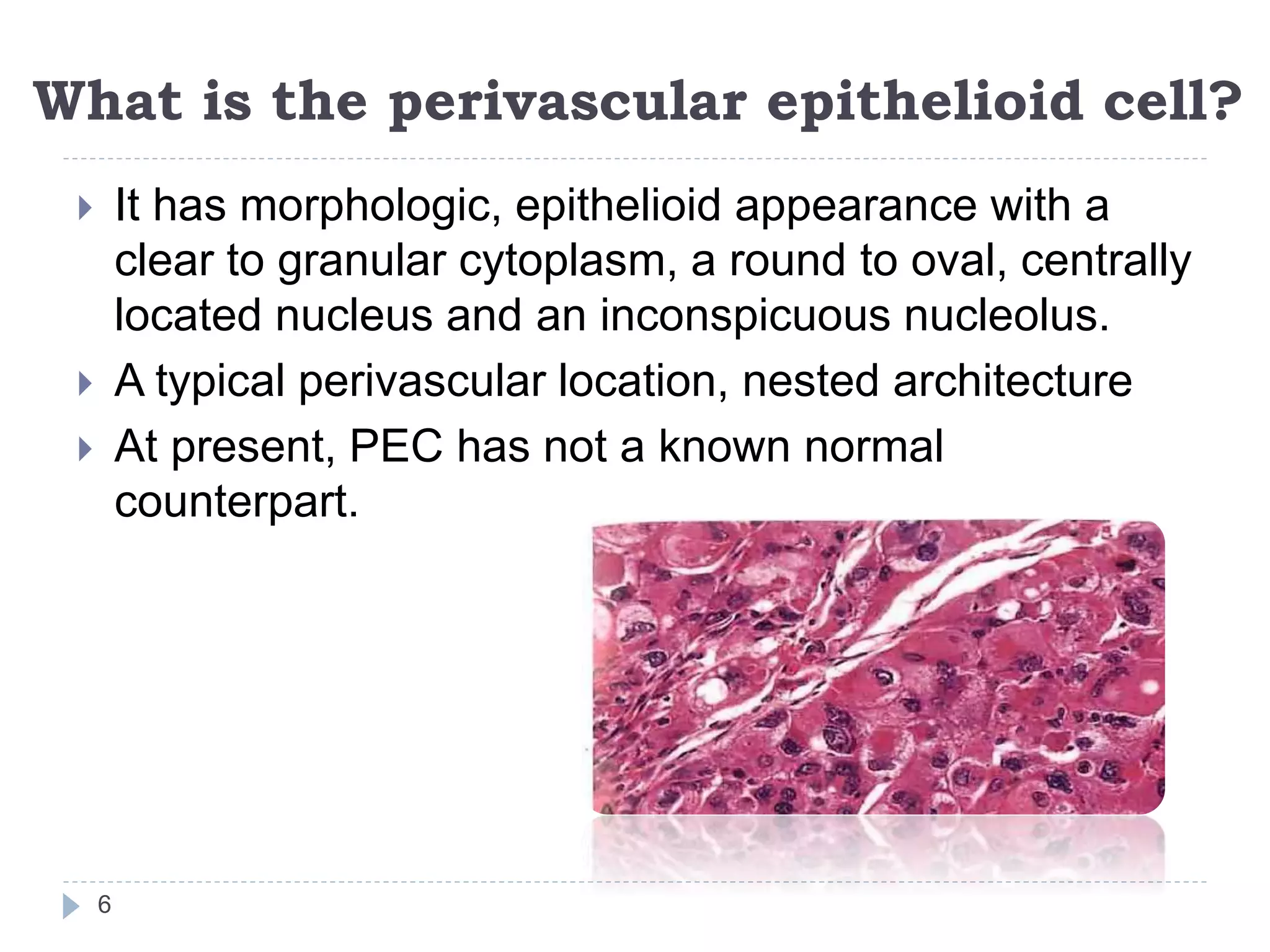
Presenter-Dr Shreya Prabhu
Moderator-Dr Jyothi A Raj
1
 Perivascular epithelioid cell tumors are
mesenchymal neoplasms defined by the
presence of histologically and
immunohistochemically distinctive perivascular
epithelioid cells
 Growing family of tumors
2
Tumor family includes:
 Angiomyolipoma (renal and extrarenal),
 Clear cell "sugar" tumor (lung and extrapulmonary)
 Lymphangioleiomyomatosis
 Tumors of falciform ligament/ ligamentum teres, skin,
uterus and other viscera and soft tissue
3
A number of spindled and epithelioid neoplasms that
have been referred to by a variety of names, including
 Primary extrapulmonary sugar tumor (PEST)
 Clear cell myomelanocytic tumor
 Abdominopelvic sarcoma of perivascular epithelioid
cells
4
ANATOMIC LOCATIONS:
 Retroperitoneum
 Lung
 Uterus,cervix
 Colon
 Pancreas
 Heart
 Adrenal gland
 Breast
 Biliary tract
 Urinary bladder
 Skull base
 Liver
 Skin
 Nasopharynx
 Upper airway
 Bone
 Soft tissues
 Eye
5
What is the perivascular epithelioid cell?
 It has morphologic, epithelioid appearance with a
clear to granular cytoplasm, a round to oval, centrally
located nucleus and an inconspicuous nucleolus.
 A typical perivascular location, nested architecture
 At present, PEC has not a known normal
counterpart.
6
7
 PEC can have a variable histologic appearance
 Epithelioid
 Elongated and spindle shaped
 Vacuolated (adipocyte-like)
 Typically
 Epithelioid cells- in an immediate perivascular
location
 Spindled cells- often away from vessels
 Typical feature
 Focal association with blood vessel wall
 Radial arrangement of tumor cells
 Subendothelial growth
PATHOGENESIS:
8
 Loss of TSC genes on chromosomes 9q34 (TSC1)
and 16p13 (TSC2)
 Loss of hamartin and tuberin expression, the
products of TSC1 and TSC2 genes
 Results in loss of control of cell signaling related to the
mtor pathway, with a variety of downstream effects
(genes which have a role in the regulation of the Rheb/mTOR/p70S6K
pathway)
 Progesterone receptor positivity has been described
in PEC with spindle morphology
 Suggesting a possible role of progesterone in this
morphologic modulation
 Cytogenetics- show trisomy 7 or 8
 It is thought that PEC can modulate its morphology
and immunophenotype
 Show epithelioid feature with a strong positivity for
HMB45
 Show muscular features with a spindle shape and a
stronger positivity for actin
 Show cytoplasmic vacoules acquiring the feature of an
adipocyte
9
10
CYTOLOGY
11
 Small and large cohesive clusters of bland
epithelioid to spindle cells with oval or elongated
nuclei, indistinct nucleoli, finely vacuolated
cytoplasm
 Delicate transgressing vessels
 Background naked nuclei
 Rare intranuclear pseudoinclusion
12
Sheets of bland-appearing spindle cells with
oval to elongated nuclei, indistinct nucleoli,
and finely vacuolated cytoplasm
Cohesive clusters of bland-appearing
epithelioid cells with oval to elongated nuclei,
indistinct nucleoli and scant to moderate
basophilic cytoplasm
13
IHC
PEComas express
myogenic and
melanocytic
markers:
• HMB45,
• HMSA-1,
• MelanA/Mart1,
• Microophtalmia
transcription
factor (Mitf),
• Actin
• Desmin
14
ULTRASTRUCTURAL ANALYSIS PEC contains
 Abundant
cytoplasmic
glycogen
 Microfilament
bundles with
electron-dense
condensation
 Numerous
mithochondria
 Membrane-
bound dense
granules
 Presence of
premelanosom
es
15
MALIGNANT POTENTIAL:
16
 Prevalence of aggressive biological behaviour (
including both local recurrence and metastasis) is
extremely variable, ranging from 5% to 66%
 Malignant PEComa can be a very aggressive
disease leading to multiple metastases and death as
expected with a high-grade sarcoma
 Malignant PEComas-NOS- MC originating in the
uterus and others arising in the jejunum, prostate,
pelvis, skull base, broad ligament, and somatic soft
tissue
 Metastatic sites- Liver, lymph nodes, lung and bone
17
18
ANGIOMYOLIPOMA
19
 Benign neoplasm composed of fat, smooth muscle
and blood vessel in varying proportion.
 Arising most commonly in one or both kidneys as a
solitary or multicentric mass
20
Extrarenal sites of AML include
 Liver
 Nasal cavity
 Oral cavity
 Colon
 Lung
 Skin
21
CLINICAL FEATURES:
 Females> males
 Age- 5th decade
 Abdominal or flank pain, hematuria, or chills and
fever asymptomatic
 Sudden, severe flank pain and shock are caused by
rupture of the tumor and massive perirenal or
retroperitoneal hemorrhage
22
 ~1/3rd of patients present with manifestations of
Tuberous sclerosis-
 Hyperpigmented spots
 Shagreen patches
 Periungual fibromas,
 Angiofibromas to renal cysts
 Cardiac rhabdomyoma
 Gliosis and calcification of the cerebral cortex with mental
deficiency
23
 PEComas of the kidney include :-
1. Classic AML
2. Microscopic AML (so-called microhamartoma)
3. Intraglomerular lesions
4. Cystic AML
5. Epithelioid AML
6. Oncocytoma-like AML
7. Lymphangiomyomatosis of the renal sinus
24
GROSSLY
 Yellow to gray mass,
 Size-few centimeters to 20 cm or more (average
9cm)
 Large lesions may become attached to the
diaphragm or liver
 Focal hemorrhage in about half of the cases
 Well delineated from the surrounding renal
parenchyma
25
26
Microscopically, composed of :
 Mature adipose tissue with some variations in cellular
size and nuclear appearance
 Convoluted thick-walled blood vessels with little or no
abnormal elastica and frequent hyalinization of the
media
 Irregularly arranged sheets and interlacing bundles of
smooth muscle (spindle or ovoid) often with a
prominent perivascular arrangement
27
28
29
31
Lipomatous component in angiomyolipoma can be dominant in areas
containing variable numbers of scattered or clustered smooth muscle–like cells
32
Vascular and perivascular features in angiomyolipoma.
(A–C) Tumor cells forming perivascular whorls.
(D) Vascular myointimal proliferation in an angiomyolipoma vessel
33
Smooth muscle-like spindle cell element in typical angiomyolipoma
34
Epithelioid angiomyolipoma shows a sharp demarcation between the typical
spindle cell and the epithelioid element.
(B–D) The epithelioid cells have abundant cytoplasm, and some cells are
multinucleated and may resemble ganglion cells or histiocytes
Oncocytoma-like
angiomyolipomas
• Rare tumours
consisting of a
homogeneous
population of
polygonal cells
with strongly
eosinophilic
cytoplasm
• In patients with
and without
tuberous
sclerosis
35
MICROSCOPIC
AML
• Small nodules
with some
features of
angiomyolipoma
• Often composed
predominantly of
epithelioid smooth
muscle cells
• The proportions of
spindle cells and
adipocytes
increase as the
lesions grow
larger
36
MALIGNANT POTENTIAL:
37
 Typical renal AML virtually always acts in a clinically
benign fashion, even those with atypical features
 No evidence that the presence of regional or
systemic lymph node involvement, perirenal satellite
tumors, or angiomyolipomatous growth in other
organs reflects malignant potential
38
 Presence of three or more of the following features is
predictive of malignant behavior:
(1) ≥70% atypical epithelioid cells (polygonal cells with
prominent nucleoli and nuclear size exceeding twice the
size of adjacent nuclei)
(2) ≥2 mitotic figures/10 hpf
(3) Atypical mitotic figures
(4) Necrosis
 Metastatic potential, especially those cases showing
necrosis and/or marked nuclear atypia
39
D/D:
 Liposarcoma
 Leiomyosarcoma-The myoid cells of AML tend to be
plumper and paler than those found in
leiomyosarcoma
 Renal cell carcinoma
40
Angiomyolipoma
with an area
composed of
atypical spindle-
shaped smooth
muscle cells
arranged in
fascicles. These
areas could be
confused for
leiomyosarcoma
41
Angiomyolipoma
composed of
uniform small
epithelioid smooth
muscle cells, feature
that could be
mistaken for renal
cell carcinoma
42
HMB-45
immunoreactivity
in smooth
muscle
43
TREATMENT
44
Surgical and excision
 Partial nephrectomy
 Total nephrectomy in large tumors
LUNG
PEComas of the lung include :-
 Lymphangioleiomyomatosis
 Clear-cell “sugar” tumor
45
LYMPHANGIOMYOMA AND
LYMPHANGIOMYOMATOSIS
46
 Lymphangiomyomatosis (LAM) is a rare disease
characterized by a proliferation of perivascular
epithelioid cells around lymphatics and lymph nodes
of the mediastinum, retroperitoneum, and the
pulmonary interstitium.
 Lymphangiomyoma- Localized lesions, mass like
involvement of lymph node
 Lymphangiomyomatosis- Extensive lesions
involving large segments of the lymphatic chain with
or without pulmonary involvement are designated.
47
CLINICAL FINDING
 >Females, ~40 years
 Progressive dyspnea (constant presence of chylous
pleural effusion or to pulmonary involvement)
 Pneumothorax, hemoptysis, chylous ascites, and
chyluria
48
Markedly dilated lymphatic vessels
suggesting proximal obstruction
49
Massive (chylous) effusion is present
Coarse reticular nodular infiltrate with
bulla
Numerous thin-walled cysts are noted
PATHOLOGIC FINDING
 Red to gray spongy masses that preferentially
replace the thoracic duct and mediastinal lymph
nodes.
 Form solitary or multiple masses that usually
measure <5 cm but can be >10 cm
 In dramatic cases- the entire lymphatic chain from
neck to inguinal region is transformed into multiple
confluent masses
 Chylous effusion/ chylous cysts is encountered
 In some instances the pleural surfaces are noted to
“weep” fluid, suggesting the presence of numerous
abnormal communications between the lymphatics
and the pleural surface.50
51
When the process affects the lungs as well, the organ has a honeycomb
appearance with formation of numerous blebs or bullae.
MICROSCOPY
 Microscopic nodules in the walls of the peripheral
airways involving the interstitial lymphatics
 Foci of lymphangiomyoma are seen perivascularly,
peribronchially, and interstitially, often associated
with emphysematous cyst formation
 The smooth muscle cells resemble those of
angiomyolipoma and have a “hollow” vacuolated,
clear cell appearance
52
53
Lymphangiomatosis involving large
lymphatic channels
Lymphangiomyo
ma
A)
Lymphangiomyoma
with distinctive
partially vacuolated
smooth mucle
surrounding
endothelium lined
spaces
B) Showing focal
positivity with HMB-
45
55
 The primary lesion-
 Haphazard proliferation of smooth muscle cells that
surround arterioles, venules, and lymphatics and which
diffusely thicken the alveolar septa
 Secondary changes-
 Bulla formation, as a result of air trapping by obstructed
bronchioles, and hemorrhage and hemosiderin
deposition as a result of venule destruction
56
D/D-
Metastatic
leiomyosarcoma
60
CLEAR CELL SUGAR TUMOR
61
 It was originally described in the lung
 Presents as a solitary pulmonary lesion
 Usually incidental radiologic findings manifesting as
spherical “coin” lesions in the lung periphery
62
63
 The pulmonary clear cell tumor forms a
circumscribed mass <1 cm to 3 cm (rarely >5 cm),
with a pushing border
 Tumor often infiltrates between small airway
epithelia, especially on its periphery, and infiltrative
margin is not a sign of malignancy
MICROSCOPY
 Perivascular tumor cells may have radial
arrangement around lumen; epithelioid cells (closest
to vessel) and spindle cells (remote from vessel) with
clear to granular eosinophilic cytoplasm; small,
central, round / oval nuclei with small nucleoli; often
multinucleated giant cells
 May have malignant features with marked atypia,
mitotic activity and necrosis
64
65
(A) Sharp demarcation to
lung tissue
(B) Tumor cells are epithelioid and
have eosinophilic to clear cytoplasm
66
(C) Tumor cells clustered around
blood vessels
(D) Focal calcification and stromal
sclerosis can be present
PEComa-NOS
67
Excluding AML, LAM and Pulmonary
CCST
 Group of rare, morphologically and
immunophenotypically similar tumors arising at a
variety of visceral and soft tissue sites, termed as
‘Perivascular epithelioid cell tumors–not
otherwise specified’.
 These tumors arising at a variety of visceral
(commonly gastrointestinal, gynecologic, and
genitourinary) and soft tissue (commonly
retroperitoneal, abdominopelvic, and cutaneous)
sites has been collectively termed non-AML, non-
LAM, non-CCST PEComas; or PEComas other than
AML, LAM, or CCST; or PEComas–not otherwise
specified (PEComas-NOS).68
CYTOGENETICS
 Few cases of pecoma have been karyotyped
 A specific cytogenetic aberration has not been
identified
 The presence of a t(3;10), losses on chromosomes
19, 16p, 17p,1p, and 18p and gains on
chromosomes X, 12q, 3q, 5, and 2q
 Show rearrangement of the TFE3 gene and
expression of TFE3 protein by
immunohistochemistry
69
CLINICAL FEATURES
 PEComas-NOS reported in almost every body site
 The uterus is the most prevalent reported site of
involvement
 Virtually arise in any age, peak in 4th decade
 F:M-7:1
70
PATHOLOGIC FEATURES
 Grossly, circumscribed, tan to gray with variable firm
and friable areas
 Tumor size- 4-6 cm
 Cut surface -white-tan to gray red, myxoid with focal
areas of hemorrhage and necrosis
 Biphasic tumors with epithelioid and spindle cell
components
71
72
The cut surface has a variegated appearance, including focal areas of necrosis
SOFT TISSUE PEComa
Pecoma of soft tissue composed
predominantly of nests of epithelioid
cells with cleared out cytoplasm
73
Higher magnification of nests of
epithelioid cells in pecoma of
soft tisssue
74
Cells with spindled morphology
in a soft tissue PEComa
Soft tissue PEComa with uniform
appearing nuclei
76
(A,B) A mesenteric example with spindled cells and partly clear cytoplasm. Focal
nuclear atypia is present.
(C,D) A pelvic PEComa that also involves a lymph node. The tumor is composed
of spindled cells with mildly eosinophilic to clear cytoplasm and vessel walls with
sclerosis
77
A malignant PEComa of the thigh contains significant mitotic activity
and bizarre multinucleated giant cells
Highly cellular smears in a malignant pecoma
78
HMB-45 immunoreactivity
79
Nuclear immunoreactivity for MiTF
80
D/D:
81
 Gastrointestinal stromal tumor (GIST)
 Clear cell carcinomas (e.g., cervix, vagina, renal cell
carcinoma) - express cytokeratins
 Leiomyosarcomas
82
83
84
REFERENCES:
85
1. Fletcher C.D.M., Unni K.K., Mertens F. (Eds.): World Health Organization
Classification of Tumours. Pathology and Genetics of Tumours of Soft Tissue and
Bone. IARC Press: Lyon 2002
2. Rosai, Juan, and Lauren Vedder Ackerman. Rosai And Ackerman's Surgical
Pathology.24,soft tissue, kidney, lung; 11th ed. Edinburgh: Mosby Elsevier, 2011.
3. Fletcher C, Carrie Y. Inwards, André M. Oliveira. Diagnostic histopathology of
tumors. Philadelphia, PA: Saunders/Elsevier; 2013; (p 1884-1890)
4. Kumar v, Abbas K A, Aster C J. Robbins and cotran pathological basis of disease.
Bones, joints, and soft tissue. 9th ed. New delhi : Elsevier ; 2014
5. Andrew H, Thomas: High yield pathology, bone and soft tissue pathology
6. Weiss, Sharon W. Enzinger And Weiss's Soft Tissue Tumors. St. Louis :Mosby,
2001. Print.
7. Sternberg SS, Antonioli DA, Carter D, Mills SE, Oberman HA . Diagnostic Surgical
Pathology. 3rd edition, Vol. 2.Philadelphia, PA, USA: Lippincott, Williams & Wilkins,
1999
8. Nicolas et al, Fine Needle Aspiration Cytology of Clear Cell ‘‘Sugar’’ Tumor
(PEComa) of the Lung: Report of a Case; Diagnostic Cytopathology, Vol 36, No 2
9. Martignoni et al, PEComas: the past, the present and the future; Virchows Arch
(2008) 452:119–132, Springer-Verlag 2007
86

Perivascular epithelioid cell neoplasm (PEComas)

  • 1.
  • 2.
     Perivascular epithelioidcell tumors are mesenchymal neoplasms defined by the presence of histologically and immunohistochemically distinctive perivascular epithelioid cells  Growing family of tumors 2
  • 3.
    Tumor family includes: Angiomyolipoma (renal and extrarenal),  Clear cell "sugar" tumor (lung and extrapulmonary)  Lymphangioleiomyomatosis  Tumors of falciform ligament/ ligamentum teres, skin, uterus and other viscera and soft tissue 3
  • 4.
    A number ofspindled and epithelioid neoplasms that have been referred to by a variety of names, including  Primary extrapulmonary sugar tumor (PEST)  Clear cell myomelanocytic tumor  Abdominopelvic sarcoma of perivascular epithelioid cells 4
  • 5.
    ANATOMIC LOCATIONS:  Retroperitoneum Lung  Uterus,cervix  Colon  Pancreas  Heart  Adrenal gland  Breast  Biliary tract  Urinary bladder  Skull base  Liver  Skin  Nasopharynx  Upper airway  Bone  Soft tissues  Eye 5
  • 6.
    What is theperivascular epithelioid cell?  It has morphologic, epithelioid appearance with a clear to granular cytoplasm, a round to oval, centrally located nucleus and an inconspicuous nucleolus.  A typical perivascular location, nested architecture  At present, PEC has not a known normal counterpart. 6
  • 7.
    7  PEC canhave a variable histologic appearance  Epithelioid  Elongated and spindle shaped  Vacuolated (adipocyte-like)  Typically  Epithelioid cells- in an immediate perivascular location  Spindled cells- often away from vessels  Typical feature  Focal association with blood vessel wall  Radial arrangement of tumor cells  Subendothelial growth
  • 8.
    PATHOGENESIS: 8  Loss ofTSC genes on chromosomes 9q34 (TSC1) and 16p13 (TSC2)  Loss of hamartin and tuberin expression, the products of TSC1 and TSC2 genes  Results in loss of control of cell signaling related to the mtor pathway, with a variety of downstream effects (genes which have a role in the regulation of the Rheb/mTOR/p70S6K pathway)  Progesterone receptor positivity has been described in PEC with spindle morphology  Suggesting a possible role of progesterone in this morphologic modulation  Cytogenetics- show trisomy 7 or 8
  • 9.
     It isthought that PEC can modulate its morphology and immunophenotype  Show epithelioid feature with a strong positivity for HMB45  Show muscular features with a spindle shape and a stronger positivity for actin  Show cytoplasmic vacoules acquiring the feature of an adipocyte 9
  • 10.
  • 11.
    CYTOLOGY 11  Small andlarge cohesive clusters of bland epithelioid to spindle cells with oval or elongated nuclei, indistinct nucleoli, finely vacuolated cytoplasm  Delicate transgressing vessels  Background naked nuclei  Rare intranuclear pseudoinclusion
  • 12.
    12 Sheets of bland-appearingspindle cells with oval to elongated nuclei, indistinct nucleoli, and finely vacuolated cytoplasm Cohesive clusters of bland-appearing epithelioid cells with oval to elongated nuclei, indistinct nucleoli and scant to moderate basophilic cytoplasm
  • 13.
  • 14.
    IHC PEComas express myogenic and melanocytic markers: •HMB45, • HMSA-1, • MelanA/Mart1, • Microophtalmia transcription factor (Mitf), • Actin • Desmin 14
  • 15.
    ULTRASTRUCTURAL ANALYSIS PECcontains  Abundant cytoplasmic glycogen  Microfilament bundles with electron-dense condensation  Numerous mithochondria  Membrane- bound dense granules  Presence of premelanosom es 15
  • 16.
    MALIGNANT POTENTIAL: 16  Prevalenceof aggressive biological behaviour ( including both local recurrence and metastasis) is extremely variable, ranging from 5% to 66%  Malignant PEComa can be a very aggressive disease leading to multiple metastases and death as expected with a high-grade sarcoma  Malignant PEComas-NOS- MC originating in the uterus and others arising in the jejunum, prostate, pelvis, skull base, broad ligament, and somatic soft tissue  Metastatic sites- Liver, lymph nodes, lung and bone
  • 17.
  • 18.
  • 19.
  • 20.
     Benign neoplasmcomposed of fat, smooth muscle and blood vessel in varying proportion.  Arising most commonly in one or both kidneys as a solitary or multicentric mass 20
  • 21.
    Extrarenal sites ofAML include  Liver  Nasal cavity  Oral cavity  Colon  Lung  Skin 21
  • 22.
    CLINICAL FEATURES:  Females>males  Age- 5th decade  Abdominal or flank pain, hematuria, or chills and fever asymptomatic  Sudden, severe flank pain and shock are caused by rupture of the tumor and massive perirenal or retroperitoneal hemorrhage 22
  • 23.
     ~1/3rd ofpatients present with manifestations of Tuberous sclerosis-  Hyperpigmented spots  Shagreen patches  Periungual fibromas,  Angiofibromas to renal cysts  Cardiac rhabdomyoma  Gliosis and calcification of the cerebral cortex with mental deficiency 23
  • 24.
     PEComas ofthe kidney include :- 1. Classic AML 2. Microscopic AML (so-called microhamartoma) 3. Intraglomerular lesions 4. Cystic AML 5. Epithelioid AML 6. Oncocytoma-like AML 7. Lymphangiomyomatosis of the renal sinus 24
  • 25.
    GROSSLY  Yellow togray mass,  Size-few centimeters to 20 cm or more (average 9cm)  Large lesions may become attached to the diaphragm or liver  Focal hemorrhage in about half of the cases  Well delineated from the surrounding renal parenchyma 25
  • 26.
  • 27.
    Microscopically, composed of:  Mature adipose tissue with some variations in cellular size and nuclear appearance  Convoluted thick-walled blood vessels with little or no abnormal elastica and frequent hyalinization of the media  Irregularly arranged sheets and interlacing bundles of smooth muscle (spindle or ovoid) often with a prominent perivascular arrangement 27
  • 28.
  • 29.
  • 30.
    31 Lipomatous component inangiomyolipoma can be dominant in areas containing variable numbers of scattered or clustered smooth muscle–like cells
  • 31.
    32 Vascular and perivascularfeatures in angiomyolipoma. (A–C) Tumor cells forming perivascular whorls. (D) Vascular myointimal proliferation in an angiomyolipoma vessel
  • 32.
    33 Smooth muscle-like spindlecell element in typical angiomyolipoma
  • 33.
    34 Epithelioid angiomyolipoma showsa sharp demarcation between the typical spindle cell and the epithelioid element. (B–D) The epithelioid cells have abundant cytoplasm, and some cells are multinucleated and may resemble ganglion cells or histiocytes
  • 34.
    Oncocytoma-like angiomyolipomas • Rare tumours consistingof a homogeneous population of polygonal cells with strongly eosinophilic cytoplasm • In patients with and without tuberous sclerosis 35
  • 35.
    MICROSCOPIC AML • Small nodules withsome features of angiomyolipoma • Often composed predominantly of epithelioid smooth muscle cells • The proportions of spindle cells and adipocytes increase as the lesions grow larger 36
  • 36.
    MALIGNANT POTENTIAL: 37  Typicalrenal AML virtually always acts in a clinically benign fashion, even those with atypical features  No evidence that the presence of regional or systemic lymph node involvement, perirenal satellite tumors, or angiomyolipomatous growth in other organs reflects malignant potential
  • 37.
    38  Presence ofthree or more of the following features is predictive of malignant behavior: (1) ≥70% atypical epithelioid cells (polygonal cells with prominent nucleoli and nuclear size exceeding twice the size of adjacent nuclei) (2) ≥2 mitotic figures/10 hpf (3) Atypical mitotic figures (4) Necrosis  Metastatic potential, especially those cases showing necrosis and/or marked nuclear atypia
  • 38.
  • 39.
    D/D:  Liposarcoma  Leiomyosarcoma-Themyoid cells of AML tend to be plumper and paler than those found in leiomyosarcoma  Renal cell carcinoma 40
  • 40.
    Angiomyolipoma with an area composedof atypical spindle- shaped smooth muscle cells arranged in fascicles. These areas could be confused for leiomyosarcoma 41
  • 41.
    Angiomyolipoma composed of uniform small epithelioidsmooth muscle cells, feature that could be mistaken for renal cell carcinoma 42
  • 42.
  • 43.
    TREATMENT 44 Surgical and excision Partial nephrectomy  Total nephrectomy in large tumors
  • 44.
    LUNG PEComas of thelung include :-  Lymphangioleiomyomatosis  Clear-cell “sugar” tumor 45
  • 45.
  • 46.
     Lymphangiomyomatosis (LAM)is a rare disease characterized by a proliferation of perivascular epithelioid cells around lymphatics and lymph nodes of the mediastinum, retroperitoneum, and the pulmonary interstitium.  Lymphangiomyoma- Localized lesions, mass like involvement of lymph node  Lymphangiomyomatosis- Extensive lesions involving large segments of the lymphatic chain with or without pulmonary involvement are designated. 47
  • 47.
    CLINICAL FINDING  >Females,~40 years  Progressive dyspnea (constant presence of chylous pleural effusion or to pulmonary involvement)  Pneumothorax, hemoptysis, chylous ascites, and chyluria 48
  • 48.
    Markedly dilated lymphaticvessels suggesting proximal obstruction 49 Massive (chylous) effusion is present Coarse reticular nodular infiltrate with bulla Numerous thin-walled cysts are noted
  • 49.
    PATHOLOGIC FINDING  Redto gray spongy masses that preferentially replace the thoracic duct and mediastinal lymph nodes.  Form solitary or multiple masses that usually measure <5 cm but can be >10 cm  In dramatic cases- the entire lymphatic chain from neck to inguinal region is transformed into multiple confluent masses  Chylous effusion/ chylous cysts is encountered  In some instances the pleural surfaces are noted to “weep” fluid, suggesting the presence of numerous abnormal communications between the lymphatics and the pleural surface.50
  • 50.
    51 When the processaffects the lungs as well, the organ has a honeycomb appearance with formation of numerous blebs or bullae.
  • 51.
    MICROSCOPY  Microscopic nodulesin the walls of the peripheral airways involving the interstitial lymphatics  Foci of lymphangiomyoma are seen perivascularly, peribronchially, and interstitially, often associated with emphysematous cyst formation  The smooth muscle cells resemble those of angiomyolipoma and have a “hollow” vacuolated, clear cell appearance 52
  • 52.
  • 53.
    A) Lymphangiomyoma with distinctive partially vacuolated smoothmucle surrounding endothelium lined spaces B) Showing focal positivity with HMB- 45 55
  • 54.
     The primarylesion-  Haphazard proliferation of smooth muscle cells that surround arterioles, venules, and lymphatics and which diffusely thicken the alveolar septa  Secondary changes-  Bulla formation, as a result of air trapping by obstructed bronchioles, and hemorrhage and hemosiderin deposition as a result of venule destruction 56
  • 55.
  • 56.
  • 57.
     It wasoriginally described in the lung  Presents as a solitary pulmonary lesion  Usually incidental radiologic findings manifesting as spherical “coin” lesions in the lung periphery 62
  • 58.
    63  The pulmonaryclear cell tumor forms a circumscribed mass <1 cm to 3 cm (rarely >5 cm), with a pushing border  Tumor often infiltrates between small airway epithelia, especially on its periphery, and infiltrative margin is not a sign of malignancy
  • 59.
    MICROSCOPY  Perivascular tumorcells may have radial arrangement around lumen; epithelioid cells (closest to vessel) and spindle cells (remote from vessel) with clear to granular eosinophilic cytoplasm; small, central, round / oval nuclei with small nucleoli; often multinucleated giant cells  May have malignant features with marked atypia, mitotic activity and necrosis 64
  • 60.
    65 (A) Sharp demarcationto lung tissue (B) Tumor cells are epithelioid and have eosinophilic to clear cytoplasm
  • 61.
    66 (C) Tumor cellsclustered around blood vessels (D) Focal calcification and stromal sclerosis can be present
  • 62.
  • 63.
    Excluding AML, LAMand Pulmonary CCST  Group of rare, morphologically and immunophenotypically similar tumors arising at a variety of visceral and soft tissue sites, termed as ‘Perivascular epithelioid cell tumors–not otherwise specified’.  These tumors arising at a variety of visceral (commonly gastrointestinal, gynecologic, and genitourinary) and soft tissue (commonly retroperitoneal, abdominopelvic, and cutaneous) sites has been collectively termed non-AML, non- LAM, non-CCST PEComas; or PEComas other than AML, LAM, or CCST; or PEComas–not otherwise specified (PEComas-NOS).68
  • 64.
    CYTOGENETICS  Few casesof pecoma have been karyotyped  A specific cytogenetic aberration has not been identified  The presence of a t(3;10), losses on chromosomes 19, 16p, 17p,1p, and 18p and gains on chromosomes X, 12q, 3q, 5, and 2q  Show rearrangement of the TFE3 gene and expression of TFE3 protein by immunohistochemistry 69
  • 65.
    CLINICAL FEATURES  PEComas-NOSreported in almost every body site  The uterus is the most prevalent reported site of involvement  Virtually arise in any age, peak in 4th decade  F:M-7:1 70
  • 66.
    PATHOLOGIC FEATURES  Grossly,circumscribed, tan to gray with variable firm and friable areas  Tumor size- 4-6 cm  Cut surface -white-tan to gray red, myxoid with focal areas of hemorrhage and necrosis  Biphasic tumors with epithelioid and spindle cell components 71
  • 67.
    72 The cut surfacehas a variegated appearance, including focal areas of necrosis SOFT TISSUE PEComa
  • 68.
    Pecoma of softtissue composed predominantly of nests of epithelioid cells with cleared out cytoplasm 73 Higher magnification of nests of epithelioid cells in pecoma of soft tisssue
  • 69.
    74 Cells with spindledmorphology in a soft tissue PEComa Soft tissue PEComa with uniform appearing nuclei
  • 70.
    76 (A,B) A mesentericexample with spindled cells and partly clear cytoplasm. Focal nuclear atypia is present. (C,D) A pelvic PEComa that also involves a lymph node. The tumor is composed of spindled cells with mildly eosinophilic to clear cytoplasm and vessel walls with sclerosis
  • 71.
    77 A malignant PEComaof the thigh contains significant mitotic activity and bizarre multinucleated giant cells
  • 72.
    Highly cellular smearsin a malignant pecoma 78
  • 73.
  • 74.
  • 75.
    D/D: 81  Gastrointestinal stromaltumor (GIST)  Clear cell carcinomas (e.g., cervix, vagina, renal cell carcinoma) - express cytokeratins  Leiomyosarcomas
  • 76.
  • 77.
  • 78.
  • 79.
    REFERENCES: 85 1. Fletcher C.D.M.,Unni K.K., Mertens F. (Eds.): World Health Organization Classification of Tumours. Pathology and Genetics of Tumours of Soft Tissue and Bone. IARC Press: Lyon 2002 2. Rosai, Juan, and Lauren Vedder Ackerman. Rosai And Ackerman's Surgical Pathology.24,soft tissue, kidney, lung; 11th ed. Edinburgh: Mosby Elsevier, 2011. 3. Fletcher C, Carrie Y. Inwards, André M. Oliveira. Diagnostic histopathology of tumors. Philadelphia, PA: Saunders/Elsevier; 2013; (p 1884-1890) 4. Kumar v, Abbas K A, Aster C J. Robbins and cotran pathological basis of disease. Bones, joints, and soft tissue. 9th ed. New delhi : Elsevier ; 2014 5. Andrew H, Thomas: High yield pathology, bone and soft tissue pathology 6. Weiss, Sharon W. Enzinger And Weiss's Soft Tissue Tumors. St. Louis :Mosby, 2001. Print. 7. Sternberg SS, Antonioli DA, Carter D, Mills SE, Oberman HA . Diagnostic Surgical Pathology. 3rd edition, Vol. 2.Philadelphia, PA, USA: Lippincott, Williams & Wilkins, 1999 8. Nicolas et al, Fine Needle Aspiration Cytology of Clear Cell ‘‘Sugar’’ Tumor (PEComa) of the Lung: Report of a Case; Diagnostic Cytopathology, Vol 36, No 2 9. Martignoni et al, PEComas: the past, the present and the future; Virchows Arch (2008) 452:119–132, Springer-Verlag 2007
  • 80.

Editor's Notes

  • #14 A) Large, irregular, cohesive clusters of columnar and spindle-shaped cells in background of single naked nuclei (Pap) (B) several large clusters with open cribriform-like spaces (Pap) (C) spindle-shaped and polygonal cells with round to oval nuclei and vacuolated ill-defined cytoplasm (Pap) (D) cell block shows neoplastic cells arranged in a nested and insular pattern surrounding sinusoid-like vessels; inset shows sinusoid-like vessel amidst tumor
  • #16 The spindle cells in PEComas are characterized by prominent smooth muscle– specific filaments, whereas the epithelioid component does not usually contain high numbers of such filaments
  • #30 About 15% are composed of cords of cells in dense collagenous stroma (Sclerosing stroma)
  • #52 The emphysematous change has been attributed to activation of the matrix metalloproteinase system in this disease
  • #74 arranged into nests, fascicles, and occasionally sheets, often with a radial arrangement around blood vessels
  • #76 one may encounter giant cells with a central eosinophilic zone surrounded by a peripheral clear zone, reminiscent of the spider cells seen in adult rhabdomyoma