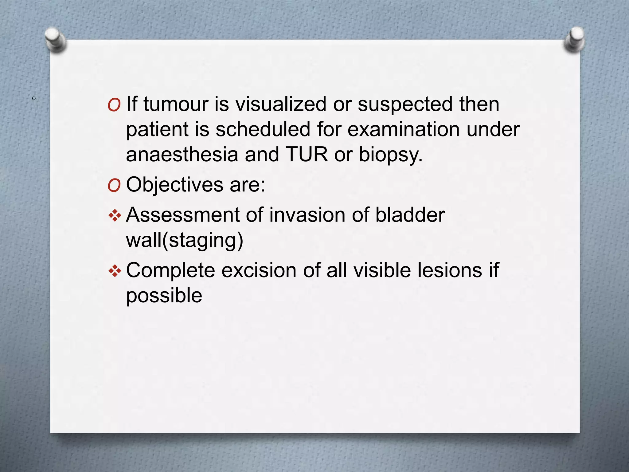
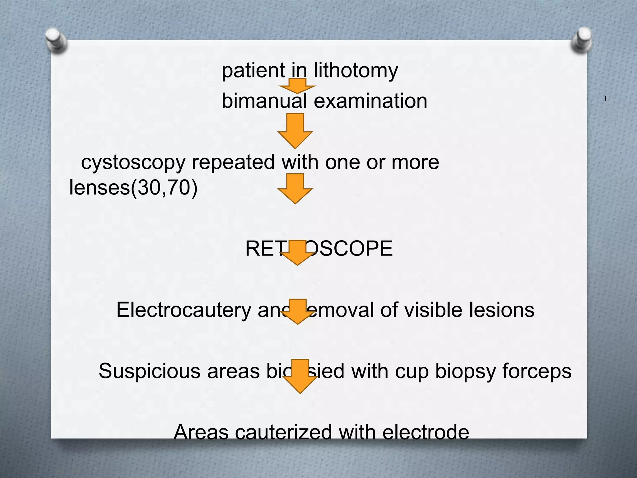
The urinary bladder is a hollow muscular organ that temporarily stores urine before expulsion. It has an apex, fundus, body, neck, and urethra. Blood supply comes from branches of the internal iliac arteries. Lymphatic drainage occurs to internal and external iliac lymph nodes. The bladder wall has layers of transitional epithelium, lamina propria, and muscularis propria. Bladder cancer is the second most common genitourinary cancer, with smoking being the main risk factor. Symptoms include hematuria. Diagnosis involves cystoscopy, biopsy, and imaging to stage the tumor. Treatment depends on tumor stage and grade but may include transurethral resection and intravesical
This document discusses bladder tumors, including:
1. Urothelial (transitional cell) carcinomas are the most common type and can be papillary or flat. Low grade tumors have mild dysplasia while high grade tumors are poorly differentiated.
2. Other tumor types include squamous cell carcinoma, adenocarcinoma, and sarcomas. Immunohistochemistry helps distinguish tumor types.
3. Tumors are staged based on depth of invasion from non-invasive papillary tumors to muscle invasive or locally advanced cancers. Treatment depends on grade and stage.
This document provides an overview of approaches to testicular tumors. It discusses updates to classifications including changing ITGCN to GCNIS. A new classification system is presented that divides tumors into GCT derived from GCNIS, GCT unrelated to GCNIS, sex cord stromal tumors, and other rare tumors. Factors like age, medical history, tumor site, and gross appearance can provide clues before histological examination. Histological patterns including cells with pale cytoplasm, glandular/tubular patterns, microcystic patterns, and oxyphilic cells can indicate tumor types.
This document discusses bladder cancer and provides information on epidemiology, etiology, pathophysiology, classification, clinical features, and histopathology of benign and malignant bladder tumors. It is from the Department of Urology at Govt Royapettah Hospital and Kilpauk Medical College in Chennai, India. The document lists the moderators and their academic titles. It then covers topics such as the higher prevalence of bladder cancer in men compared to women, risk factors including smoking and occupational exposures, genetic factors, pathogenesis, WHO and other classification systems, clinical features of non-muscle invasive and muscle invasive bladder cancers, histopathology of benign lesions and different grades of bladder tumors.
This document discusses testicular tumors, including their etiology, classification, clinical presentation, diagnosis, staging, and treatment. Some key points:
- Testicular cancer is the most common cancer in men ages 15-35 and has a high cure rate with early detection and treatment.
- Risk factors include cryptorchidism, prior testicular cancer, infertility, and genetic factors. Carcinoma in situ is a precursor to most germ cell tumors.
- Tumors are classified as seminomas or non-seminomas. Staging involves tumor markers, imaging, and pathology to determine extent of disease.
- Treatment involves radical orchidectomy followed by radiotherapy for seminomas or chemotherapy for
This document discusses the World Health Organization classification of ovarian tumors. It outlines the major categories of epithelial tumors, the most common type of ovarian tumors, comprising 60% of cases. Within epithelial tumors, it describes the histological features and classifications of serous, mucinous, endometrioid, clear cell, Brenner, and seromucinous tumors. It notes that serous and mucinous cystadenomas are the most prevalent epithelial tumors, together accounting for 30% of ovarian cancers. Details are provided on the benign, borderline, and malignant subtypes for each tumor type based on histological appearance.
This document summarizes applications of artificial intelligence in pathology. It discusses machine learning and deep learning techniques used for tasks like cancer detection and classification from histopathology images. Examples are given of using AI for breast, lung, prostate, brain, ovarian and cervical cancer analysis from whole slide images and digital pathology. Applications in immunohistochemistry, genetic mutation prediction and tumor detection for molecular analysis are also summarized.
Perivascular epithelioid cell lesions (PEComas) are a collection of rare mesenchymal tumors composed of distinctive perivascular epithelioid cells. PEComas can occur in many anatomic locations and have been given various names. The defining cell, the perivascular epithelioid cell, has an epithelioid appearance with clear to granular cytoplasm and expresses myogenic and melanocytic markers. PEComas are related to genetic alterations in the TSC1 and TSC2 genes and have a strong female predominance. The document goes on to describe specific PEComa subtypes that can occur in the kidney, lung, liver, pancreas, uterus, bladder,
This document discusses testicular cancer, including its epidemiology, classification, investigations, and treatment. It notes that testicular cancer is most common in young men aged 15-35 years old and has a high cure rate. The main types are seminomas and non-seminomas. Staging involves tumor markers, ultrasound, CT scan, and radical orchiectomy. For high-risk patients, retroperitoneal lymph node dissection may be used. Modern treatment is multimodal and based on the patterns of metastasis, with chemotherapy and nerve-sparing surgery improving outcomes.
The document summarizes the Bethesda system for reporting cervical cytology. It discusses the history and updates of the Bethesda system in 1988, 1991, 2001, and 2014. The 2014 Bethesda system provides a standardized terminology for reporting Pap test results. It includes sections for specimen type, adequacy, interpretation/result, and adjunctive testing. Under interpretation/result, it outlines categories for negative, epithelial cell abnormalities, and malignant neoplasms. It also describes normal cervical cell types and non-neoplastic findings that can be reported.
The document discusses several pediatric neoplasms that appear as small round blue cell tumors due to their primitive histological features. These include neuroblastoma, Wilms tumor, rhabdomyosarcoma, Ewing's sarcoma, medulloblastoma, retinoblastoma, and lymphoma. For each tumor, the document outlines characteristics such as common age of diagnosis, clinical features, histopathological appearance under the microscope, immunohistochemistry profiles, genetics where relevant, and important prognostic factors. Differential diagnosis of these small round blue cell tumors in children is provided for accurate diagnosis and treatment.
This document discusses the cytopathology of metastatic neoplasms. It covers metastatic neoplasms in lymph nodes and lungs, including patterns of spread and cytological features that can indicate the primary site. Diagnosis of primary and secondary lymphomatous effusions is also addressed. Key points include that lymph nodes and lungs are common sites of metastasis, cytology can provide clues to the primary site through cell morphology and patterns, and immunohistochemistry is often needed to confirm diagnosis and primary site.
The document discusses the anatomy, physiology, and development of the breast from embryological development through adulthood, as well as several benign clinical conditions that can present in the breast including mastalgia, nipple discharge, breast abscesses, cysts, fibroadenomas, and gynecomastia. It provides details on the histology, presentation, workup, and treatment for each benign condition.
This document summarizes common testicular tumors, including their classification, histology, and characteristics. Germ cell tumors are divided into seminomas and non-seminomatous germ cell tumors. The main types described are seminoma, embryonal carcinoma, teratoma, choriocarcinoma, and yolk sac tumor. Sex cord-stromal tumors include Leydig cell tumor and Sertoli cell tumor. Details are provided on histological patterns, patient demographics, clinical presentations, and pathogenesis for each tumor type.
Carcinoma of unknown primary IHC ApproachDeeksha Sikri
1. Immunohistochemistry (IHC) remains the gold standard for diagnosing carcinomas of unknown primary site (CUPs). IHC uses antibodies to keratins and organ-specific markers to determine the cell line of differentiation and potentially identify the primary site.
2. Key IHC markers that can help identify primary sites include TTF-1 for lung cancer, CK7/CK20 patterns for gastrointestinal cancers, PAX-8 for ovarian cancer, PSA for prostate cancer, and GCDFP-15 for breast cancer. ER and WT-1 can also help distinguish cancers arising in the female reproductive system.
3. While individual markers may not be entirely specific, using antibody panels and
Myofibroblasts are specialized cells that play an important role in wound healing, fibrosis, and tissue remodeling. They have characteristics of both fibroblasts and smooth muscle cells. Myofibroblasts secrete growth factors, cytokines, and extracellular matrix proteins that promote wound contraction, tissue repair, and the formation of new tissue. They are formed through the differentiation of various cell types and help regulate inflammation and the proliferation of epithelial, vascular and other cells through paracrine signaling. Myofibroblasts are vital for normal tissue development and wound healing but excessive accumulation can lead to fibrosis.
This document discusses several pathologies that can affect the male genital tract, including the testis and epididymis. It describes congenital anomalies like cryptorchidism and complications like infertility. Specific conditions covered include torsion of the testis, atrophy, hydrocele, varicocele, tuberculosis, and granulomatous inflammation. For each condition, the document provides details on etiology, histological findings, clinical presentation, diagnosis and management.
Cervical cancer arises from the transformation zone of the cervix. Risk factors include HPV infection, early age of first intercourse, multiple sexual partners, and smoking. It typically spreads locally first through direct extension, then can metastasize via lymph nodes or hematogenously to distant sites like lungs and liver. Screening via Pap smears can detect pre-cancerous changes and has reduced cervical cancer rates in developed nations by 75% over 50 years. Vaccination against HPV also prevents infection and future cancer development. Treatment and prognosis depends on the stage, with early localized disease having the best outcomes.
The document discusses the role of immunohistochemistry (IHC) in head and neck pathology. IHC uses antibodies to identify antigens in tissues and can help diagnose cancers and determine tumor type. It plays an important role in pathology subspecialties like oncology, neuropathology, and hematopathology. The document provides details on the principle of IHC, various tissue and tumor markers used in IHC, and how IHC can help diagnose lesions in the head and neck region like oral cancers, salivary gland tumors, melanomas, sarcomas, and lymphomas.
This document discusses squamous cell carcinoma of the anal canal. It describes the anatomy of the anal canal and defines the anal canal, transitional zone, and anal margin. Risk factors for anal cancer include HPV infection and HIV/AIDS. Combined modality treatment including chemotherapy with mitomycin and 5-fluorouracil alongside radiation therapy is an effective standard of care based on clinical trials showing improved local control and survival compared to radiation alone.
This is a powerpoint presentation of Immunohistochemistry of lesions of prostate. This presentation will be helpful for postgraduate pathology students and practitioners alike. We are also on youtube. Please visit our channel at https://www.youtube.com/channel/UCwjkzK-YnJ-ra4HMOqq3Fkw
This document discusses serum tumor markers, which are molecules that can be detected in blood, body fluids, or tissue that are produced by or in response to cancer cells. It describes several commonly used tumor markers, including their history, clinical uses, and interpretations. The key tumor markers discussed are alpha-fetoprotein (AFP) for hepatocellular carcinoma and germ cell tumors, carcinoembryonic antigen (CEA) for colorectal cancer, CA-125 for ovarian cancer, human chorionic gonadotropin (hCG) for gestational trophoblastic tumors, prostate-specific antigen (PSA) for prostate cancer, CA 19-9 for pancreatic cancer, and CA 15-3
The document discusses cytology of various bone lesions. It covers classification of bone tumors and describes cytological features of inflammatory conditions like osteomyelitis. It also discusses osteoid forming lesions such as fracture callus and osteoblastoma. Cartilage forming tumors described include chondroma, chondromyxoid fibroma and osteochondroma. Giant cell containing lesions and cystic bone lesions are also mentioned. The document provides cytological details of various bone tumors like osteosarcoma, chondrosarcoma and chondroblastoma through multiple case studies. It highlights differential diagnoses and ancillary techniques used in evaluation of bone lesions.
Soft tissue pathology is rapidly changing
Novel molecular findings
Tumors previously known under one rubric are reclassified with relative frequency
Lesions that for decades were thought to be reactive now are discovered to possess gene rearrangements
Features of previously unknown or incompletely described tumors are coalesced and synthesized into new entities
Relative rarity of soft tissue tumors only adds to the challenge
of keeping abreast of all of these advances
Male genital system and lower urinary tract and Sexually Transmitted DiseasesChito Disomangcop
This document summarizes the male genital system and lower urinary tract. It describes common conditions such as hypospadias and epispadias, balanitis and balanoposthitis, phimosis, and carcinoma in situ of the penis. It also discusses cryptorchidism, epididymitis, orchitis, testicular torsion, and the most common testicular neoplasms including seminoma, embryonal carcinoma, yolk sac tumor, choriocarcinoma, and teratoma. Finally, it provides an overview of the prostate and conditions such as prostatitis.
Digital Pathology, FDA Approval and Precision MedicineJoel Saltz
Digital pathology platforms combined with machine learning can improve the consistency and quality of clinical decision making by precisely scoring known criteria from pathology images and predicting treatment outcomes and cancer types. Researchers are developing tools to extract features from pathology images, link these features to molecular data and clinical outcomes, and use these integrated datasets to gain new insights into cancer and select the best interventions. The SEER Virtual Tissue Repository aims to enable population-level cancer research by creating a linked collection of de-identified clinical data and whole slide images from pathology samples that can be analyzed using computational methods.
This document provides information on carcinoma of the stomach, including:
- Risk factors include H. pylori infection, diet, genetics, smoking.
- Types include intestinal and diffuse. Staging uses TNM and other classifications.
- Common symptoms are weight loss, abdominal pain, vomiting. Investigations include endoscopy and biopsy.
- Treatment depends on stage but commonly includes surgery such as gastrectomy along with lymph node dissection. Endoscopic resection may be used for early stages. Adjuvant therapy is sometimes used for later stages.
This document provides an overview of colon cancer, including its anatomy, epidemiology, risk factors, clinical features, screening, pathology, staging, diagnostic workup, treatment options, and management. Some key points include that colon cancer is the third leading cause of cancer deaths, adenocarcinoma makes up 98% of cases, and risk factors include age, diet low in fiber/high in fat, personal or family history, genetic factors, inflammatory bowel disease, and radiation exposure. Treatment involves surgery as the main treatment, with chemotherapy as an adjuvant for late stage or high-risk cases.
This document provides an overview of bladder cancer presented by Dr. Vikas Kumar. Some key points:
- Bladder cancer is the 9th most common cancer worldwide and the 13th most common cause of death. Risk factors include smoking, occupational exposures, infections, and genetic factors.
- At initial presentation, 80% of bladder cancers are non-muscle invasive. Staging involves evaluating the extent of primary tumor invasion and spread to lymph nodes and distant organs.
- Diagnosis involves cystoscopy, urine cytology, and imaging tests. Random bladder biopsies are also recommended to detect cancers that cannot be seen.
- For non-muscle invasive cancers, the main treatment is transure
Renal pathology lecture 4 Tumors of kidney and urinary tract. Sufia Husain 2020Sufia Husain
This document provides an overview of tumors of the kidney and urinary tract. It begins by outlining the objectives and key topics to be covered, which include benign kidney tumors, renal cell carcinoma, Wilms tumor, and transitional cell and squamous carcinomas of the bladder. The document then covers these topics in detail over several sections, describing the histology, risk factors, clinical features, and characteristics of each tumor type. The major tumor types discussed are renal oncocytoma, angiomyolipoma, renal cell carcinoma (clear cell and papillary subtypes), Wilms tumor, and transitional cell neoplasms of the bladder.
1. The document discusses various types of pancreatic cysts including pseudocysts, congenital cysts, and neoplastic cystic tumors.
2. It outlines benign cystic neoplasms like serous cystadenomas and malignant mucinous cystic neoplasms.
3. Pancreatic ductal adenocarcinoma is discussed as the fourth leading cause of cancer death which often has KRAS and p16 mutations and a desmoplastic response.
The document discusses the anatomy and histology of the bladder and various benign and malignant tumors that can occur. It provides details on:
1. The anatomy of the bladder including its shape, surfaces and location in the pelvis.
2. The normal histology of the bladder wall and urothelial lining.
3. Various benign tumors of the bladder including epithelial metaplasia, leukoplakia, inverted papilloma, nephrogenic adenoma, leiomyoma, cystitis cystica and glandularis.
4. Risk factors, pathology, clinical features and staging of urothelial cancer of the bladder, which represents the majority of bladder cancers.
Testicular tumors-Cassification, Biomarkers and Staging by Dr RajeshRajesh Sinwer
This document discusses testicular tumors, including:
- Germ cell tumors are the most common type, comprising 95% of cases. Seminomas and non-seminomatous germ cell tumors are the main subtypes.
- Important biomarkers for testicular cancer include AFP, HCG, LDH, and PLAP. Elevated levels can indicate the presence of a non-seminoma.
- Staging is important and is based on whether the cancer is confined to the testis or has spread to lymph nodes or other organs. Spread beyond the retroperitoneum is considered stage III.
- Diagnostic workup involves imaging like ultrasound, CT, MRI and PET scans
Colorectal cancer is the third most common cancer in the United States. The risk increases with age, with over 90% of cases being diagnosed in patients over 50 years old. Colorectal cancer can develop from pre-cancerous polyps through a process known as the adenoma-carcinoma sequence. Genetic and environmental factors can contribute to the development of colorectal cancer. Staging systems such as Dukes staging and TNM staging are used to determine the prognosis and appropriate treatment.
This document summarizes colorectal carcinoma, including risk factors, pathology, spread, staging, clinical features, diagnosis, differential diagnosis, and treatment options. It notes that colorectal carcinoma is the fourth most common cancer in females and second most common in males after lung cancer. Risk factors include advanced age, diet high in animal fat, genetic factors, and conditions like familial adenomatous polyposis or inflammatory bowel disease. Diagnosis involves examinations like sigmoidoscopy, colonoscopy, or barium enema to detect tumors. Treatment depends on tumor location and staging but may include surgery such as anterior resection or abdominoperineal resection with or without radiation, as well as palliative procedures.
The document provides an overview of testicular cancers. It discusses the anatomy and development of the testis. It describes the different types of testicular cancers including seminomas, non-seminomatous germ cell tumors, and sex cord-stromal tumors. It provides details on epidemiology, risk factors, pathological classification, and characteristics of specific types of tumors such as embryonal carcinoma, yolk sac tumor, and choriocarcinoma.
The document describes the anatomy, blood supply, innervation, and common cancers of the urinary bladder. It discusses the following key points:
- The bladder wall has four layers - serous, muscular, submucosal, and mucosal coats. The detrusor muscle in the muscular layer allows the bladder to expand and contract.
- The main arteries supplying the bladder are branches from the internal iliac arteries. Lymph drainage is to the external and internal iliac and sacral nodes.
- Over 90% of bladder cancers are transitional cell carcinomas. Risk factors include smoking, occupational exposures, schistosomiasis infection, and certain drugs.
-
Ca ovary staging etiology pathogenesisNilesh Kucha
This document describes the anatomy, blood supply, lymphatic drainage, and classification of ovarian cancers. It discusses the various histological subtypes of epithelial ovarian cancer, sex cord-stromal tumors, and germ cell tumors. It also covers the epidemiology, risk factors, pathogenesis, patterns of metastasis, and WHO classification of ovarian tumors. The majority of malignant ovarian neoplasms are epithelial ovarian cancers, which typically present at an advanced stage in postmenopausal women.
This document discusses imaging in testicular malignancies. It begins with an overview of testicular tumors, noting they comprise 1% of cancers in men and have various risk factors. Ultrasound is highlighted as the primary imaging method, being able to distinguish intra- from extra-testicular lesions and detect tumors with 100% sensitivity. Germ cell tumors, comprising 90-95% of cases, are then classified and characteristics of seminomas and non-seminomatous germ cell tumors are outlined. The document concludes with descriptions of tumor staging using the TNM system and serum tumor markers.
1. ca ovary staging etiology pathogenesis.pptxVivek Ghosh
This document discusses carcinoma of the ovary, including its etiopathogenesis, staging, and evaluation. It begins with the anatomy and blood supply of the ovaries. It then covers the risk factors, pathogenesis, patterns of metastasis, histology, clinical presentation, diagnostic workup including imaging, and challenges with screening for early detection of ovarian cancer. The key points are that epithelial ovarian cancer typically presents at an advanced stage and has spread through the peritoneal cavity. Diagnosis involves imaging such as ultrasound, CT, or MRI to identify an adnexal mass and stage the cancer. Blood markers such as CA-125 are also evaluated but have limitations for early detection.
This document discusses carcinoma of the endometrium, including its anatomy, risk factors, histological subtypes, staging, and pre-treatment workup. It notes that carcinoma of the endometrium is the most common gynecological cancer in developed countries. The pre-treatment workup involves a complete history, physical exam, blood tests, imaging of the pelvis and abdomen, and an endometrial biopsy to establish a diagnosis. Staging utilizes the FIGO system and stratifies patients based on tumor characteristics, lymph node involvement, and distant metastases.
- Transitional cell carcinoma accounts for 90% of primary bladder tumors and arises from the bladder epithelium. Squamous cell carcinoma and adenocarcinoma make up the remaining cases.
- Risk factors for bladder cancer include occupational exposures to chemicals and dyes, smoking, and Schistosoma haematobium infection.
- Diagnosis involves cystoscopy to visualize the bladder. Treatment depends on tumor stage and grade, ranging from transurethral resection for noninvasive papillary tumors to radical cystectomy for invasive cancers.
This document summarizes transitional cell carcinoma of the pelvicalices and ureter. It discusses the pathology, etiology, clinical findings, diagnosis using urine cytology and retrograde brush biopsy, and radiologic appearance on plain films and excretory urography. Transitional cell carcinoma accounts for over 90% of cancers in these locations. Radiology plays a critical role in detection, evaluation, and monitoring of the disease. Urographic findings can include filling defects, caliceal obliteration, and hydronephrosis.
Level 3 NCEA - NZ: A Nation In the Making 1872 - 1900 SML.pptHenry Hollis
The History of NZ 1870-1900.
Making of a Nation.
From the NZ Wars to Liberals,
Richard Seddon, George Grey,
Social Laboratory, New Zealand,
Confiscations, Kotahitanga, Kingitanga, Parliament, Suffrage, Repudiation, Economic Change, Agriculture, Gold Mining, Timber, Flax, Sheep, Dairying,
ISO/IEC 27001, ISO/IEC 42001, and GDPR: Best Practices for Implementation and...PECB
Denis is a dynamic and results-driven Chief Information Officer (CIO) with a distinguished career spanning information systems analysis and technical project management. With a proven track record of spearheading the design and delivery of cutting-edge Information Management solutions, he has consistently elevated business operations, streamlined reporting functions, and maximized process efficiency.
Certified as an ISO/IEC 27001: Information Security Management Systems (ISMS) Lead Implementer, Data Protection Officer, and Cyber Risks Analyst, Denis brings a heightened focus on data security, privacy, and cyber resilience to every endeavor.
His expertise extends across a diverse spectrum of reporting, database, and web development applications, underpinned by an exceptional grasp of data storage and virtualization technologies. His proficiency in application testing, database administration, and data cleansing ensures seamless execution of complex projects.
What sets Denis apart is his comprehensive understanding of Business and Systems Analysis technologies, honed through involvement in all phases of the Software Development Lifecycle (SDLC). From meticulous requirements gathering to precise analysis, innovative design, rigorous development, thorough testing, and successful implementation, he has consistently delivered exceptional results.
Throughout his career, he has taken on multifaceted roles, from leading technical project management teams to owning solutions that drive operational excellence. His conscientious and proactive approach is unwavering, whether he is working independently or collaboratively within a team. His ability to connect with colleagues on a personal level underscores his commitment to fostering a harmonious and productive workplace environment.
Date: May 29, 2024
Tags: Information Security, ISO/IEC 27001, ISO/IEC 42001, Artificial Intelligence, GDPR
-------------------------------------------------------------------------------
Find out more about ISO training and certification services
Training: ISO/IEC 27001 Information Security Management System - EN | PECB
ISO/IEC 42001 Artificial Intelligence Management System - EN | PECB
General Data Protection Regulation (GDPR) - Training Courses - EN | PECB
Webinars: https://pecb.com/webinars
Article: https://pecb.com/article
-------------------------------------------------------------------------------
For more information about PECB:
Website: https://pecb.com/
LinkedIn: https://www.linkedin.com/company/pecb/
Facebook: https://www.facebook.com/PECBInternational/
Slideshare: http://www.slideshare.net/PECBCERTIFICATION
A Visual Guide to 1 Samuel | A Tale of Two HeartsSteve Thomason
These slides walk through the story of 1 Samuel. Samuel is the last judge of Israel. The people reject God and want a king. Saul is anointed as the first king, but he is not a good king. David, the shepherd boy is anointed and Saul is envious of him. David shows honor while Saul continues to self destruct.
Chapter wise All Notes of First year Basic Civil Engineering.pptxDenish Jangid
Chapter wise All Notes of First year Basic Civil Engineering
Syllabus
Chapter-1
Introduction to objective, scope and outcome the subject
Chapter 2
Introduction: Scope and Specialization of Civil Engineering, Role of civil Engineer in Society, Impact of infrastructural development on economy of country.
Chapter 3
Surveying: Object Principles & Types of Surveying; Site Plans, Plans & Maps; Scales & Unit of different Measurements.
Linear Measurements: Instruments used. Linear Measurement by Tape, Ranging out Survey Lines and overcoming Obstructions; Measurements on sloping ground; Tape corrections, conventional symbols. Angular Measurements: Instruments used; Introduction to Compass Surveying, Bearings and Longitude & Latitude of a Line, Introduction to total station.
Levelling: Instrument used Object of levelling, Methods of levelling in brief, and Contour maps.
Chapter 4
Buildings: Selection of site for Buildings, Layout of Building Plan, Types of buildings, Plinth area, carpet area, floor space index, Introduction to building byelaws, concept of sun light & ventilation. Components of Buildings & their functions, Basic concept of R.C.C., Introduction to types of foundation
Chapter 5
Transportation: Introduction to Transportation Engineering; Traffic and Road Safety: Types and Characteristics of Various Modes of Transportation; Various Road Traffic Signs, Causes of Accidents and Road Safety Measures.
Chapter 6
Environmental Engineering: Environmental Pollution, Environmental Acts and Regulations, Functional Concepts of Ecology, Basics of Species, Biodiversity, Ecosystem, Hydrological Cycle; Chemical Cycles: Carbon, Nitrogen & Phosphorus; Energy Flow in Ecosystems.
Water Pollution: Water Quality standards, Introduction to Treatment & Disposal of Waste Water. Reuse and Saving of Water, Rain Water Harvesting. Solid Waste Management: Classification of Solid Waste, Collection, Transportation and Disposal of Solid. Recycling of Solid Waste: Energy Recovery, Sanitary Landfill, On-Site Sanitation. Air & Noise Pollution: Primary and Secondary air pollutants, Harmful effects of Air Pollution, Control of Air Pollution. . Noise Pollution Harmful Effects of noise pollution, control of noise pollution, Global warming & Climate Change, Ozone depletion, Greenhouse effect
Text Books:
1. Palancharmy, Basic Civil Engineering, McGraw Hill publishers.
2. Satheesh Gopi, Basic Civil Engineering, Pearson Publishers.
3. Ketki Rangwala Dalal, Essentials of Civil Engineering, Charotar Publishing House.
4. BCP, Surveying volume 1
This presentation was provided by Rebecca Benner, Ph.D., of the American Society of Anesthesiologists, for the second session of NISO's 2024 Training Series "DEIA in the Scholarly Landscape." Session Two: 'Expanding Pathways to Publishing Careers,' was held June 13, 2024.
2. ANATOMY
O Bladder is a hollow muscular organ that
serves as the reservoir of urine.
O Adult bladder capacity =400-600 ml
O It plays two imp. roles :
temporary storage of urine
assists in expulsion of urine
O When empty, bladder lies behind the
pubic symphysis and is a pelvic organ and
when full its rises above the symphysis
and can be palpated and percussed.
3.
4. I
O Bladder has a apex anteriorly, fundus or base
posteroinferioly and body located between
fundus and apex.
O Neck formed by convergence of fundus and two
inferolateral surfaces and continues with urethra
O urine enters bladder through right and left
ureters
internally these orifices are marked by the
trigone
O Blood supply is by
branches of internal iliac(the superior and
inferior vesical arteries) in males
vaginal arteries in females obviously
5. v
O Venous drainage is achieved by vesical
venous plexus which drains into internal iliac
veins.
O Lymphatic drainage:
superolateral aspect drains into external
iliac lymph nodes
neck and fundus drain into internal iliac,
sacral and common iliac nodes
6. histology
O The normal urothelium is composed of three to seven layers
of transitional layers of transitional cell epithelium resting on
basement membrane.
O The epithelial cells vary in appearance; the basal cells
resting on basement membrane and luminal cells are larger
umbrella like cells that are bound together by tight junctions.
O Beyond basement membrane is loose connective tissue,
lamina propria with smooth muscle fibers.
O Then comes muscularis propria which consists of three
layers: inner longitudinal ,middle circular and outer
longitudinal
O Serosa is thin connective tissue layer which blood vessels
O Adventitia which serves as bladder outer layer in areas
where there is no serosa
7.
8.
9. Bladdercarcinomaincidence
O Bladder cancer is the 2nd most common cancer
of genitourinary tract.
O The incidence is higher in whites than in african-
american.
O The average age at diagnosis is 65yrs.
O By that age, 75% of bladder cancers are
localized to bladder; 25% will have spread to
regional lymph nodes or distant sites.
O It accounts 7% of new cases in men and 2% of
new cases in women
10. Risk factors
O Cigarette smoking accounts for 65% of cases in
men and 20-30% in women.
O Smokers have two to three fold greater risk than
non-smokers and this is due secretion of alpha
and beta naphthylamine in the urine.
O Occupational exposure accounts for 15-35%
cases in men and 1-6% in women. Workers in
dye, chemical, rubber, petroleum, leather and
printing industries have higher risk.
O Patients who have received cyclophaosamide for
malignant diseases are at high risk.
11.
12. l
O Physical trauma to urothelium induced by
infection , instrumentation and calculi inc. risk of
malignancy.
O Arsenic in drinking water has been implicated as
a causative agent for bladder cancer.
O Ingestion of artificial sweeteners has been
proposed to be a risk factor but several studies
has denied that.
13.
14.
15.
16. pathogenesis
O The exact genetic events leading to development
of bladder cancer are unknown but are likely to
be multiple and involve activation of oncogenes.
O Loss of genetic material on chromosome 9
appears in pt. with low stage, low grade and high
grade, high stage disease.
O 11p which contains c-Ha-ras-proto oncogene
detected in 40% of bladder cancers.
O Inc. expression of c-Ha-ras protein, p21 has been
detected in dysplastic and high grade tumours.
17. v
O Deletion of 17p detected in over 60% in all
invasive bladder cancers.
O TP53 alterations represent most common
finding in human cancers, deletion of this
chromosome in imp. Finding in CIS and
muscle invasive tumours
O Fibroblast growth factor 3(FGFr3) is found in
>60% papilloma's and low grade tumours.
O RAS mutations are found in both low grade ,
high grade and muscle invasive tumours.
O Muscle invasive bladder cancers have high
mutation rate. Common mutated genes are
TP53, ERCC2, FGFr3,ERBB2,KDM6A etc.
18. m
O ERCC2 are more commonly found in
smokers.
O Bladder cancers that arise from luminal
cells tend to be papillary and have better
prognosis.
O Cancers that arise from basal cells tend to
have worst prognosis but appear to be
more responsive to chemotherapy.
19. histopathology
OOf all bladder cancers, 95% are
transitional cell carcinomas and
about 5% of adenocarcinoma,
squamous cell carcinoma,
neuroendocrine tumours and other
histological subtypes.
20. papilloma/PUNLMP
O WHO recognizes papilloma as papillary
tumour with a fine fibro vascular stalk
supporting epithelial layer of transitional
cells with normal thickness and cytology.
O Also termed as papillary urothelial
neoplasms of low malignant potential
which are rare benign conditions which
don’t require aggressive treatment.
24. Transitional cell carcinomas
O 90% of all cancers
O Appear as papillary exophytic
lesions(superficial) or sessile or
ulcerated(invasive)lesions.
O These carcinomas can be formed purely
of urothelial cells or have a minor
histological variant. These variants could
be nested, microcystic, signet ring,
squamous, micro papillary, lipid rich, clear
cells or giant cells; be of mullerian type.
25.
26.
27. Non-urothelial cell carcinomas
1. Adenocarcinomas:
<2% cases are mucus secreting have
glandular colloid or signet ring pattern. They
arise on the floor of bladder,
adenocarcinomas arising from the urachus
occur at dome.
Both cancers are localized at the time of
diagnosis but muscle invasion in usually
present.
28.
29. k
2. Squamous cell carcinoma:
5-10% cases. Invasive bladder cancer
associated with h/o chronic inf., chronic catheter
use, vesical calculi and also with Schistosoma
haematobium bcoz it accounts for 60% of all
bladder cancers in Egypt, parts of Africa and
middle east.
Often nodular and invasive at the time of
diagnosis and appear as poorly differentiated
neoplasm composed of polygonal cells and
intercellular bridges.
30.
31. Squamous cell carcinoma of the urinary
bladder showing well-formed keratin pearls
(a) and corresponding immunoreactivity
with p16 (b).
32. l
3.Undifferentiated carcinomas:
rare<2%. No epithelial element.
Neuroendocrine features with small ones
being aggressive and present with
metastasis.
O Carcinoma in situ:
flat anaplastic epithelium which lacks
normal cytology and contain hyperchromatic
nuclei with prominent nucleoli.
33.
34. Rare epithelial and non epithelial cancers
O Rare epithelial cancers:
• Adenoma
• Carcinoid tumour
• Carcinoscarcoma
• Melanomas
O Epithelial cancers:
• Pheochromocytoma
• Lymphoma
• choriosarcoma
35. Most common tumour metastatic to the bladder
according to incidence
O Melanoma
O Lymphoma
O Stomach
O Breast
O Kidney
O Lung
O liver
36. symptoms
O Haematuria is a presenting symptom in
85-90%. It maybe gross or microscopic,
intermittent or constant.
O Accompanied by symptoms of vesical
irritability :frequency, urgency, dysuria.
O Advance disease include bone pain from
bone metastases or flank pain from
retroperitoneal metastases
O Extensive tumours can present with pain
radiating to buttocks and thighs.
37.
38. SIGNS
O Pt. with large volumes or invasive tumours may
have wall thickening or palpable mass –detected
on bimanual examination under anaesthesia.
O If bladder not mobile may suggest fixation to
adjacent structures by direct invasion
O Hepatomegaly and supraclavicular
lymphadenopathy(signs of metastasis disease)
O Lymphedema from occlusive lymphadenopathy
O Painful nodules with ulceration on skin if
metastases.
39. investigations
O CBC : anaemia may be present due to blood
loss or replacement of bone marrow by
metastatic disease
O Urinalysis:
Haematuria most common
Pyuria due to concomitant urinary tract
infection.
Azotaemia maybe noted in pts with ureteral
occlusion.
O Urinary cytology: exfoliated cells from both
normal and neoplastic urothelium can be
identified in voided urine. High grade and CIS
have +ve cytology.
40. l
O Other markers: bladder tumour antigen BTA,
NMP22 assay, NMP22 bladder chektest, the
immunocyst test, Cxbladder test. Cx bladder test
detects specific mRNA in the urine to predict the
likelihood of cancer
O CT/MRI: To detect the extend of cancer invasion
and to detect enlarged nodes.
CT SCAN is more accurate for evaluation of
entire abdominal cavity, renal parenchyma and
ureters in pt.
With haematuria.
MRI using 3T scanners appears to be particularly
helpful in detecting lymphadenopathy but small
pelvic lymph nodes are always missed that’s why
PET CT can be used to detect microscopic nodal
41.
42.
43. i
OChest x-ray and bone scan: to
complete staging bcoz some
invasive cancers metastasize to
lungs and bones.
OCystoscopy
Obiopsy
44.
45. Cystourethroscopy and tumour resection
O The diagnosis and initial staging of bladder cancer is
made by cystoscopy and transurethral resection
O Cystoscopy can be flexible or rigid
O Flexible cystoscopy is associated with less discomfort
and only requires local anaesthesia
O Non muscle invasive, low grade tumours appear as
single or multiple papillary lesions
O Higher grade lesions are larger and sessile
O CIS appear as flat areas of erythema and mucosal
irregularity.
O Use of fluorescent cystoscopy with blue light can
detect lesions by as much as 20%
46.
47. o
O If tumour is visualized or suspected then
patient is scheduled for examination under
anaesthesia and TUR or biopsy.
O Objectives are:
Assessment of invasion of bladder
wall(staging)
Complete excision of all visible lesions if
possible
48. l
patient in lithotomy
bimanual examination
cystoscopy repeated with one or more
lenses(30,70)
RETROSCOPE
Electrocautery and removal of visible lesions
Suspicious areas biopsied with cup biopsy forceps
Areas cauterized with electrode
49.
50.
51. treatment
A. Intravesical chemotherapy: immuno or
chemotherapeutic agents can be
instilled into the bladder via catheter,
thereby avoiding the morbidity of
systemic administration in most cases
O Prophylactically to reduceTumor cell
transplantation
OAlso therapeutically to reduce risk of
reoccurrence and progression for low risk
tumours
52.
53. i
O Mitomycin C: antitumor, antibiotic, alkylating agent that
inhibits DNA synthesis. Its given 40mg in 40ml of sterile
water or saline given once a week for 6 wks.
S.E: in 10-43% frequency, urgency, dysuria, rash on
palms and genital
O Gemcitabine: used in pts not responding to BCG.
These agents are well tolerated and is a cost effective
alternative to mitomycin C
O BCG:MYOBACTERIUM BOVIS, very effective
therapeutically and prophylactically. Most efficacious
agent in treatment of CIS. Complete responses in 36-
71% pts. The most common induction regimen is
weekly for 6 wks and 6 wks with no BCG.
S.E: mucosal ulceration and granuloma formation
Should be discontinued in pts. With BCG sepsis(high
fever, chills, confusion, hypotension and resp. failure,
jaundice)
54. B. surgery
O TURB: initial form of treatment for all bladder
cancers. Pts with single, low grade non invasive
tumours treated with TURB alone. Superficial
disease but high risk features should be treated
with TUR followed by selective dose of
intravesical therapy. Careful follow-up of pts with
superficial disease is mandatory bcoz disease
with recur in 30-80% of pts. Disease status after 3
months is very imp for the risk for recurrence after
initial resection.
pts. With low risk tumours who are free of 3 month
recurrence repeat cystoscopy after 1 yr and in pts
with high risk tumours after 3 months its necessary.
55.
56. l
O PARTIAL CYSTECTOMY: for solitary, infiltrating
tumours(T1-T3) localized along the posterior lateral
wall and dome of the bladder. Patients with CIS and
those with lymph node metastasis don’t respond to this
treatment.
O RADICAL CYSTECTOMY: removal of anterior pelvic
organs: in men, bladder with surrounding fat and
peritoneal attachment, prostrate, and seminal vesicles.
In women, bladder with its surrounding fat and
peritoneal attachment, cervix, uterus, ant. Vaginal vault,
urethra and ovaries.
Gold standard for muscle invasive cancers.
recurrence after surgery usually occurs after 3 yrs. The
risk of urethral tumour occurrence in men is 6.1-10.6%.
Urethrectomy was once performed in all pts undergoing
cystectomy but now bladder replacement is acceptable
procedure.
57. l
C. RADIOTHERAPY: external beam irradiation
(5000-7000cGy) over a period of 5-8wks. Alternative
to radical cystectomy in well selected pts with
invasive bladder carcinoma. Recurrence is common,
occurring in 33-68%. 5 yrs. survival rate for T2-T3
stage.
D. IMMUNOTHERAPY: although initial choice for
advance bladder cancer is chemotherapy but such
cancers have shown response to immunotherapy
specifically programmed death ligand 1(PD-L1)
atezolizumab. This class helps rejuvenate the
immune response to tumour by eliminating
checkpoints that serve to suppress tumour. Cancers
with high mutational load respond best
58.
59. chemotherapy
O Almost 15% pts presenting with bladder cancer are
found to have regional or distant metastases.
O 30-40% develop distant metastasis even after
radical cystectomy or radiotherapy.
O Combination of chemotherapeutic agents cisplatin,
active agent when used alone and produces
response in 30%
O Methotrexate, doxorubicin, vinblastine,
cyclophosphamide, gemcitabine, 5-fluorouracil.
O Regimen of methotrexate, vinblastine, doxorubicin
and cisplatin(MVAC) commonly used in pts with
advanced bladder caners.
60. l
carcinoma in situ
BCG( weekly for 6 wks) plus maintain for
3yrs
cystoscopy at 3 and 6 months
Persistent carcinoma in situ at 6 months
O YES= radical cystectomy or clinical trial
or salvage intravesical therapy
O NO= continue BCG, continue surveillance
cystoscopy every 3 months for 2 yrs. and
then every 6 months for 2 yrs. and then
annually
61. Treatment selection
O Management of low grade NMIBC characterized by primary,
single, papillary Ta under 3cm can be treated by TUR alone
and instillation intravesical chemotherapy.
O Pts with intermediate NMIBC can be treated with TUR with
instillation of intervesical chemotherapy with addition chemo
or immunotherapy with BCG.
O High risk NMIBC treated with intravesical immunotherapy
after complete and careful TUR.
O Some patients with larger tumours, variant histology and
angiolymphatic invasion maybe candidates for radical
cystectomy.
O Pts with(T2-T3) are candidates for partial or radical
cystectomy and radiation and systemic chemotherapy
O Pts with T4b treated with systemic chemotherapy
O Pts with distant metastasis should be treated with systemic
chemotherapy followed by selective use of irradiation or
surgery
O Pt with recurrence after use of BCG treated with cystectomy