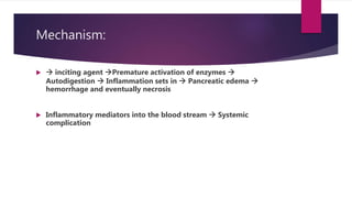
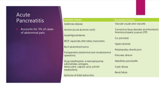
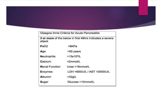
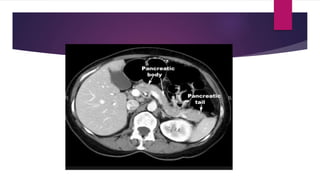
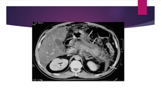
2. Introduction
Inflammation of the parenchyma
Divided into A/c & C/c.
A/c presents with acute abdominal pain with elevated
enzyme levels
Underlying mechanism???
A/c pancreatitis subdivided into Mild and Severe
3. Chronic pancreatitis:
Continuing inflammatory disease characterized by irreversible
morphological change permanent loss of function.
4. Mechanism:
inciting agent Premature activation of enzymes
Autodigestion Inflammation sets in Pancreatic edema
hemorrhage and eventually necrosis
Inflammatory mediators into the blood stream Systemic
complication
6. Hereditary pancreatitis is a rare condition associated with
mutation of cationic trypsin gene.
They suffer from c/c pancreatitis from teenage and by 8th decade
have high chance of developing Ca.
7. Clinical presentation
Pain is cardinal symptom
Develops quickly and reaches peak within minutes to hours and
then plateaus at that level for hours to days
Pain is constant, refractory to analgesics
Radiates to the back
Mimics Perforation, angina or biliary colic
Nausea and repeated vomiting with retching may be present.
Hiccoughs d/t distended Stomach irritation of diaphragm.
10. Imaging:
X-ray abdomen: Non specific signs like Colon Cut off sign,
renal halo.
Occasionally calcified gall stones or pancreatic calcification may
be seen
CXR: Pleural effusion.
In severe cases diffuse alveolar interstitial shadowing d/t ARDS
USG: Not quiet useful but can establish Gall stones as the cause
of pancreatitis
11. Assessment of severity:
Outcome of the patient depends on the severity of the
condition
Hence assessment of severity is paramount
12.
13.
14. CT: Not necessary if only a mild attack
CT advised when
Diagnostic uncertainty
Severe acute pancreatitis to diff from interstitial and necrotizing
pancreatitis
Patients with MODS and progressive sepsis
Localized complication is suspected
18. Management
Acute Mild Pancreatitis:
Conservative with IV Fluids and non-invasive monitoring and NG
aspiration + NPO
Abx not indicated
Good analgesia.
19. A/c Severe Pancreatitis Management:
ICU admission
Analgesia
Aggressive fluid management
Invasive monitoring with Arterial line and CVP, urine output and ABG
Bloods for hematological and biochemical parameters
20. Abx prophylaxis. (3rd gen cephalosporins or carbapenem)
CT scan if organ failure ensues or if clinical deterioration noted
ERCP if gall stone related pancreatitis to be done within 72 hrs
Supportive care
BP Fall ???
Raised creatinine???
Respiratory distress ???
22. Local Complications:
Acute Fluid collection: Occurs early in severe cases. Usually no
intervention needed.
If large, guided drainage.
Sterile and infected pancreatic necrosis:
CT to find out extent of necrosis
If sterile No local intervention
If Infected CT guided percutaneous drainage
Pancreatic necrosectomy
Retroperitoneal approach
23. If necrotic material reaccumulates
Closed continuous lavage
Closed drainage
Open packing
Closure and relaparotomy
24. Pancreatic abscess:
Any collection d/t Pseudocyst or acute pancreatic collection
Percutaneous drainage Pus sent for C&S
Pancreatic ascites:
Generalized, peritoneal, enzyme rich fluid caused d/t disruption of
pancreatic duct.
Paracentesis amylase rich turbid fluid
Decrease pancreatic secretion by ???
25. Pancreatic effusion
Hemorrhage: Pseudo-aneurysm
Portal and splenic vein thrombosis
Sudden rise in platelet should rise suspicion.
Should be screened for pro coagulant status
For elevated platelets Anti platelets should be started.
26. Pseudocyst of Pancreas
Amylase rich fluid collected in wall of fibrous or granulation
tissue.
Formation requires about 4 weeks.
Usually forms as a result of a/c pancreatitis.
Not unusual to have a communication with the pancreatic
duct.
Diagnosis???
Treatment???