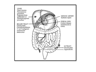
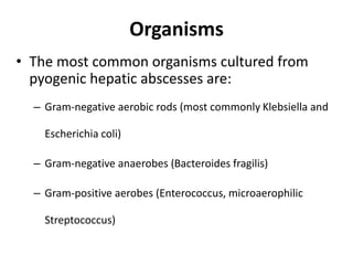
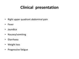
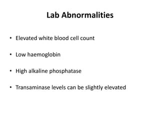
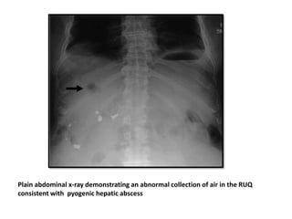
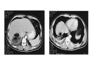
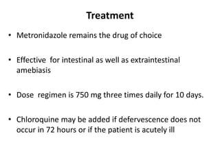
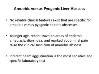
Liver Abscess
Team Lister
Dr.Sampoorna Vinod
Dr.Syed Alim
Dr.Sanghita Paul
Dr.Sneha Kataria
Dr.Rupali Rastogi
Dr.Sonal Yadav
Dr.Shubham Gaur
Dr.Shashank Lodawara
• Liver parenchyma is constantly exposed to a low level of enteric
bacteria through the portal blood flow
• However, liver infections are rare
• Liver is the largest repository of the reticuloendothelial system
and is therefore able to cope with this constant barrage.
• When the inoculum exceeds the capacity for control, infection
and abscess occur
• Pyogenic hepatic abscesses constitute over 80% of liver abscesses,
the rest primarily being amoebic in nature
Pyogenic Liver Abscess
Causes
• Transportation of virulent organisms through portal
system from the GI tract
• Trauma
• Spread of infection from the biliary tract (35% cases)
• Blood-borne infection via the hepatic artery
• Extension from a contiguous disease process
Organisms
• The most common organisms cultured from
pyogenic hepatic abscesses are:
– Gram-negative aerobic rods (most commonly Klebsiella and
Escherichia coli)
– Gram-negative anaerobes (Bacteroides fragilis)
– Gram-positive aerobes (Enterococcus, microaerophilic
Streptococcus)
Clinical presentation
• Right upper quadrant abdominal pain
• Fever
• Jaundice
• Nausea/vomiting
• Diarrhoea
• Weight loss
• Progressive fatigue
Lab Abnormalities
• Elevated white blood cell count
• Low haemoglobin
• High alkaline phosphatase
• Transaminase levels can be slightly elevated
Radiological Investigations
• Ultrasonography: sensitivity is 85% to 90%
• Computed tomography (CT) scan: sensitivity is
approximately 95%
• Magnetic resonance imaging
• Chest X ray: may display an elevated right hemi diaphragm,
pleural effusion, or extra luminal air fluid level
Plain abdominal x-ray demonstrating an abnormal collection of air in the RUQ
consistent with pyogenic hepatic abscess
Treatment
• First, the abscess must be managed, most often with a
drainage procedure.
• Antimicrobial therapy is essential in the treatment of
pyogenic abscesses.
• Second, the initiating process must be identified and
managed to ensure that recurrence is avoided.
Guiding principle for surgical management prevail:
Diagnosis, drug(s), and drainage.
• Once the diagnosis of a single or multiple liver abscess is made,
broad-spectrum parenteral antibiotics should be started.
• Antibiotics alone is given in:
– multiple small abscesses
– low risk of abscess rupture
– lack of toxemia
Clinical response is gauged by defervescence , fall in
leukocytosis , and resolution of symptoms
• Imaging with ultrasonography or CT can be used to assess
resolution of abscess(es).
• Lack of improvement after a reasonable course (10 to 14 days)
indicates failure of treatment.
• Oral antibiotics should be continued for at least 4 weeks after
discontinuance of parenteral antibiotics.
• Worsening of fever, leukocytosis, and symptoms at any time also
indicates failure of treatment and immediately qualifies the
patient for a more aggressive treatment regimen involving a
drainage procedure
• Percutaneous aspiration
Aspirated fluid should be sent for aerobic and anaerobic cultures
• Percutaneous catheter drainage :
– In whom aspiration fails
– Coagulopathy
– Lack of a safe or appropriate access route
– Multiple macroscopic abscesses
In 10% to 15% of cases, percutaneous drainage fails and
intraoperative drainage is required.
• Operative drainage
Indications:
– patients who require laparotomy for the underlying problem
– those in whom percutaneous catheter drainage fails
– patients with contraindications to percutaneous drainage
• Management of Underlying disease
• Sphincterotomy , biliary-enteric bypass, or resection of
liver for ductal obstruction
• Cholecystectomy
• Evacuation of the hematoma (hepatic trauma)
Amoebic Liver Abscess
Pathogenesis
• Cysts containing the parasite are transmitted via the fecal and oral
route.
• Trophozoites are released in the intestinal tract after ingestion of
cysts and then reside primarily in the large bowel.
• Amoebic abscesses in the liver form when the amoebic
trophozoites invade through the colonic mucosa and spread via
venules or lymphatics from the colon to the liver.
• E. histolytica may live within the lumen of the colon and may or
may not be invasive.
• liver is the most common extra intestinal site of amoebic invasion
USG showing: peripheral location, rounded shape with poor rim and internal echoes
CT scan of amebic abscess,peripherally located,round, rim is nonenhancing but
shows peripheral edema (black arrows) with extension into the intercostal
space (white arrow)
Treatment
• Metronidazole remains the drug of choice
• Effective for intestinal as well as extraintestinal
amebiasis
• Dose regimen is 750 mg three times daily for 10 days.
• Chloroquine may be added if defervescence does not
occur in 72 hours or if the patient is acutely ill
Percutaneous aspiration of amoebic abscesses
– bacterial suprainfection is suspected
– pyogenic liver abscess is suspected
– abscess is large and left sided (segments 2 and 3) so that the
risk of rupture into the pericardium is significant.
Laparotomy is indicated for ruptured amebic abscesses into
the pericardium
Rupture into the pleura or pericardium may be treated with
amoebicides and pleuracentesis or pericardiocentesis as
necessary
If laparotomy is performed, a midline incision should be used
Amoebic versus Pyogenic Liver Abscess
• No reliable clinical features exist that are specific for
amoebic versus pyogenic hepatic abscesses
• Younger age, recent travel to areas of endemic
amebiasis, diarrhoea, and marked abdominal pain
raise the clinical suspicion of amoebic abscess
• Indirect haem agglutination is the most sensitive and
specific laboratory test
Pre and post op management
• Give albendazole 400mg 3 weeks before
surgery
• Give albendazole 400mg 4 to 6 weeks post
surgery.
• For E. multiloccularis, post-op albendazole
400mg for 2 years.
THANK YOU

liverabscess.pptxjaahdhdhdndyebshdndjdns

  • 1.
    Liver Abscess Team Lister Dr.SampoornaVinod Dr.Syed Alim Dr.Sanghita Paul Dr.Sneha Kataria Dr.Rupali Rastogi Dr.Sonal Yadav Dr.Shubham Gaur Dr.Shashank Lodawara
  • 2.
    • Liver parenchymais constantly exposed to a low level of enteric bacteria through the portal blood flow • However, liver infections are rare • Liver is the largest repository of the reticuloendothelial system and is therefore able to cope with this constant barrage. • When the inoculum exceeds the capacity for control, infection and abscess occur • Pyogenic hepatic abscesses constitute over 80% of liver abscesses, the rest primarily being amoebic in nature
  • 3.
  • 4.
    Causes • Transportation ofvirulent organisms through portal system from the GI tract • Trauma • Spread of infection from the biliary tract (35% cases) • Blood-borne infection via the hepatic artery • Extension from a contiguous disease process
  • 6.
    Organisms • The mostcommon organisms cultured from pyogenic hepatic abscesses are: – Gram-negative aerobic rods (most commonly Klebsiella and Escherichia coli) – Gram-negative anaerobes (Bacteroides fragilis) – Gram-positive aerobes (Enterococcus, microaerophilic Streptococcus)
  • 7.
    Clinical presentation • Rightupper quadrant abdominal pain • Fever • Jaundice • Nausea/vomiting • Diarrhoea • Weight loss • Progressive fatigue
  • 8.
    Lab Abnormalities • Elevatedwhite blood cell count • Low haemoglobin • High alkaline phosphatase • Transaminase levels can be slightly elevated
  • 9.
    Radiological Investigations • Ultrasonography:sensitivity is 85% to 90% • Computed tomography (CT) scan: sensitivity is approximately 95% • Magnetic resonance imaging • Chest X ray: may display an elevated right hemi diaphragm, pleural effusion, or extra luminal air fluid level
  • 10.
    Plain abdominal x-raydemonstrating an abnormal collection of air in the RUQ consistent with pyogenic hepatic abscess
  • 12.
    Treatment • First, theabscess must be managed, most often with a drainage procedure. • Antimicrobial therapy is essential in the treatment of pyogenic abscesses. • Second, the initiating process must be identified and managed to ensure that recurrence is avoided. Guiding principle for surgical management prevail: Diagnosis, drug(s), and drainage.
  • 13.
    • Once thediagnosis of a single or multiple liver abscess is made, broad-spectrum parenteral antibiotics should be started. • Antibiotics alone is given in: – multiple small abscesses – low risk of abscess rupture – lack of toxemia Clinical response is gauged by defervescence , fall in leukocytosis , and resolution of symptoms
  • 14.
    • Imaging withultrasonography or CT can be used to assess resolution of abscess(es). • Lack of improvement after a reasonable course (10 to 14 days) indicates failure of treatment. • Oral antibiotics should be continued for at least 4 weeks after discontinuance of parenteral antibiotics. • Worsening of fever, leukocytosis, and symptoms at any time also indicates failure of treatment and immediately qualifies the patient for a more aggressive treatment regimen involving a drainage procedure
  • 15.
    • Percutaneous aspiration Aspiratedfluid should be sent for aerobic and anaerobic cultures • Percutaneous catheter drainage : – In whom aspiration fails – Coagulopathy – Lack of a safe or appropriate access route – Multiple macroscopic abscesses In 10% to 15% of cases, percutaneous drainage fails and intraoperative drainage is required.
  • 16.
    • Operative drainage Indications: –patients who require laparotomy for the underlying problem – those in whom percutaneous catheter drainage fails – patients with contraindications to percutaneous drainage
  • 17.
    • Management ofUnderlying disease • Sphincterotomy , biliary-enteric bypass, or resection of liver for ductal obstruction • Cholecystectomy • Evacuation of the hematoma (hepatic trauma)
  • 18.
  • 19.
    Pathogenesis • Cysts containingthe parasite are transmitted via the fecal and oral route. • Trophozoites are released in the intestinal tract after ingestion of cysts and then reside primarily in the large bowel. • Amoebic abscesses in the liver form when the amoebic trophozoites invade through the colonic mucosa and spread via venules or lymphatics from the colon to the liver. • E. histolytica may live within the lumen of the colon and may or may not be invasive. • liver is the most common extra intestinal site of amoebic invasion
  • 20.
    USG showing: peripherallocation, rounded shape with poor rim and internal echoes
  • 21.
    CT scan ofamebic abscess,peripherally located,round, rim is nonenhancing but shows peripheral edema (black arrows) with extension into the intercostal space (white arrow)
  • 22.
    Treatment • Metronidazole remainsthe drug of choice • Effective for intestinal as well as extraintestinal amebiasis • Dose regimen is 750 mg three times daily for 10 days. • Chloroquine may be added if defervescence does not occur in 72 hours or if the patient is acutely ill
  • 23.
    Percutaneous aspiration ofamoebic abscesses – bacterial suprainfection is suspected – pyogenic liver abscess is suspected – abscess is large and left sided (segments 2 and 3) so that the risk of rupture into the pericardium is significant. Laparotomy is indicated for ruptured amebic abscesses into the pericardium Rupture into the pleura or pericardium may be treated with amoebicides and pleuracentesis or pericardiocentesis as necessary If laparotomy is performed, a midline incision should be used
  • 24.
    Amoebic versus PyogenicLiver Abscess • No reliable clinical features exist that are specific for amoebic versus pyogenic hepatic abscesses • Younger age, recent travel to areas of endemic amebiasis, diarrhoea, and marked abdominal pain raise the clinical suspicion of amoebic abscess • Indirect haem agglutination is the most sensitive and specific laboratory test
  • 25.
    Pre and postop management • Give albendazole 400mg 3 weeks before surgery • Give albendazole 400mg 4 to 6 weeks post surgery. • For E. multiloccularis, post-op albendazole 400mg for 2 years.
  • 26.