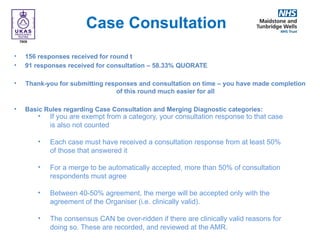
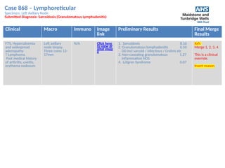
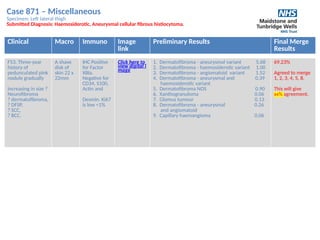
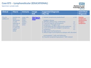
South East England General Histopathology EQA Scheme
Case Discussion Round t
Wednesday 30th
November, 2022
THANK YOU FOR WAITING
The meeting will start at 12:00pm
Mute your mic
if you’re not
speaking
3
Use the “raise hand”
Or “chat” feature
to raise questions
or share ideas
4
Wait for the
Chair person to call
on you before you
unmute your mic
Meeting Etiquette
6
Remember…
Everyone can see
your chat comments
1
If your camera
is on, everyone
can see you
2
Agenda
1. Welcome & Introduction of Scheme Staff
2. Meeting Terms of Reference
3. Case and Preliminary Score Review
a) Case 865-874
b) Educational Cases – 875 - 876
4. Questions / comments
2. Meeting Terms of Reference
• This meeting is held between the end of case consultation and
results being issued and now replaces the additional final week of
the case consultation.
• This meeting is an educational exercise; an opportunity to explain
the reasons behind scoring and merging or why cases were
excluded.
• For clarity, this is not an opportunity to alter merging decisions, as
participants have that opportunity during the “Case Consultation”
period.
• An additional CPD point will be awarded to those who attend, and it
will be added to the annual certificate. Please note you have to stay
for >50% of the meeting to gain this point (attendance times are
monitored automatically by Teams)
• We always welcome any feedback – good or bad – you may have
about today.
3. Round t Review
Case Consultation
• 156 responses received for round t
• 91 responses received for consultation – 58.33% QUORATE
• Thank-you for submitting responses and consultation on time – you have made completion
of this round much easier for all
• Basic Rules regarding Case Consultation and Merging Diagnostic categories:
• If you are exempt from a category, your consultation response to that case
is also not counted
• Each case must have received a consultation response from at least 50%
of those that answered it
• For a merge to be automatically accepted, more than 50% of consultation
respondents must agree
• Between 40-50% agreement, the merge will be accepted only with the
agreement of the Organiser (i.e. clinically valid).
• The consensus CAN be over-ridden if there are clinically valid reasons for
doing so. These are recorded, and reviewed at the AMR.
Case 865 – Respiratory
Specimen: Right lower lobe
Submitted Diagnosis: Pulmonary Hamartoma
Submitted
Clinical Macro Immuno Image
link
Preliminary Results Final Merge
Results
M51. 38mm
RLL Mass,
enlarging and
faintly FDG
avid. Asthma
Two cores 6 & 8
mm
N/A Click here
to view
digital im
age
1. Chondroid / pulmonary hamartoma 9.53
2. Lipoma 0.07
3. Cartilaginous hamartoma 0.14
4. Chondroma 0.07
5. Chondrosarcoma 0.07
6. Enchondroma 0.07
7. Benign-Chronic inflammation 0.07
32.94%
Agreed to
merge 1,3,4.
This will give xx
% agreement.
Case 866 – Endocrine
Specimen: Thyroid
Submitted Diagnosis: Hyalinising trabecular tumour
Clinical Macro Immuno Image
link
Preliminary Results Final
Merge
Results
F41. Left
hemithyroidectomy
for Thy3f thyroid
nodule (not
included in
the slide).
Incidental finding in
one tissue block
A thyroid lobe
weighing 66g and
measuring
79x60x36mm.
The main tan
nodule measures
55x48x37mm
(not included in
the slide).
Incidental finding
(lesion not seen
grossly).
TTF-1(+),
Thyroglobulin (+),
HBME-1 (-),
Calcitonin (-), DPAS
stain:
Positive
intratrabecular
hyalin
Click her
e to vie
w digita
l image
1. Hyalinising adenoma and thyroiditis 4.28
2. Hashitmoto thyroiditis with 0.33
micropapillary carcinoma
3. Papillary carcinoma 0.09
4. Hyalinising adenoma 4.91
(thyroiditis not mentioned)
5. Papillary microcarcinoma 0.07
6. Chronic Thyroiditis (Hashimoto) 0.07
7. Tall cell variant micropapillary 0.26
adenocarcinoma
79.78%
Agreed to
merge
1, 4
This will
give xx%
agreement
Case 867 – Gynae
Specimen: Loop biopsy of cervix
Submitted Diagnosis: Mesonephric/gartner's duct remnants
Clinical Macro Immuno Image
link
Preliminary Results Final Merge
Results
F27. Cervical
smear showed
severe
dyskaryosis.
Colposcopy -
?
microinvasive
Loop biopsy 23 x 20
x 13 including the
os.
Immuno
shows
positivity
for CD10
and low
Ki67 index
Click here
to view
digital im
age
1. Mesonephric hyperplasia / Remnants 8.84
2. Mesonephric adenocarcinoma 0.41
3. Minimal deviation adenocarcinoma 0.54
4. Papillary endocervicitis 0.07
5. Microglandular adenosis 0.07
6. Endocervical polyp 0.07
7. Metastatic carcinoma 0.01
79.07%
Agreed not to
merge any other
categories.
This will give xx
% agreement.
Case 868 – Lymphoreticular
Specimen: Left Axillary Node
Submitted Diagnosis: Sarcoidosis (Granulomatous Lymphadenitis)
Clinical Macro Immuno Image
link
Preliminary Results Final Merge
Results
F75. Hypercalcemia
and widespread
adenopathy
? Lymphoma.
Past medical history
of arthritis, uveitis,
erythema nodosum
Left axillary
node biopsy.
Three cores 13-
17mm
N/A Click here
to view di
gital imag
e
1. Sarcoidosis 8.16
2. Granulomatous lymphadenitis 0.50
DD incl sarcoid / infectious / Crohns etc
3. Non-caseating granulomatous 1.27
inflammation NOS
4. Lofgren Syndrome 0.07
Xx%
Merge 1, 2, 3, 4
This is a clinical
override.
Insert reason
Case 869 – Breast
Specimen: Breast
Submitted Diagnosis: High grade DCIS (solid and cribriform, with comedo necrosis).
Multiple foci of invasive NST carcinoma in other blocks.
Clinical Macro Immuno Image
link
Preliminary Results Final
Merge
Results
F44. Left skin
sparing
mastectomy
Mastectomy
specimen weighing
270g and
measuring 12 x 11 x
4cm.
Extensive ill-
defined area of
grittiness in UOQ
measuring 9.5cm
maximally.
N/A Click here t
o view digit
al image
1. DCIS - High Grade 7.16
2. DCIS - intermediate grade 0.20
3. Pleomorphic LCIS 0.07
with comedo necrosis
4. DCIS NOS 1.42
5. DCIS - High grade ? microinvasion 0.34
6. Microinvasive ductal carcinoma 0.31
and DCIS
7. Invasive ductal carcinoma and DCIS 0.07
8. DCIS. Comedo necrosis 0.27
9. Grade 3 DCIS with comedo necrosis 0.14
10. DCIS-like-invasive carcinoma. (IHC) 0.03
52.33%
Agreed to
merge
1, 2, 4, 8, 9.
This will give
Xx %
agreement.
Case 870 – GU
Specimen: Bladder
Submitted Diagnosis: Cystitis Glandularis
Clinical Macro Immuno Image
link
Preliminary Results Final Merge
Results
M36. Small
lesion
? inverted
papilloma of
bladder
trigone
One piece of pale
tissue measuring
3mm
N/A Click here t
o view digit
al image
1. Cystitis Glandularis / cystica 9.79
2. Prostatic type polyp 0.07
3. Nephrogenic adenoma 0.07
4. Prostatic acinar and Glandular 0.07
metaplasia & Von Brunn’s nests
69.88%
Agreed not to
merge any other
categories.
This will give xx%
agreement.
Case 871 – Miscellaneous
Specimen: Left lateral thigh
Submitted Diagnosis: Haemosiderotic, Aneurysmal cellular fibrous histiocytoma.
Clinical Macro Immuno Image
link
Preliminary Results Final Merge
Results
F53. Three-year
history of
pedunculated pink
nodule gradually
increasing in size ?
Neurofibroma
? dermatofibroma,
? DFSP,
? SCC,
? BCC.
A shave
disk of
skin 22 x
22mm
IHC Positive
for Factor
XIIIa.
Negative for
CD34, S100,
Actin and
Desmin. Ki67
is low <1%
Click here to
view digital i
mage
1. Dermatofibroma - aneurysmal variant 5.68
2. Dermatofibroma - haemosiderotic variant 1.00
3. Dermatofibroma - angiomatoid variant 1.52
4. Dermatofibroma - aneurysmal and 0.39
haemosiderotic variant
5. Dermatofibroma NOS 0.90
6. Xanthogranuloma 0.06
7. Glomus tumour 0.13
8. Dermatofibroma - aneurysmal 0.26
and angiomatoid
9. Capillary haemangioma 0.06
69.23%
Agreed to merge
1, 2, 3, 4, 5, 8.
This will give
xx% agreement.
Case 872 – GI
Specimen: Right hemicolectomy
Submitted Diagnosis: Fat necrosis secondary to acute pancreatitis
Clinical Macro Immuno Image
link
Preliminary Results Final Merge
Results
F38. Laparotomy
for haemorrhagic
pancreatitis
The caecum and
ascending colon are
dilated and their
surface shows
adherent fibrin and
blood. The mucosa
appears
oedematous.
N/A Click here
to view
digital im
age
1. Pancreatitis with fat necrosis 6.34
2. Mesenteric fat necrosis 0.39
3. Amoebic colitis 0.03
4. Fat necrosis 2.99
(role of pancreas not mentioned)
5. Steatonecrosis 0.03
6. Ischaemia 0.08
8. Lipomatous proliferation 0.06
9. Colonic paralytic ileus 0.03
10. Large bowel pseudo obstruction 0.04
56.67%
Agreed to merge
1, 2, 4, 5.
This will give xx%
agreement.
Case 873 – Skin
Specimen: Skin
Submitted Diagnosis: Sebacous hyperplasia
Clinical Macro Immuno Image
link
Preliminary Results Final Merge
Results
M66. ?BCC tip of
nose
Disc of skin 8 x
7mm, depth
2mm. There is
a raised, pale
nodule
3mm
across
N/A Click her
e to vie
w digita
l image
1. Sebaceous hyperplasia 8.51
2. Sebaceous adenoma 1.23
3. Sebaceous trichofolliculoma 0.13
4. Sebaceous adenoma in background 0.06
of hyperplasia
5. Sebaceous naevus 0.06
32.22%
Agreed to merge
1, 2, 4.
This will give xx%
agreement.
Case 874 – Lymphoreticular
Specimen: Nose biopsy
Submitted Diagnosis: Mantle Cell Lymphoma. Clondality results showed clonal IG gene,
consistent with mantle cell lymphoma
Clinical Macro Immuno Image
link
Preliminary Results Final Merge
Results
M74.
nasopharyng
eal lesion. ?
Lymphoma.
Asymmetric
tonsil L>R.
Biopsy from
post nasal
space.
Tissue
aggregates
15 x 15 x
4mm
Positive: CD20, PAX-5,
CD5, Cyclin D1, SOX-11,
BCL-2, weak CD43.
Scattered - CD10, BCL-6,
CD21, CD23.
Negative: CD30, EBER-
ISH, AE1/AE3. No light
chain restriction.
Click here to
view digital i
mage
1. Mantle Cell Lymphoma 9.86
2. Small lymphocytic lymphoma 0.07
3. Diffuse large B cell lymphoma 0.07
- activated subtype (germinal centre)
93.83%
Agreed not to
merge any other
categories.
This will give
xx% agreement.
Case 875 – Lymphoreticular (EDUCATIONAL)
Specimen: Lymph node
Clinical Macro Immuno Image
link
Suggested Diagnosis
(Top 10)
Submitted
Diagnosis
F54. Rt
axillary LN
Nodular tan
tissue
20x10x10m
m with
tattooing at
one end.
Cut surface
is solid and
yellow
white
Large cells:
CD45+,
CD30-,
MUM1-,
CD20+,
CD15-
Click here to
view digital i
mage
1. Nodular lymphocyte predominant x 114
Hodgkins disease
2. DIFFUSE LARGE B-CELL LYMPHOMA x 6
3. T-cell rich large B-cell lymphoma x 3
4. Lymphoma ? Hodgkin type x 2
5. T-cell/Histiocyte-Rich Large B-Cell Lymphoma x 2
6. T-cell rich B-cell Non-Hodgkin’s lymphoma x 2
7. Lymphoma x 2
8. High grade B cell lymphoma
9. Diffuse large B cell lymphoma subtype with abundant
nonneoplastic T cells and histiocytes
10. Reactive lymphoid hyperplasia (? EBV-related)
Nodular
lymphocyte
predominant
Hodgkins
Lymphoma
Case 876 – Lymphoreticular (EDUCATIONAL)
Specimen: Bone Marrow Trephine
Clinical Macro Immuno Image
link
Suggested Diagnosis
(Top 10)
Submitted
Diagnosis
F42. Splenomegaly +/-
lymphadenopathy
A1 = Core
measuring
7mm
A2 = Clot 5
x 4 x 2 mm
CD68 positive Digital Imag
e A1
Digital Imag
e A2
1. Gaucher’s disease x 60
2. Storage disease / disorder x 38
3. Langerhans cell histiocytosis x 12
4. Erdheim-Chester disease x 12
5. Niemann-Pick Disease
(Acid Sphingomyelinase Deficiency) x 7
6. Rosai-Dorfman disease x 5
7. Crystal storing histiocytosis x 5
8. Histiocytosis, ? LCH x 5
9. Leishmaniasis x 4
Suggestive of
Gauches's disease
4. Questions
Comments
Suggestions
Feedback
Thank you for attending. This presentation can be found on the EQA website from next week.

Case-Discussion-Presentation-Round-t-FINAL.pptx

  • 1.
    South East EnglandGeneral Histopathology EQA Scheme Case Discussion Round t Wednesday 30th November, 2022 THANK YOU FOR WAITING The meeting will start at 12:00pm
  • 2.
    Mute your mic ifyou’re not speaking 3 Use the “raise hand” Or “chat” feature to raise questions or share ideas 4 Wait for the Chair person to call on you before you unmute your mic Meeting Etiquette 6 Remember… Everyone can see your chat comments 1 If your camera is on, everyone can see you 2
  • 3.
    Agenda 1. Welcome &Introduction of Scheme Staff 2. Meeting Terms of Reference 3. Case and Preliminary Score Review a) Case 865-874 b) Educational Cases – 875 - 876 4. Questions / comments
  • 4.
    2. Meeting Termsof Reference
  • 5.
    • This meetingis held between the end of case consultation and results being issued and now replaces the additional final week of the case consultation. • This meeting is an educational exercise; an opportunity to explain the reasons behind scoring and merging or why cases were excluded. • For clarity, this is not an opportunity to alter merging decisions, as participants have that opportunity during the “Case Consultation” period. • An additional CPD point will be awarded to those who attend, and it will be added to the annual certificate. Please note you have to stay for >50% of the meeting to gain this point (attendance times are monitored automatically by Teams) • We always welcome any feedback – good or bad – you may have about today.
  • 6.
    3. Round tReview
  • 7.
    Case Consultation • 156responses received for round t • 91 responses received for consultation – 58.33% QUORATE • Thank-you for submitting responses and consultation on time – you have made completion of this round much easier for all • Basic Rules regarding Case Consultation and Merging Diagnostic categories: • If you are exempt from a category, your consultation response to that case is also not counted • Each case must have received a consultation response from at least 50% of those that answered it • For a merge to be automatically accepted, more than 50% of consultation respondents must agree • Between 40-50% agreement, the merge will be accepted only with the agreement of the Organiser (i.e. clinically valid). • The consensus CAN be over-ridden if there are clinically valid reasons for doing so. These are recorded, and reviewed at the AMR.
  • 8.
    Case 865 –Respiratory Specimen: Right lower lobe Submitted Diagnosis: Pulmonary Hamartoma Submitted Clinical Macro Immuno Image link Preliminary Results Final Merge Results M51. 38mm RLL Mass, enlarging and faintly FDG avid. Asthma Two cores 6 & 8 mm N/A Click here to view digital im age 1. Chondroid / pulmonary hamartoma 9.53 2. Lipoma 0.07 3. Cartilaginous hamartoma 0.14 4. Chondroma 0.07 5. Chondrosarcoma 0.07 6. Enchondroma 0.07 7. Benign-Chronic inflammation 0.07 32.94% Agreed to merge 1,3,4. This will give xx % agreement.
  • 9.
    Case 866 –Endocrine Specimen: Thyroid Submitted Diagnosis: Hyalinising trabecular tumour Clinical Macro Immuno Image link Preliminary Results Final Merge Results F41. Left hemithyroidectomy for Thy3f thyroid nodule (not included in the slide). Incidental finding in one tissue block A thyroid lobe weighing 66g and measuring 79x60x36mm. The main tan nodule measures 55x48x37mm (not included in the slide). Incidental finding (lesion not seen grossly). TTF-1(+), Thyroglobulin (+), HBME-1 (-), Calcitonin (-), DPAS stain: Positive intratrabecular hyalin Click her e to vie w digita l image 1. Hyalinising adenoma and thyroiditis 4.28 2. Hashitmoto thyroiditis with 0.33 micropapillary carcinoma 3. Papillary carcinoma 0.09 4. Hyalinising adenoma 4.91 (thyroiditis not mentioned) 5. Papillary microcarcinoma 0.07 6. Chronic Thyroiditis (Hashimoto) 0.07 7. Tall cell variant micropapillary 0.26 adenocarcinoma 79.78% Agreed to merge 1, 4 This will give xx% agreement
  • 10.
    Case 867 –Gynae Specimen: Loop biopsy of cervix Submitted Diagnosis: Mesonephric/gartner's duct remnants Clinical Macro Immuno Image link Preliminary Results Final Merge Results F27. Cervical smear showed severe dyskaryosis. Colposcopy - ? microinvasive Loop biopsy 23 x 20 x 13 including the os. Immuno shows positivity for CD10 and low Ki67 index Click here to view digital im age 1. Mesonephric hyperplasia / Remnants 8.84 2. Mesonephric adenocarcinoma 0.41 3. Minimal deviation adenocarcinoma 0.54 4. Papillary endocervicitis 0.07 5. Microglandular adenosis 0.07 6. Endocervical polyp 0.07 7. Metastatic carcinoma 0.01 79.07% Agreed not to merge any other categories. This will give xx % agreement.
  • 11.
    Case 868 –Lymphoreticular Specimen: Left Axillary Node Submitted Diagnosis: Sarcoidosis (Granulomatous Lymphadenitis) Clinical Macro Immuno Image link Preliminary Results Final Merge Results F75. Hypercalcemia and widespread adenopathy ? Lymphoma. Past medical history of arthritis, uveitis, erythema nodosum Left axillary node biopsy. Three cores 13- 17mm N/A Click here to view di gital imag e 1. Sarcoidosis 8.16 2. Granulomatous lymphadenitis 0.50 DD incl sarcoid / infectious / Crohns etc 3. Non-caseating granulomatous 1.27 inflammation NOS 4. Lofgren Syndrome 0.07 Xx% Merge 1, 2, 3, 4 This is a clinical override. Insert reason
  • 12.
    Case 869 –Breast Specimen: Breast Submitted Diagnosis: High grade DCIS (solid and cribriform, with comedo necrosis). Multiple foci of invasive NST carcinoma in other blocks. Clinical Macro Immuno Image link Preliminary Results Final Merge Results F44. Left skin sparing mastectomy Mastectomy specimen weighing 270g and measuring 12 x 11 x 4cm. Extensive ill- defined area of grittiness in UOQ measuring 9.5cm maximally. N/A Click here t o view digit al image 1. DCIS - High Grade 7.16 2. DCIS - intermediate grade 0.20 3. Pleomorphic LCIS 0.07 with comedo necrosis 4. DCIS NOS 1.42 5. DCIS - High grade ? microinvasion 0.34 6. Microinvasive ductal carcinoma 0.31 and DCIS 7. Invasive ductal carcinoma and DCIS 0.07 8. DCIS. Comedo necrosis 0.27 9. Grade 3 DCIS with comedo necrosis 0.14 10. DCIS-like-invasive carcinoma. (IHC) 0.03 52.33% Agreed to merge 1, 2, 4, 8, 9. This will give Xx % agreement.
  • 13.
    Case 870 –GU Specimen: Bladder Submitted Diagnosis: Cystitis Glandularis Clinical Macro Immuno Image link Preliminary Results Final Merge Results M36. Small lesion ? inverted papilloma of bladder trigone One piece of pale tissue measuring 3mm N/A Click here t o view digit al image 1. Cystitis Glandularis / cystica 9.79 2. Prostatic type polyp 0.07 3. Nephrogenic adenoma 0.07 4. Prostatic acinar and Glandular 0.07 metaplasia & Von Brunn’s nests 69.88% Agreed not to merge any other categories. This will give xx% agreement.
  • 14.
    Case 871 –Miscellaneous Specimen: Left lateral thigh Submitted Diagnosis: Haemosiderotic, Aneurysmal cellular fibrous histiocytoma. Clinical Macro Immuno Image link Preliminary Results Final Merge Results F53. Three-year history of pedunculated pink nodule gradually increasing in size ? Neurofibroma ? dermatofibroma, ? DFSP, ? SCC, ? BCC. A shave disk of skin 22 x 22mm IHC Positive for Factor XIIIa. Negative for CD34, S100, Actin and Desmin. Ki67 is low <1% Click here to view digital i mage 1. Dermatofibroma - aneurysmal variant 5.68 2. Dermatofibroma - haemosiderotic variant 1.00 3. Dermatofibroma - angiomatoid variant 1.52 4. Dermatofibroma - aneurysmal and 0.39 haemosiderotic variant 5. Dermatofibroma NOS 0.90 6. Xanthogranuloma 0.06 7. Glomus tumour 0.13 8. Dermatofibroma - aneurysmal 0.26 and angiomatoid 9. Capillary haemangioma 0.06 69.23% Agreed to merge 1, 2, 3, 4, 5, 8. This will give xx% agreement.
  • 15.
    Case 872 –GI Specimen: Right hemicolectomy Submitted Diagnosis: Fat necrosis secondary to acute pancreatitis Clinical Macro Immuno Image link Preliminary Results Final Merge Results F38. Laparotomy for haemorrhagic pancreatitis The caecum and ascending colon are dilated and their surface shows adherent fibrin and blood. The mucosa appears oedematous. N/A Click here to view digital im age 1. Pancreatitis with fat necrosis 6.34 2. Mesenteric fat necrosis 0.39 3. Amoebic colitis 0.03 4. Fat necrosis 2.99 (role of pancreas not mentioned) 5. Steatonecrosis 0.03 6. Ischaemia 0.08 8. Lipomatous proliferation 0.06 9. Colonic paralytic ileus 0.03 10. Large bowel pseudo obstruction 0.04 56.67% Agreed to merge 1, 2, 4, 5. This will give xx% agreement.
  • 16.
    Case 873 –Skin Specimen: Skin Submitted Diagnosis: Sebacous hyperplasia Clinical Macro Immuno Image link Preliminary Results Final Merge Results M66. ?BCC tip of nose Disc of skin 8 x 7mm, depth 2mm. There is a raised, pale nodule 3mm across N/A Click her e to vie w digita l image 1. Sebaceous hyperplasia 8.51 2. Sebaceous adenoma 1.23 3. Sebaceous trichofolliculoma 0.13 4. Sebaceous adenoma in background 0.06 of hyperplasia 5. Sebaceous naevus 0.06 32.22% Agreed to merge 1, 2, 4. This will give xx% agreement.
  • 17.
    Case 874 –Lymphoreticular Specimen: Nose biopsy Submitted Diagnosis: Mantle Cell Lymphoma. Clondality results showed clonal IG gene, consistent with mantle cell lymphoma Clinical Macro Immuno Image link Preliminary Results Final Merge Results M74. nasopharyng eal lesion. ? Lymphoma. Asymmetric tonsil L>R. Biopsy from post nasal space. Tissue aggregates 15 x 15 x 4mm Positive: CD20, PAX-5, CD5, Cyclin D1, SOX-11, BCL-2, weak CD43. Scattered - CD10, BCL-6, CD21, CD23. Negative: CD30, EBER- ISH, AE1/AE3. No light chain restriction. Click here to view digital i mage 1. Mantle Cell Lymphoma 9.86 2. Small lymphocytic lymphoma 0.07 3. Diffuse large B cell lymphoma 0.07 - activated subtype (germinal centre) 93.83% Agreed not to merge any other categories. This will give xx% agreement.
  • 18.
    Case 875 –Lymphoreticular (EDUCATIONAL) Specimen: Lymph node Clinical Macro Immuno Image link Suggested Diagnosis (Top 10) Submitted Diagnosis F54. Rt axillary LN Nodular tan tissue 20x10x10m m with tattooing at one end. Cut surface is solid and yellow white Large cells: CD45+, CD30-, MUM1-, CD20+, CD15- Click here to view digital i mage 1. Nodular lymphocyte predominant x 114 Hodgkins disease 2. DIFFUSE LARGE B-CELL LYMPHOMA x 6 3. T-cell rich large B-cell lymphoma x 3 4. Lymphoma ? Hodgkin type x 2 5. T-cell/Histiocyte-Rich Large B-Cell Lymphoma x 2 6. T-cell rich B-cell Non-Hodgkin’s lymphoma x 2 7. Lymphoma x 2 8. High grade B cell lymphoma 9. Diffuse large B cell lymphoma subtype with abundant nonneoplastic T cells and histiocytes 10. Reactive lymphoid hyperplasia (? EBV-related) Nodular lymphocyte predominant Hodgkins Lymphoma
  • 19.
    Case 876 –Lymphoreticular (EDUCATIONAL) Specimen: Bone Marrow Trephine Clinical Macro Immuno Image link Suggested Diagnosis (Top 10) Submitted Diagnosis F42. Splenomegaly +/- lymphadenopathy A1 = Core measuring 7mm A2 = Clot 5 x 4 x 2 mm CD68 positive Digital Imag e A1 Digital Imag e A2 1. Gaucher’s disease x 60 2. Storage disease / disorder x 38 3. Langerhans cell histiocytosis x 12 4. Erdheim-Chester disease x 12 5. Niemann-Pick Disease (Acid Sphingomyelinase Deficiency) x 7 6. Rosai-Dorfman disease x 5 7. Crystal storing histiocytosis x 5 8. Histiocytosis, ? LCH x 5 9. Leishmaniasis x 4 Suggestive of Gauches's disease
  • 20.
    4. Questions Comments Suggestions Feedback Thank youfor attending. This presentation can be found on the EQA website from next week.