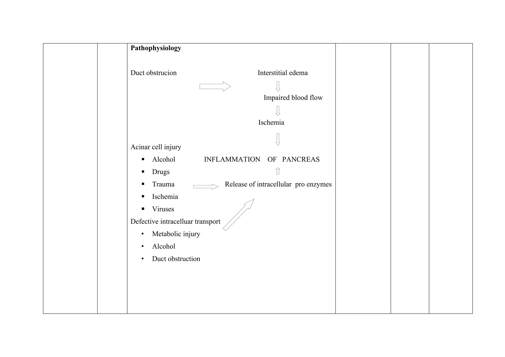
Lesson plan on
pancreatitis
Specific
objectives
Time Content Teacher’s
activity
Learner’s
activity
Evaluation
 Pancreas is a long ,flat gland that located behind the stomach
in upper abdomen.
 The pancreas produce enzymes that help in digestion and
hormones that help in metabolism
 Enzymes secreted from pancreas reach the intestine through
pancreatic duct and help digest the food.
 The hormones, insulin and glucagon are released into the
blood stem which helps to regulate the blood sugar level
Definition
Inflammation of the pancreas
Classification
 Acute pancreatitis- single episode of pancreatitis in a
previously normal gland
 Acute relapsing pancreatitis- recurrent attacks of pancreatitis
with normalcy in the intervals between attacks and without
permanent functional damage .
 Chronic pancreatitis- irreversible destruction of the gland with
constant pain
 Chronic relapsing pancreatitis- recurrent attacks of pain with
frequent pain intervals with progressive functional damage of
the pancreas.
Causes of pancreatitis
1.Gall stones and choledocholithiasis –presence of gall stones in the
biliary tract and biliary duct, causes reflux of bile in to the pancreatic
duct at the common bile duct .Reflux of bile causes obstruction of
the pancreatic duct flow ,which results in increased pressure with in
the pancreatic duct leading to inflammation .
2.Alcohol –prolonged alcohol intake causes protein precipitation in
the pancreatic juice, which damages the pancreatic parenchyma.
- alcohol also cause inflammation of the duodenum that
produces some degree of bile duct obstruction
-alcohol stimulates pancreatic secretion and also at the
same time increases sphincteric tone at the ampullary region .Thus
increases secretion against an unyielding sphincter.
3.Metabolic factors
 hyperlipideemia
 hypercalcemia
 hemochroatosis
4. Vascular factors
atherosclerosis ,vasculitis
5. Post operative pancreatitis-
 surgical procedures such as common bile duct
exploration, passing of a long armed T- tube at
sphincter of oddi causes pancreatic injury and
inflammation.
 gastrectomy may cause injury to the head of
pancreas ,spleenectomy may result in operative injury
to the tail of pancreas.
6. Toxins-methyl alcohol, Zinc oxide, cholinesterase inhbitors
7.Viral diseases -mumps,echovirusinfection,coxsackievirus,
mononucleosis.
8. Drugs- Corticosteroids, phenformin,azathioprin,
chlorthiazide, frusemide
9. Investigative procedures- ERCP
10.Others –Trauma, Scorpion stings
Pathophysiology
Duct obstrucion Interstitial edema
Impaired blood flow
Ischemia
Acinar cell injury
 Alcohol INFLAMMATION OF PANCREAS
 Drugs
 Trauma Release of intracellular pro enzymes
 Ischemia
 Viruses
Defective intracelluar transport
• Metabolic injury
• Alcohol
• Duct obstruction
Pancreatitis
Activated enzymes
 Interstitial inflammation and edema
 Proteolysis
 Lipolysis free fat binds with ca to form fat necrosis
 Hemorrhage Activation of PAF Thrombus in
mesenteric vessels
Forms of pancreatitis
 Oedematous –mildest form,whole or part of organ become
oedematus
 Hemorrhagic –areas of hemorrhage in the gland. The
retroperitoneal tissues around the pancreas are engorged with
blood stained fluid.
 Necrotising and gangrenous pancreatitis-fluid may
accumulate in the lesser sac and when the condition
gradually resolves in acute pancreatitis, fibrosis occur which
walls off such collection of fluid within the lesser sac to form
PSEUDOCYST.With migration of bacteria in to such fluid
collection leads to ABSCESS FORMATION.
Clinical features
 Pain
• Distressing type of pain
 In oedematous pancreatitis ,penetrating upper abdominal pain
following heavy meal, pain frequently located in the mid
epigastrium and often radiate to the back or even to the flanks.
 In hemorrhagic and necrotising pancreatitis –cramping and
excruciating pain.
 Sometimes pain in the right or left quadrant due to
involvement of the head or tail of pancreas.
 Persistent and repated vomiting,vomiting even in empty
stomach
 Retching
 Low grade fever
 Epigastric tenderness
 Guarding
 Involuntary rigidity of epigastric region
 Signs of shock-due to loss of fluid, Myocardial Depressant
Factor(MDF) released from pancreas .
 Mild jaundice
 Carpopedal spasm due to hpocalcemia
 Abdominal distension
 Paralytic ileus –seen in the beginning in the duodenum and
proximal jejunum
 Cullen’s sign-discoloration of the skin,that varies from slate
blue to mottled yellowish brown colour due to ecchymosis
and extravasated blood
 Grey turners' sign-discoloration in the loins
Diagnosis
Blood examination
• Serum amylase-increased serum amlase values.
• Serum lipase –elevated serum lipase values
• Serum lactescence-specific indicator of acute pancreatitis
• Hyperlipidemia-.This is observed when circulating
triglyceride values exceed 500mg/100 ml
• Hyperglycemia-due to relative hypoinsulinemia
• Hypocalcemia
• Liver function test- shows elevated liver enzymes
• WBC- above 10,000 cells/cumm
Urine examination
 Increased excretion of urinary amylase
 Renal clearance of amylase-
urine amylase X serum creatinine X 100
serum amylase urine creatinine
Normal ratio – 1 to 4%,greater than 6 indicate pancreatitis
 ECG –shows variations due to electrolyte imbalance.
 X ray of abdomen
 sentinel loop sign(paralytic loop of jejunum)
 distension of duodenum,transverse colon
 cuff off sign( air filled distended transverse colon)
• Barium x ray
• USG
• CT scan
• MRI
ERCP (Endoscopic Retrograde Cholangio pancreatography)
Management
1.Conservative management
• Management of shock and electrolyte imbalance- IV
administration o fluids ,blood transfusion. Hypo calcemia
corrected by IV calcium gluconate
• NPO with gastric aspiration
 Pain relief- Demerol(50 to 100 mg IV) combined with
papaverine or nitroglycerine.
• Antibiotics – Imipenem,norfloxacin,amphotericin,quinolone
• Non absorbable liquid antacid – combination of magnesium
silicate and aluminium hydroxide
• H2 receptor antagonist- rantidine,famotidine
• Somatostatin –potent inhibitor of pancreatic exosecretion
• Cholecystokinin receptor antagonist-proglumide
• Antienzyme preparations
• Lexipafant(PAF antagonist)-prevents local damage
andmesenteric vessel occlusion
• Insulin therapy in case of chronic pancreatitis
2.Peritoneal lavage
• Done in patients who do not show progress with conservative
management and to reduce systemic complications of
pancreatitis
• A peritoneal lavage cathether is introduced in the lower
midline or left lower quadrant
• 2 litres of peritoneal dialysis solution run in gravity during 30
minutes.fluid drained over 90 minutes.
• Cycle repeated every 2 hours for 72 hours
3. Correction of associated biliary tract diseases
4.Treatment of secondary pancreatic infections
5. Surgical management
Surgical management
a) Ampullary dilatations
b) Drainage procedures
c) Excisional procedures
1.Ampullary dilatation- choice of removal of gall bladder stones
I. Endoscopic sphincterotomy-use of endoscopic procedures to
cut the muscles of sphincter of odii.this facilitates the free
flow of bile and pancreatic juice into the intestine.
II. Endoscopic papillary ballon dilation –procedure that makes
major duodenal papilla orifice widen larger than stones using
dilation ballon cathether
III. Transduodenal sphincteroplasty-a permanent wide open stoma
is created between the common bile duct and the duodenum.
2.Drainage procedures
I. Dural’s pancreatojejunostomy- A limited pancreatectomy is
performed along with end to end anastomosis of Roux-en-Y
loop of jejunum with the cut end of the pancreatic duct .This
is suitable,if there is single proximal obstruction in the head of
the gland
II. Peustow’s longitudinal pancreatojejunostomy- done in
patients with multiple strictures of pancreatic duct with
dilated pockets and pancreatic calcifications.Defunctioned
limb of jejunum is anastomosed with the pancreatic duct side
to side longtudially and intestinal continuity is reestablished
by a Roux-en-Y jejunostomy
3.Excision procedures
I.Pancreatoduodenectomy (modified Whipple operation)-
Surgical removal of headofpancreas,duodenum,proximal
jejunum,gall bladder and ocassionally part of the stomach.
II. Distal subtotal pancreatectomy –Removal of the body and tail of
the pancreas. Sometimes spleen along with its artery and vein and
lymph nodes removed
III. Total pancreatomy-Surgical removal of all the parts of pancreas.
Done in patients with chronic pancreatitis, where other treatment
methods are unsuccessful
IV.Cholecystectomy-surgical removal of gallbladder
Nursing management
Nursing Diagnosis
 Pain and discomfort related to edema, distention of the
pancreas, and peritoneal irritation
 Ineffective breathing pattern related to severe pain, pulmonary
infiltrates, pleural effusion and atelactasis
 Imbalanced nutrition: less than body requirements related to
inadequacy dietary intake, impaired absorption, reduced food
intake, and increased metabolic demands.
 Activity intolerance related to fatigue
 Impaired skin integrity resulting from poor nutritional status,
bed rest
 Ineffective coping related to pain and discomfort
Pre operative nursing management
Nutrition
 Avoid oral intake to inhibit pancreatic stimulation and
secretion of pancreatic enzymes.
 Total parenteral nutrition is administered to assist with
metabolic stress.
Maintain fluid and electrolyte balance.
 Assess fluid and electrolyte status (e.g. skin turgor, mucous
membranes, intake and output); and provide replacement
therapy as indicated.
Promote adequate nutrition.
 Assess nutritional status; monitor glucose levels; monitor IV
therapy, provide a high-carbohydrate, low-protein, low-fat
diet when tolerate; and instruct the client to avoid spicy foods.
Maintain optimal respiratory status.
 Place the client in semi-Fowler’s position to decrease pressure
on the diaphragm.
 Teach the client coughing and deep-breathing techniques.
 Maintain patent nasogastric suctioning to relieve nausea and
vomiting, decrease painful abdominal distention, and remove
hydrochloric acid.
 Monitor for complications, which may include fluid and
electrolyte disturbances, pancreatic necrosis, shock, and multiple
organ failure.
 Administer prescribed medications, which may include opioid or
nonopioid analgesics, histamine receptor antagonists, and proton-
pump inhibitors.
 Anti thrombotic prophylaxis
 Pre operative counselling –reduce fear and anxiety, enhance post
operative recovery and discharge
Intensive monitoring
 The purpose of early intensive monitoring is to detect the
post-operative complications.
 The patients are kept on a continuous monitoring of the vital
signs for the first 2-3 hours and then every hourly
 Pulse oximetry, respiratory rate, blood pressure, blood
glucose level, abdominal distention, body temperature are
monitored.
Post-operative nausea and vomiting (PONV)
 Antiemetic drugs such as Ondansetron, granisetron,
dolasetron.
 Good therapeutic communication with the patients
reassurance and a positive rapport, psychological support is
provided to reduce the risk of PONV
Post-operative pain management
 Non-pharmacological techniques include distraction, physical
therapy, TENS (Transcutaneous Electrical Nerve
Stimulation), acupressure, etc.
 Pharmacological interventions include
 Non opioids -paracetamol, Non-steroidal anti-inflammatory
drugs (NSAIDs), clonidine
 Opioids -morphine, meperidine, and hydromorphone
 Local anesthetics
Post-operative nutrition
 Enteral and parenteral nutrition together with the supplements
such as omega-3 fatty acids, arginine, glutamine and selenium
with probiotics and prebiotics meet the nutritional
requirement
 The use of nasogastric/jejunal tube or percutaneous
endoscopic jejunostomy can be beneficial in patients with
high risk of post-operative complications
Early mobilization
 Early mobilization after the surgery helps to improve
functional independence, psychological well-being, level of
consciousness, cardiovascular and respiratory system.
 It also helps to avoid post operative pain and paralytic ileus
 Early mobilization decreases the risk of thrombo embolic
function and pulmonary complications which ultimately helps
in the wound healing process.
 Inadequate pain control, catheters, continuous intake of
intravenous fluid can be the cause of failure in early
mobilization process
Wound care
 The wound usually heals in 2-3 weeks
 The wound kept clean and dry
 Taking shower, using swimming pool right after the surgery
can be harmful and infectious to the wound
 Eating nutritious food high in protein, vitamin c and zinc
helps in wound healing process.
Fluid balance
• Intravenous infusion of 0.9% sodium chloride, 5% dextrose or
Hartman’s solution (Ringer’s lactate solution) helps in
maintaining post operative fluid balance
• Excessive blood loss needs to be restored with blood
transfusion
Catheters and drainage
 Early removal of urinary catheters significantly reduces the
chance of urinary tract infection, pain and uncomfortable
feeling to the patient
 Drainingtubes are kept in the surgical site to remove excess
fluid or blood after the surgery.
 Draining tubes are taken out by health professionals once the
excess fluid/blood stops coming out or are less than 30ml
Post operative health education/home care
Insulin therapy
 Monitor blood sugar levels regularly.
 Follow a diabetic diet and need to take pancreatic enzymes
which help digest food.
 In total pancreatectomy, life long insulin therapy is required.
Care of incision
 The incision normally heals over several weeks.
 There may be swelling and a feeling of firmness around the
area of the incision that can last for several months.
 Avoid wearing tight restrictive clothing.
 Staples used to close the incision will be removed 7-14 days
after surgery.
 Usually after 48 hours gently wash over the incision with
soap and water using a clean cloth. Use a clean towel to
gently pat the incision dry.
 Do not apply ointments or powders to your incision(s) unless
indicated
Diet
 Always eat a well-balanced diet
 Appetite is decreased at first, but it will improve later.
 Eat small, more frequent meals
 Stay well hydrated.
 Nutritional supplements such as Boost or Ensure used to
increase caloric and protein intake.
 Alternative supplements for diabetic patients.
Activity
 Walk as much as possible.
 Gradually increase the length of time and the distance
 Can climb stairs.
 Do not drive while taking narcotics
 Do not lift, pull, or push anything greater than 10 pounds
(about a gallon of milk) for at least 4 weeks.
 Avoid activities that would tense or strain abdominal muscles
for at least 4 weeks
Pain management
 NSAIDs (non-steroidal anti-inflammatory drugs) such as
ibuprofen ,naproxen or acetaminophen are most helpful for
pain experienced after surgery.
 Do not drive a car or drink alcohol while taking narcotics.
 Narcotics can cause constipation.
 Stool softeners, fiber (fruits, bran, vegetables), and extra fluid
intake help in preventing constipation
When to seek health care
 Fever greater than 101.5 degrees Fahrenheit
 Difficulty in breathing or yellow colour sputum production
 Incision becomes red or begins to drain fluid
 Difficulty in urinating
 Unable to eat or drink, have ongoing nausea or vomiting, or
abdomen becomes significantly distended and can't pass gas
or have a bowel movement.
Lesson plan on pancreatitis for nurses .docx

Lesson plan on pancreatitis for nurses .docx

  • 1.
  • 2.
    Specific objectives Time Content Teacher’s activity Learner’s activity Evaluation Pancreas is a long ,flat gland that located behind the stomach in upper abdomen.  The pancreas produce enzymes that help in digestion and hormones that help in metabolism  Enzymes secreted from pancreas reach the intestine through pancreatic duct and help digest the food.  The hormones, insulin and glucagon are released into the blood stem which helps to regulate the blood sugar level Definition Inflammation of the pancreas Classification  Acute pancreatitis- single episode of pancreatitis in a previously normal gland  Acute relapsing pancreatitis- recurrent attacks of pancreatitis with normalcy in the intervals between attacks and without permanent functional damage .  Chronic pancreatitis- irreversible destruction of the gland with constant pain  Chronic relapsing pancreatitis- recurrent attacks of pain with
  • 3.
    frequent pain intervalswith progressive functional damage of the pancreas. Causes of pancreatitis 1.Gall stones and choledocholithiasis –presence of gall stones in the biliary tract and biliary duct, causes reflux of bile in to the pancreatic duct at the common bile duct .Reflux of bile causes obstruction of the pancreatic duct flow ,which results in increased pressure with in the pancreatic duct leading to inflammation . 2.Alcohol –prolonged alcohol intake causes protein precipitation in the pancreatic juice, which damages the pancreatic parenchyma. - alcohol also cause inflammation of the duodenum that produces some degree of bile duct obstruction -alcohol stimulates pancreatic secretion and also at the same time increases sphincteric tone at the ampullary region .Thus increases secretion against an unyielding sphincter. 3.Metabolic factors  hyperlipideemia  hypercalcemia  hemochroatosis 4. Vascular factors atherosclerosis ,vasculitis
  • 4.
    5. Post operativepancreatitis-  surgical procedures such as common bile duct exploration, passing of a long armed T- tube at sphincter of oddi causes pancreatic injury and inflammation.  gastrectomy may cause injury to the head of pancreas ,spleenectomy may result in operative injury to the tail of pancreas. 6. Toxins-methyl alcohol, Zinc oxide, cholinesterase inhbitors 7.Viral diseases -mumps,echovirusinfection,coxsackievirus, mononucleosis. 8. Drugs- Corticosteroids, phenformin,azathioprin, chlorthiazide, frusemide 9. Investigative procedures- ERCP 10.Others –Trauma, Scorpion stings
  • 5.
    Pathophysiology Duct obstrucion Interstitialedema Impaired blood flow Ischemia Acinar cell injury  Alcohol INFLAMMATION OF PANCREAS  Drugs  Trauma Release of intracellular pro enzymes  Ischemia  Viruses Defective intracelluar transport • Metabolic injury • Alcohol • Duct obstruction
  • 6.
    Pancreatitis Activated enzymes  Interstitialinflammation and edema  Proteolysis  Lipolysis free fat binds with ca to form fat necrosis  Hemorrhage Activation of PAF Thrombus in mesenteric vessels Forms of pancreatitis  Oedematous –mildest form,whole or part of organ become oedematus  Hemorrhagic –areas of hemorrhage in the gland. The retroperitoneal tissues around the pancreas are engorged with blood stained fluid.  Necrotising and gangrenous pancreatitis-fluid may accumulate in the lesser sac and when the condition
  • 7.
    gradually resolves inacute pancreatitis, fibrosis occur which walls off such collection of fluid within the lesser sac to form PSEUDOCYST.With migration of bacteria in to such fluid collection leads to ABSCESS FORMATION. Clinical features  Pain • Distressing type of pain  In oedematous pancreatitis ,penetrating upper abdominal pain following heavy meal, pain frequently located in the mid epigastrium and often radiate to the back or even to the flanks.  In hemorrhagic and necrotising pancreatitis –cramping and excruciating pain.  Sometimes pain in the right or left quadrant due to involvement of the head or tail of pancreas.  Persistent and repated vomiting,vomiting even in empty stomach  Retching  Low grade fever  Epigastric tenderness  Guarding  Involuntary rigidity of epigastric region  Signs of shock-due to loss of fluid, Myocardial Depressant
  • 8.
    Factor(MDF) released frompancreas .  Mild jaundice  Carpopedal spasm due to hpocalcemia  Abdominal distension  Paralytic ileus –seen in the beginning in the duodenum and proximal jejunum  Cullen’s sign-discoloration of the skin,that varies from slate blue to mottled yellowish brown colour due to ecchymosis and extravasated blood  Grey turners' sign-discoloration in the loins Diagnosis Blood examination • Serum amylase-increased serum amlase values. • Serum lipase –elevated serum lipase values • Serum lactescence-specific indicator of acute pancreatitis • Hyperlipidemia-.This is observed when circulating triglyceride values exceed 500mg/100 ml • Hyperglycemia-due to relative hypoinsulinemia • Hypocalcemia • Liver function test- shows elevated liver enzymes • WBC- above 10,000 cells/cumm
  • 9.
    Urine examination  Increasedexcretion of urinary amylase  Renal clearance of amylase- urine amylase X serum creatinine X 100 serum amylase urine creatinine Normal ratio – 1 to 4%,greater than 6 indicate pancreatitis  ECG –shows variations due to electrolyte imbalance.  X ray of abdomen  sentinel loop sign(paralytic loop of jejunum)  distension of duodenum,transverse colon  cuff off sign( air filled distended transverse colon) • Barium x ray • USG • CT scan • MRI ERCP (Endoscopic Retrograde Cholangio pancreatography) Management 1.Conservative management • Management of shock and electrolyte imbalance- IV administration o fluids ,blood transfusion. Hypo calcemia corrected by IV calcium gluconate
  • 10.
    • NPO withgastric aspiration  Pain relief- Demerol(50 to 100 mg IV) combined with papaverine or nitroglycerine. • Antibiotics – Imipenem,norfloxacin,amphotericin,quinolone • Non absorbable liquid antacid – combination of magnesium silicate and aluminium hydroxide • H2 receptor antagonist- rantidine,famotidine • Somatostatin –potent inhibitor of pancreatic exosecretion • Cholecystokinin receptor antagonist-proglumide • Antienzyme preparations • Lexipafant(PAF antagonist)-prevents local damage andmesenteric vessel occlusion • Insulin therapy in case of chronic pancreatitis 2.Peritoneal lavage • Done in patients who do not show progress with conservative management and to reduce systemic complications of pancreatitis • A peritoneal lavage cathether is introduced in the lower midline or left lower quadrant • 2 litres of peritoneal dialysis solution run in gravity during 30
  • 11.
    minutes.fluid drained over90 minutes. • Cycle repeated every 2 hours for 72 hours 3. Correction of associated biliary tract diseases 4.Treatment of secondary pancreatic infections 5. Surgical management Surgical management a) Ampullary dilatations b) Drainage procedures c) Excisional procedures 1.Ampullary dilatation- choice of removal of gall bladder stones I. Endoscopic sphincterotomy-use of endoscopic procedures to cut the muscles of sphincter of odii.this facilitates the free flow of bile and pancreatic juice into the intestine. II. Endoscopic papillary ballon dilation –procedure that makes major duodenal papilla orifice widen larger than stones using dilation ballon cathether III. Transduodenal sphincteroplasty-a permanent wide open stoma is created between the common bile duct and the duodenum. 2.Drainage procedures I. Dural’s pancreatojejunostomy- A limited pancreatectomy is performed along with end to end anastomosis of Roux-en-Y
  • 12.
    loop of jejunumwith the cut end of the pancreatic duct .This is suitable,if there is single proximal obstruction in the head of the gland II. Peustow’s longitudinal pancreatojejunostomy- done in patients with multiple strictures of pancreatic duct with dilated pockets and pancreatic calcifications.Defunctioned limb of jejunum is anastomosed with the pancreatic duct side to side longtudially and intestinal continuity is reestablished by a Roux-en-Y jejunostomy 3.Excision procedures I.Pancreatoduodenectomy (modified Whipple operation)- Surgical removal of headofpancreas,duodenum,proximal jejunum,gall bladder and ocassionally part of the stomach. II. Distal subtotal pancreatectomy –Removal of the body and tail of the pancreas. Sometimes spleen along with its artery and vein and lymph nodes removed III. Total pancreatomy-Surgical removal of all the parts of pancreas. Done in patients with chronic pancreatitis, where other treatment methods are unsuccessful IV.Cholecystectomy-surgical removal of gallbladder Nursing management
  • 13.
    Nursing Diagnosis  Painand discomfort related to edema, distention of the pancreas, and peritoneal irritation  Ineffective breathing pattern related to severe pain, pulmonary infiltrates, pleural effusion and atelactasis  Imbalanced nutrition: less than body requirements related to inadequacy dietary intake, impaired absorption, reduced food intake, and increased metabolic demands.  Activity intolerance related to fatigue  Impaired skin integrity resulting from poor nutritional status, bed rest  Ineffective coping related to pain and discomfort Pre operative nursing management Nutrition  Avoid oral intake to inhibit pancreatic stimulation and secretion of pancreatic enzymes.  Total parenteral nutrition is administered to assist with metabolic stress. Maintain fluid and electrolyte balance.  Assess fluid and electrolyte status (e.g. skin turgor, mucous membranes, intake and output); and provide replacement
  • 14.
    therapy as indicated. Promoteadequate nutrition.  Assess nutritional status; monitor glucose levels; monitor IV therapy, provide a high-carbohydrate, low-protein, low-fat diet when tolerate; and instruct the client to avoid spicy foods. Maintain optimal respiratory status.  Place the client in semi-Fowler’s position to decrease pressure on the diaphragm.  Teach the client coughing and deep-breathing techniques.  Maintain patent nasogastric suctioning to relieve nausea and vomiting, decrease painful abdominal distention, and remove hydrochloric acid.  Monitor for complications, which may include fluid and electrolyte disturbances, pancreatic necrosis, shock, and multiple organ failure.  Administer prescribed medications, which may include opioid or nonopioid analgesics, histamine receptor antagonists, and proton- pump inhibitors.  Anti thrombotic prophylaxis  Pre operative counselling –reduce fear and anxiety, enhance post operative recovery and discharge
  • 15.
    Intensive monitoring  Thepurpose of early intensive monitoring is to detect the post-operative complications.  The patients are kept on a continuous monitoring of the vital signs for the first 2-3 hours and then every hourly  Pulse oximetry, respiratory rate, blood pressure, blood glucose level, abdominal distention, body temperature are monitored. Post-operative nausea and vomiting (PONV)  Antiemetic drugs such as Ondansetron, granisetron, dolasetron.  Good therapeutic communication with the patients reassurance and a positive rapport, psychological support is provided to reduce the risk of PONV Post-operative pain management  Non-pharmacological techniques include distraction, physical therapy, TENS (Transcutaneous Electrical Nerve Stimulation), acupressure, etc.  Pharmacological interventions include  Non opioids -paracetamol, Non-steroidal anti-inflammatory drugs (NSAIDs), clonidine
  • 16.
     Opioids -morphine,meperidine, and hydromorphone  Local anesthetics Post-operative nutrition  Enteral and parenteral nutrition together with the supplements such as omega-3 fatty acids, arginine, glutamine and selenium with probiotics and prebiotics meet the nutritional requirement  The use of nasogastric/jejunal tube or percutaneous endoscopic jejunostomy can be beneficial in patients with high risk of post-operative complications Early mobilization  Early mobilization after the surgery helps to improve functional independence, psychological well-being, level of consciousness, cardiovascular and respiratory system.  It also helps to avoid post operative pain and paralytic ileus  Early mobilization decreases the risk of thrombo embolic function and pulmonary complications which ultimately helps in the wound healing process.  Inadequate pain control, catheters, continuous intake of intravenous fluid can be the cause of failure in early mobilization process
  • 17.
    Wound care  Thewound usually heals in 2-3 weeks  The wound kept clean and dry  Taking shower, using swimming pool right after the surgery can be harmful and infectious to the wound  Eating nutritious food high in protein, vitamin c and zinc helps in wound healing process. Fluid balance • Intravenous infusion of 0.9% sodium chloride, 5% dextrose or Hartman’s solution (Ringer’s lactate solution) helps in maintaining post operative fluid balance • Excessive blood loss needs to be restored with blood transfusion Catheters and drainage  Early removal of urinary catheters significantly reduces the chance of urinary tract infection, pain and uncomfortable feeling to the patient  Drainingtubes are kept in the surgical site to remove excess fluid or blood after the surgery.  Draining tubes are taken out by health professionals once the
  • 18.
    excess fluid/blood stopscoming out or are less than 30ml Post operative health education/home care Insulin therapy  Monitor blood sugar levels regularly.  Follow a diabetic diet and need to take pancreatic enzymes which help digest food.  In total pancreatectomy, life long insulin therapy is required. Care of incision  The incision normally heals over several weeks.  There may be swelling and a feeling of firmness around the area of the incision that can last for several months.  Avoid wearing tight restrictive clothing.  Staples used to close the incision will be removed 7-14 days after surgery.  Usually after 48 hours gently wash over the incision with soap and water using a clean cloth. Use a clean towel to gently pat the incision dry.  Do not apply ointments or powders to your incision(s) unless indicated Diet
  • 19.
     Always eata well-balanced diet  Appetite is decreased at first, but it will improve later.  Eat small, more frequent meals  Stay well hydrated.  Nutritional supplements such as Boost or Ensure used to increase caloric and protein intake.  Alternative supplements for diabetic patients. Activity  Walk as much as possible.  Gradually increase the length of time and the distance  Can climb stairs.  Do not drive while taking narcotics  Do not lift, pull, or push anything greater than 10 pounds (about a gallon of milk) for at least 4 weeks.  Avoid activities that would tense or strain abdominal muscles for at least 4 weeks Pain management  NSAIDs (non-steroidal anti-inflammatory drugs) such as ibuprofen ,naproxen or acetaminophen are most helpful for
  • 20.
    pain experienced aftersurgery.  Do not drive a car or drink alcohol while taking narcotics.  Narcotics can cause constipation.  Stool softeners, fiber (fruits, bran, vegetables), and extra fluid intake help in preventing constipation When to seek health care  Fever greater than 101.5 degrees Fahrenheit  Difficulty in breathing or yellow colour sputum production  Incision becomes red or begins to drain fluid  Difficulty in urinating  Unable to eat or drink, have ongoing nausea or vomiting, or abdomen becomes significantly distended and can't pass gas or have a bowel movement.