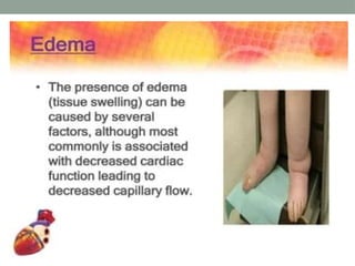
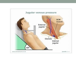
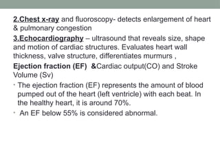
CARDIOVASCULAR
ASSESSMENT
Physical assessment
General Appearance
Vital Signs
Height and Weight
. Inspection and palpation
1-Skin • Color • Turgor • Temperature and moisture:-
2- Nails • Nails should be assessed for color, shape, thickness,
symmetry, and adherence.
• Nail abnormalities:-
a. Clubbing of the fingers is associated with decreased oxygen. In
clubbing, the distal tips of the fingers become bulbous, the nails
are thickened hard, and curved at the tip, and the nail bed feels
boggy when squeezed
b. Separation from the nail bed produces a white, yellowish, or
greenish color on the non-adherent portion of the nail.
c.Capillary refill time: A quick test to assess the adequacy of
circulation in an individual with poor cardiac output. An area of skin/ a
fingertip is pressed firmly until it becomes white; the number of
seconds for the area to turn pink again indicates capillary refill time.
Normal capillary refill takes around 2 seconds.
• 3.Inspection of neck
• Inspecting the carotid artery and jugular venous system
• With the patient in a supine position, inspect the carotid and jugular
venous systems in the neck for pulsations.
• To visualize external venous pulsations, look for pulsations in the
supraclavicular area.
• To visualize internal venous pulsations, look for pulsations at the
suprasternal notch. • Using a penlight to cast a shadow on the neck
vessels may help you visualize the pulsations
• carotids have visible pulsation, jugulars have undulated wave.
• Measuring Jugular Venous Pressure
-Position patient with the head of bed at 30 to 45-degree
angle.
- Place a ruler vertically, perpendicular to the chest at the
angle of Louis (sternal angle).
-Identify the highest level of the jugular vein pulsation; if
unable to see pulsations, use the highest level of jugular
vein distension.
- Place another ruler horizontally at the point of the highest
level of the venous pulsation
Measure the distance up from the chest wall.
• The normal JVP is less than 3 cm. A central venous
pressure can be estimated by adding 5 cm to the JVP
• Palpation
• • Palpate the Carotid and jugular veins
• Palpating the Precordium • - Identify and palpate each
cardiac site for pulsations, and thrills:
• 1. Apex (left ventricular area), or mitral area -fifth
intercostal space, midclavicular line
• 2.Aortic area, second intercostal space right sternal
border.
• 3.Pulmonic area-second intercostal space left sternal
border
• 4.tricuspid area, fourth to fifth intercostal space at left
sternal border.
AUSCULTATION
• Whoop (sometimes called a honk):
• Loud, variable intensity, musical sound heard at the apex
in late systole
• Friction rub: Creaking sound heard with pericardial
inflammation
• Murmur: Sounds made by turbulence in the heart or
blood stream
• Laboratory tests
• Creatine kinase (CK) and its isoenzyme CK-MB
• Lactic dehydrogenase
• Troponin I
• low-density lipoproteins (LDL) and high-density lipoproteins
(HDL).
• Coagulation Studies
• Partial thromboplastin time (PTT)
• • Prothrombin time (PT)
Diagnostic procedures
1.CARDIAC CYCLE EKG/ECG /Electrocardiography
A 12 lead EKG is a graphic record of the electrical forces
produced by the heart
2.Chest x-ray and fluoroscopy- detects enlargement of heart
& pulmonary congestion
3.Echocardiography – ultrasound that reveals size, shape
and motion of cardiac structures. Evaluates heart wall
thickness, valve structure, differentiates murmurs ,
Ejection fraction (EF) &Cardiac output(CO) and Stroke
Volume (Sv)
• The ejection fraction (EF) represents the amount of blood
pumped out of the heart (left ventricle) with each beat. In
the healthy heart, it is around 70%.
• An EF below 55% is considered abnormal.
5.Angiography / cardiac catherization determines coronary
lesion size, location, evaluate (L) ventricular function,
measures heart pressures
6.Exercise tolerance test
7.Radionuclide Imaging
Cardiovascular assessment for nurses.pptx

Cardiovascular assessment for nurses.pptx

  • 1.
  • 2.
    Physical assessment General Appearance VitalSigns Height and Weight . Inspection and palpation 1-Skin • Color • Turgor • Temperature and moisture:- 2- Nails • Nails should be assessed for color, shape, thickness, symmetry, and adherence. • Nail abnormalities:- a. Clubbing of the fingers is associated with decreased oxygen. In clubbing, the distal tips of the fingers become bulbous, the nails are thickened hard, and curved at the tip, and the nail bed feels boggy when squeezed b. Separation from the nail bed produces a white, yellowish, or greenish color on the non-adherent portion of the nail.
  • 7.
    c.Capillary refill time:A quick test to assess the adequacy of circulation in an individual with poor cardiac output. An area of skin/ a fingertip is pressed firmly until it becomes white; the number of seconds for the area to turn pink again indicates capillary refill time. Normal capillary refill takes around 2 seconds. • 3.Inspection of neck • Inspecting the carotid artery and jugular venous system • With the patient in a supine position, inspect the carotid and jugular venous systems in the neck for pulsations. • To visualize external venous pulsations, look for pulsations in the supraclavicular area. • To visualize internal venous pulsations, look for pulsations at the suprasternal notch. • Using a penlight to cast a shadow on the neck vessels may help you visualize the pulsations • carotids have visible pulsation, jugulars have undulated wave.
  • 8.
    • Measuring JugularVenous Pressure -Position patient with the head of bed at 30 to 45-degree angle. - Place a ruler vertically, perpendicular to the chest at the angle of Louis (sternal angle). -Identify the highest level of the jugular vein pulsation; if unable to see pulsations, use the highest level of jugular vein distension. - Place another ruler horizontally at the point of the highest level of the venous pulsation Measure the distance up from the chest wall. • The normal JVP is less than 3 cm. A central venous pressure can be estimated by adding 5 cm to the JVP
  • 10.
    • Palpation • •Palpate the Carotid and jugular veins • Palpating the Precordium • - Identify and palpate each cardiac site for pulsations, and thrills: • 1. Apex (left ventricular area), or mitral area -fifth intercostal space, midclavicular line • 2.Aortic area, second intercostal space right sternal border. • 3.Pulmonic area-second intercostal space left sternal border • 4.tricuspid area, fourth to fifth intercostal space at left sternal border.
  • 12.
    AUSCULTATION • Whoop (sometimescalled a honk): • Loud, variable intensity, musical sound heard at the apex in late systole • Friction rub: Creaking sound heard with pericardial inflammation • Murmur: Sounds made by turbulence in the heart or blood stream
  • 13.
    • Laboratory tests •Creatine kinase (CK) and its isoenzyme CK-MB • Lactic dehydrogenase • Troponin I • low-density lipoproteins (LDL) and high-density lipoproteins (HDL). • Coagulation Studies • Partial thromboplastin time (PTT) • • Prothrombin time (PT) Diagnostic procedures 1.CARDIAC CYCLE EKG/ECG /Electrocardiography A 12 lead EKG is a graphic record of the electrical forces produced by the heart
  • 15.
    2.Chest x-ray andfluoroscopy- detects enlargement of heart & pulmonary congestion 3.Echocardiography – ultrasound that reveals size, shape and motion of cardiac structures. Evaluates heart wall thickness, valve structure, differentiates murmurs , Ejection fraction (EF) &Cardiac output(CO) and Stroke Volume (Sv) • The ejection fraction (EF) represents the amount of blood pumped out of the heart (left ventricle) with each beat. In the healthy heart, it is around 70%. • An EF below 55% is considered abnormal.
  • 16.
    5.Angiography / cardiaccatherization determines coronary lesion size, location, evaluate (L) ventricular function, measures heart pressures 6.Exercise tolerance test 7.Radionuclide Imaging