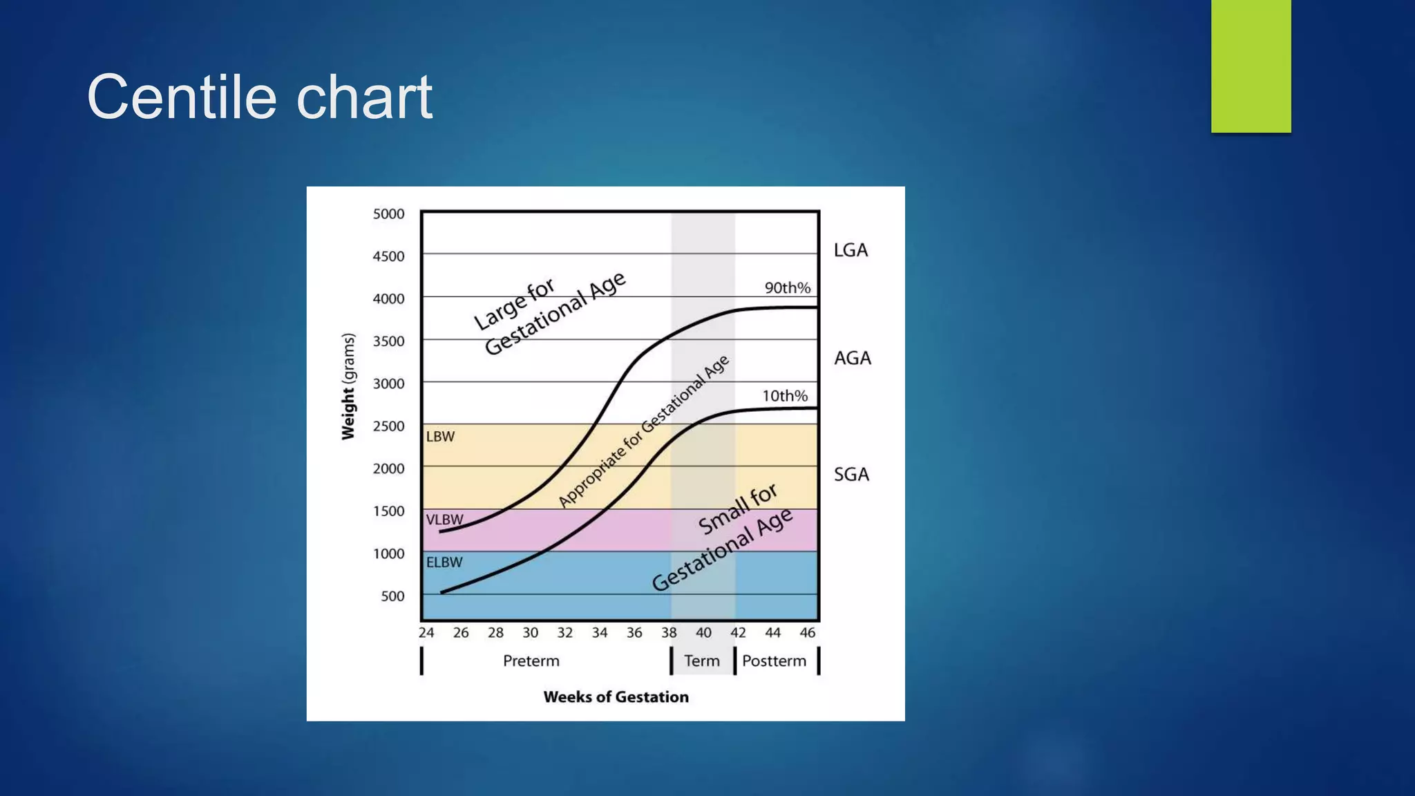
Fetal physiology & Normal Growth
cvs
 Fetal cardiac output is more ( 100ml /kg) vs 80 ml /kg in adult
 Cardiac index is 4 times higher
 Aortic pressure 30-45 mmgh increasing with increasing GA
 Blood volume of neonate = 85 ml /kg
 Low systemic and high pulmonary pressure and resistance
Fetal circulation
 The umbilical arteries arise from the caudal end of the
 dorsal fetal aorta and carry deoxygenated blood from the fetus to the placenta
 Oxygenated blood is returned to the fetus via the 2 umbilical vein to the fetal liver
 Bypass the liver joining the IVC by ductus venosus to enter the right atrium
 The ductus venosus stream passes across the right atrium through a
 physiological defect in the atrial septum called the foramen ovale, to the left
 atrium
 the blood passes through the mitral valve to the left ventricle and hence to the aorta
 50% of the blood goes to the head and upper extremities, providing high levels of oxygen
to supply the fetal heart, upper thorax and brain
 Deoxygenated blood returning from the fetal head and lower body flows through the right
atrium and ventricle and into the pulmonary artery, after which it bypasses the lungs to
enter the descending aorta via the ductus arteriosus that connects the two vessels
 desaturated blood from the right ventricle passes down the aorta to enter the umbilical
arterial circulation and be returned to the placenta for reoxygenation
Adult structure of fetal CVS remnants
Ductus arteriosus closed by increasing po2
after birth , bradykinin , and decreased PG
Umbilical arteries became the mediaL
umbilical ligament
Urachus became mediaN umbilical ligament
Umbilical vein became ligamentum teres
Ductus arteriosus
 Why its kept open in fetus ?
 Low po2 , PG (PGE2, PGI2*prostacyclin), fall In pulmonary vascular resistance
 Why it closes after birth
 Elevated po2 , bradykinin

 When its closed normally ?
 Few days after birth

 What does it became when anatomically closed ?
 Ligamentum arteriosus

 why it may be delayed to be closed ?
 Prematurity , Rubella
 Why it might be closed prematurely ?
 Prostaglandin inhibitors (indomethacin )
 What happens if still open for long time PDA ?
 Cyanosis , hypoxia, lung congestion , necrotizing enterocolitis , IVH
 What is the treatment of PDA ?
 PG inhibitors (indomethacin ) in premature babies and surgical correction in term babies
 Do we need to keep it open in some conditions ?
 Yes , with some cyanotic heart diseases using prostaglandin as in pulmonary atresia and TGA
Fetal hematology
 Hemopoiesis starts from yolk sac at 3 weeks until 10-12 weeks
from liver from 8 weeks to 18 weeks
from bone marrow from 12 weeks until birth
(at 20 weeks its almost from BM )
at birth its only BM
 Fetal blood is Gower Hb …. Until 10 weeks
 From 10 weeks its fetal Hb
 From 28 weeks starts to be converted to Hb A2
 At birth its 80 % HbF
 Became adult hemoglobin completely at 6 month age( 1% is HbF)
Fetal RBCs
 Larger
 Shorter half life (80 days )
 Less 2,3 dpg
 Resist acid and alkali
 More affinity to o2 (d.curve to left )
Immune system
 T cells from thymus at 9 weeks , spleen and lymph nodes at 12 weeks …
circulate as mature cells at 16 weeks
 B cells from liver and spleen at 12 weeks
 Passive immunization from mother is IgG and active immunity from fetus
by IgM
Fetal CNS
 From ectoderm (neural tube and crest)
 Movement from 1st trimester (body 7 weeks , limbs 9 weeks )
 Maternal perception of movement around 18-20 weeks
(24 weeks PG and 16 weeks multiparas)
 First sensation by the fetus is touch at 10 weeks with other senses after 26
weeks
 Myelination completed at 3 years old
Fetal GIT
 Gut differentiation at end of 4th week weeks
 Physiological hernia at 6 weeks till maximum 12 weeks
 Swallowing start at 14 weeks
 Suckling at 28 weeks
 Meconium formed at 10 weeks but appear in colon at 16 weeks
Fetal GIT abnormalities
 Atresia (duodenal atresia in down syndrome)
 Tracheo-esophageal fistula
 Malrotation(volvulus)
 Mackle's diverticulum
(persistent vetillointestinal duct between midgut and yolk sac)
 Omphalocele
Failure of intestine to get back after physiological hernia , central , umbilical , covered , 50 %
risk of chromosomal abnormalities
 Gastroschisis
Para-umbilical , exposed , risk of IUGR in 30 %
Fetal Liver, spleen and pancreas
 From endoderm at 4th week
 By the sixth week, the fetal liver performs haematopoiesis. This peaks at
12–
 16 weeks and continues until approximately 36 weeks
 Spleen at 5th week
 Pancreas at 4th week
FETAL LUNG
 From primitive foregut at 4 weeks GA
 Fetal breathing is seen from 12 weeks GA during REM sleep
 Pulmonary surfactant, a complex mixture of phospholipids and
 proteins that reduces surface tension at the air–liquid interface of the alveolus, is
 produced by the type II pneumocytes starting from about 30 weeks
 The predominant phospholipid in surfactant (80%) is phosphatidylcholine (lecithin)
 The production of which is enhanced by cortisol, growth restriction and prolonged
rupture of the membranes,steroids
 delayed in maternal diabetes mellitus and androgen
Fetal urine
 Start to be produced at 10-12 week
 Make a major component of amniotic fluid after 18 weeks
 Increased as in diabetic mothers ---- hydramnios
 Decreased in obstructive uropathy , potter syndrome , PCK …….
Oligohydramnios
Fetal Growth
 Multifactorial
 growth potential of the fetus
genetic as gender and aneuploids
hormonal
Perinatal infections
 intrauterine environment (placental )
As in chronic abruptio placenta , maternal vascular diseases , antepartum hemorrhage
 Maternal (+paternal)
Age , race , pre pregnancy weight , weight gain in pregnancy , maternal illness , behavioral as
smoking and alcohol intake
 Of hormonal influence IGF1 is of particular importance ….
 Role in macrosomia in Diabetic mothers !
Assessment of fetal
growth
 SFH
(low risk population)
 Ultrasound estimation of fetal weight
• (high risk population or when SFH is not reliable as in multiple gestation , obesity , fibroids, hydramnios
• Expressed by centile chart as AGA (10-90 centile )
SGA below 10th centile
LGA above the 90th centile
Centile chart
Fetal ABGS and neonatal APGAR

Lecture 6 Fetal physiology and normal growth

  • 1.
    Fetal physiology &Normal Growth
  • 2.
    cvs  Fetal cardiacoutput is more ( 100ml /kg) vs 80 ml /kg in adult  Cardiac index is 4 times higher  Aortic pressure 30-45 mmgh increasing with increasing GA  Blood volume of neonate = 85 ml /kg  Low systemic and high pulmonary pressure and resistance
  • 3.
    Fetal circulation  Theumbilical arteries arise from the caudal end of the  dorsal fetal aorta and carry deoxygenated blood from the fetus to the placenta  Oxygenated blood is returned to the fetus via the 2 umbilical vein to the fetal liver  Bypass the liver joining the IVC by ductus venosus to enter the right atrium  The ductus venosus stream passes across the right atrium through a  physiological defect in the atrial septum called the foramen ovale, to the left  atrium
  • 4.
     the bloodpasses through the mitral valve to the left ventricle and hence to the aorta  50% of the blood goes to the head and upper extremities, providing high levels of oxygen to supply the fetal heart, upper thorax and brain  Deoxygenated blood returning from the fetal head and lower body flows through the right atrium and ventricle and into the pulmonary artery, after which it bypasses the lungs to enter the descending aorta via the ductus arteriosus that connects the two vessels  desaturated blood from the right ventricle passes down the aorta to enter the umbilical arterial circulation and be returned to the placenta for reoxygenation
  • 6.
    Adult structure offetal CVS remnants Ductus arteriosus closed by increasing po2 after birth , bradykinin , and decreased PG Umbilical arteries became the mediaL umbilical ligament Urachus became mediaN umbilical ligament Umbilical vein became ligamentum teres
  • 7.
    Ductus arteriosus  Whyits kept open in fetus ?  Low po2 , PG (PGE2, PGI2*prostacyclin), fall In pulmonary vascular resistance  Why it closes after birth  Elevated po2 , bradykinin   When its closed normally ?  Few days after birth   What does it became when anatomically closed ?  Ligamentum arteriosus   why it may be delayed to be closed ?  Prematurity , Rubella
  • 8.
     Why itmight be closed prematurely ?  Prostaglandin inhibitors (indomethacin )  What happens if still open for long time PDA ?  Cyanosis , hypoxia, lung congestion , necrotizing enterocolitis , IVH  What is the treatment of PDA ?  PG inhibitors (indomethacin ) in premature babies and surgical correction in term babies  Do we need to keep it open in some conditions ?  Yes , with some cyanotic heart diseases using prostaglandin as in pulmonary atresia and TGA
  • 9.
    Fetal hematology  Hemopoiesisstarts from yolk sac at 3 weeks until 10-12 weeks from liver from 8 weeks to 18 weeks from bone marrow from 12 weeks until birth (at 20 weeks its almost from BM ) at birth its only BM  Fetal blood is Gower Hb …. Until 10 weeks  From 10 weeks its fetal Hb  From 28 weeks starts to be converted to Hb A2  At birth its 80 % HbF  Became adult hemoglobin completely at 6 month age( 1% is HbF)
  • 10.
    Fetal RBCs  Larger Shorter half life (80 days )  Less 2,3 dpg  Resist acid and alkali  More affinity to o2 (d.curve to left )
  • 11.
    Immune system  Tcells from thymus at 9 weeks , spleen and lymph nodes at 12 weeks … circulate as mature cells at 16 weeks  B cells from liver and spleen at 12 weeks  Passive immunization from mother is IgG and active immunity from fetus by IgM
  • 12.
    Fetal CNS  Fromectoderm (neural tube and crest)  Movement from 1st trimester (body 7 weeks , limbs 9 weeks )  Maternal perception of movement around 18-20 weeks (24 weeks PG and 16 weeks multiparas)  First sensation by the fetus is touch at 10 weeks with other senses after 26 weeks  Myelination completed at 3 years old
  • 13.
    Fetal GIT  Gutdifferentiation at end of 4th week weeks  Physiological hernia at 6 weeks till maximum 12 weeks  Swallowing start at 14 weeks  Suckling at 28 weeks  Meconium formed at 10 weeks but appear in colon at 16 weeks
  • 14.
    Fetal GIT abnormalities Atresia (duodenal atresia in down syndrome)  Tracheo-esophageal fistula  Malrotation(volvulus)  Mackle's diverticulum (persistent vetillointestinal duct between midgut and yolk sac)  Omphalocele Failure of intestine to get back after physiological hernia , central , umbilical , covered , 50 % risk of chromosomal abnormalities  Gastroschisis Para-umbilical , exposed , risk of IUGR in 30 %
  • 15.
    Fetal Liver, spleenand pancreas  From endoderm at 4th week  By the sixth week, the fetal liver performs haematopoiesis. This peaks at 12–  16 weeks and continues until approximately 36 weeks  Spleen at 5th week  Pancreas at 4th week
  • 16.
    FETAL LUNG  Fromprimitive foregut at 4 weeks GA  Fetal breathing is seen from 12 weeks GA during REM sleep  Pulmonary surfactant, a complex mixture of phospholipids and  proteins that reduces surface tension at the air–liquid interface of the alveolus, is  produced by the type II pneumocytes starting from about 30 weeks  The predominant phospholipid in surfactant (80%) is phosphatidylcholine (lecithin)  The production of which is enhanced by cortisol, growth restriction and prolonged rupture of the membranes,steroids  delayed in maternal diabetes mellitus and androgen
  • 17.
    Fetal urine  Startto be produced at 10-12 week  Make a major component of amniotic fluid after 18 weeks  Increased as in diabetic mothers ---- hydramnios  Decreased in obstructive uropathy , potter syndrome , PCK ……. Oligohydramnios
  • 18.
    Fetal Growth  Multifactorial growth potential of the fetus genetic as gender and aneuploids hormonal Perinatal infections  intrauterine environment (placental ) As in chronic abruptio placenta , maternal vascular diseases , antepartum hemorrhage  Maternal (+paternal) Age , race , pre pregnancy weight , weight gain in pregnancy , maternal illness , behavioral as smoking and alcohol intake  Of hormonal influence IGF1 is of particular importance ….  Role in macrosomia in Diabetic mothers !
  • 19.
    Assessment of fetal growth SFH (low risk population)  Ultrasound estimation of fetal weight • (high risk population or when SFH is not reliable as in multiple gestation , obesity , fibroids, hydramnios • Expressed by centile chart as AGA (10-90 centile ) SGA below 10th centile LGA above the 90th centile
  • 20.
  • 21.
    Fetal ABGS andneonatal APGAR