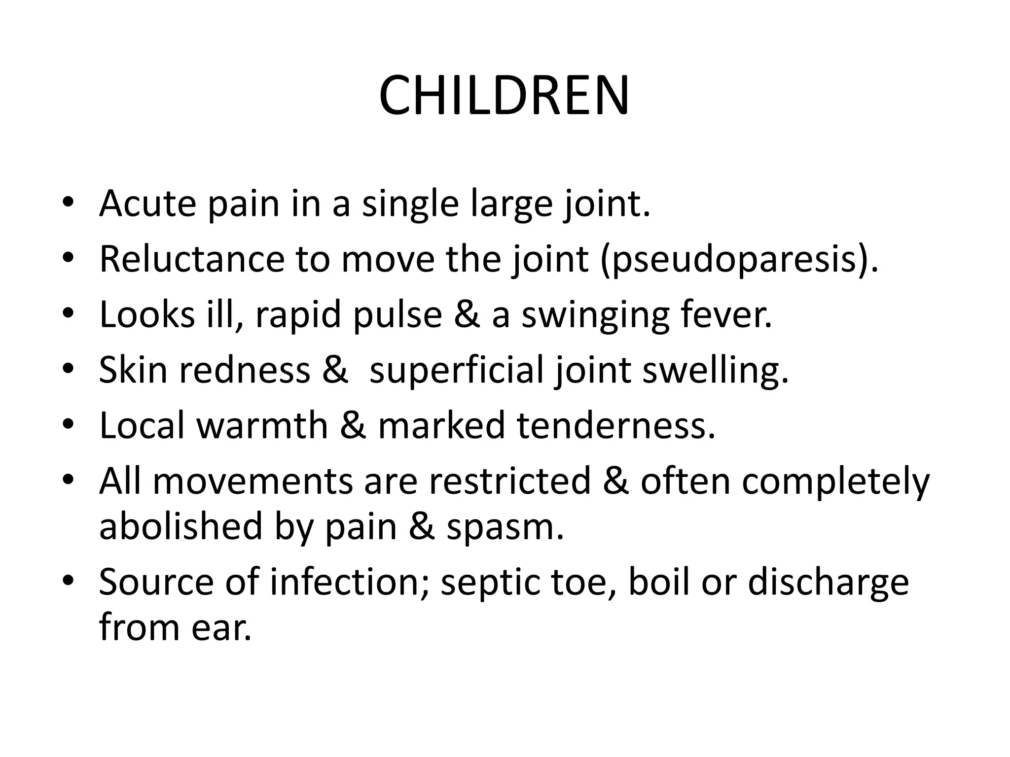
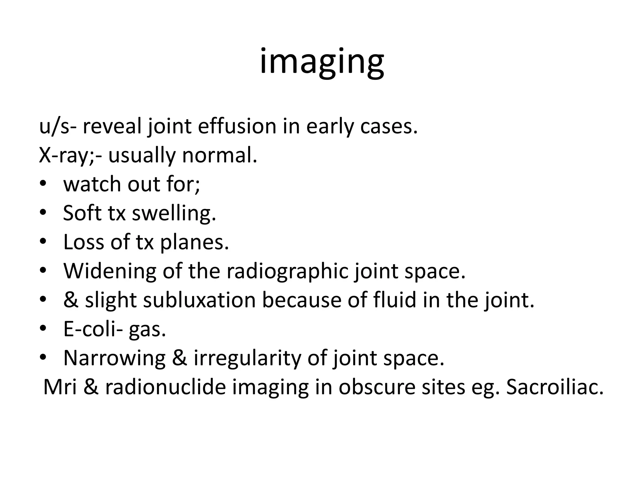
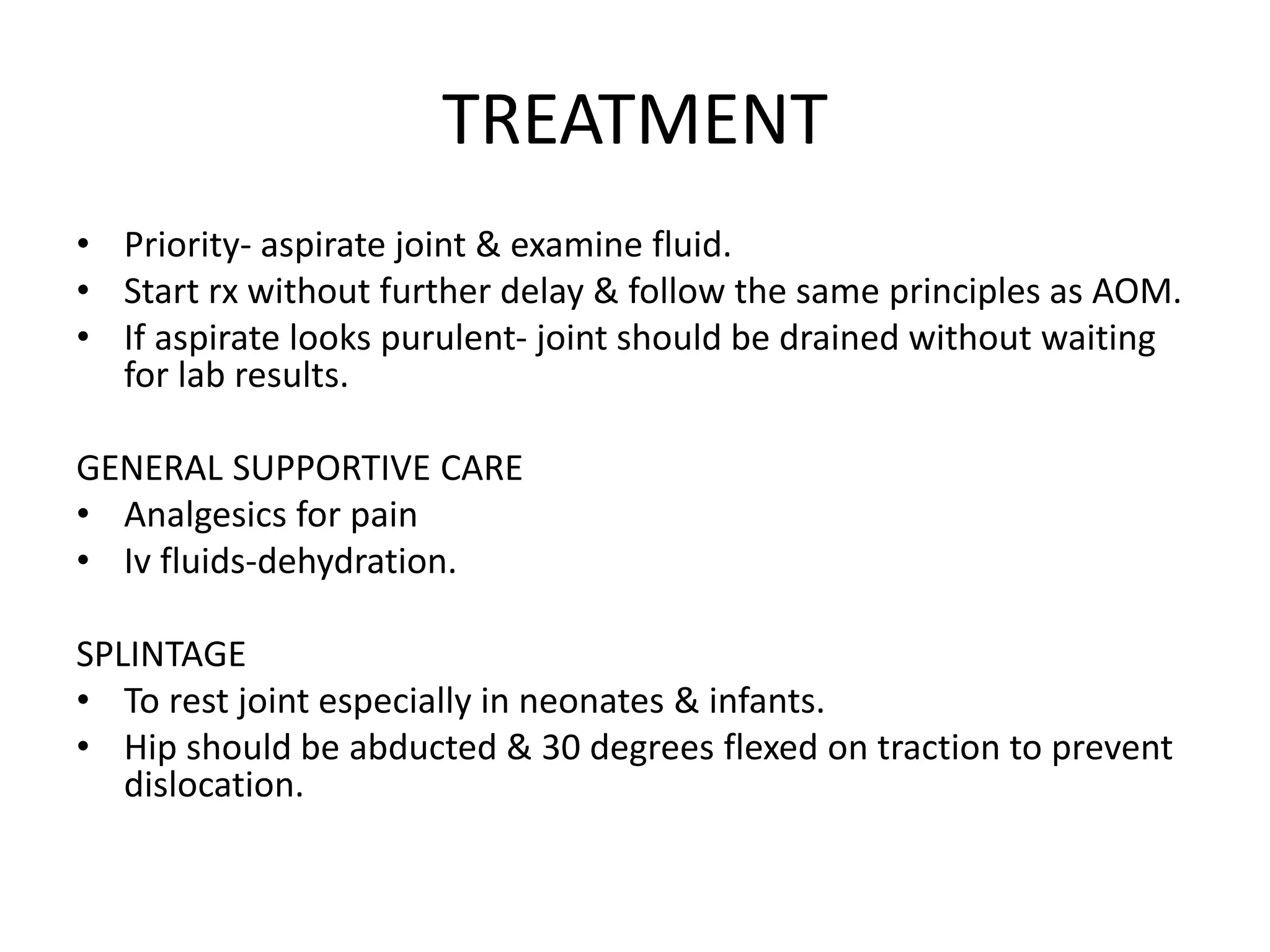
ACUTE SUPPURATIVE ARTHRITIS.
Dr. Muthoka
A jt can become infected by;
I. Direct invasion through a penetrating wound, inta-
articular injection or arthroscopy.
II. Direct spread from an adjacent bone abscess.
III. Blood spread from a distant site.
In infants infection start in the metaphyseal
bone(osteomyelitis) then spread to the joint or vice
versa.
Causal organism: staph-aureus.
In children btn 1 & 4 years old; H influenzae,
streptococcus, E-coli, & proteus.
Predisposing conditions: Rh arthritis, chronic
debilitating disorder, IV drug abuse, AIDs
PATHOLOGY
• Haematogenous spread.
• Settle in the synovial membrane.
• Acute inflammatory reaction with serous or
seropurulent exudate & an increase in synovial fluid.
• Pus appears in the joint.
• Articular cartilage is eroded & destroyed by bacterial
enzymes & proteolytic enzymes released from synovial
cells, inflammatory cells & pus.
• In infants the epiphysis may be severely damaged.
• Older children, vascular occlusion may lead to necrosis
of epiphyseal bone
• In adults the effects are usually confined to the
articular cartilage, but in the late cases there may be
extensive erosion due to synovial proliferation &
ingrowth.
• If untreated, infection spread to the underlying bone or
burst out of the joint to form an abscess & sinuses.
• With healing there may be:
1. Complete resolution & return to normal.
2. Partial loss of articular cartilage & fibrosis of the joint.
3. Loss of articular cartilage & bony ankylosis.
4. Bone destruction & permanent deformity of the joint.
CLINICAL FEATURES
• Differ according to age of the patient.
NEW BORN INFANT
• Emphasis is on septicemia rather than jt pain.
• Irritable & refuses to feed.
• Rapid pulse & fever.
• JT;
– FEEL
– Move to elicit local signs of warmth & tenderness.
– Resistance to mvt.
– Examine umbilical cord.
CHILDREN
• Acute pain in a single large joint.
• Reluctance to move the joint (pseudoparesis).
• Looks ill, rapid pulse & a swinging fever.
• Skin redness & superficial joint swelling.
• Local warmth & marked tenderness.
• All movements are restricted & often completely
abolished by pain & spasm.
• Source of infection; septic toe, boil or discharge
from ear.
Adults
• Often a superficial joint: knee, wrist, a finger,
ankle or toe that is painful swollen &
inflamed.
• Warmth & local tenderness.
• Mvts are restricted.
• Pt should be questioned & examined for
gonococcal infection or drug abuse.
• Pt on rheumatoid arthritis rx may develop a
silent joint infection.
imaging
u/s- reveal joint effusion in early cases.
X-ray;- usually normal.
• watch out for;
• Soft tx swelling.
• Loss of tx planes.
• Widening of the radiographic joint space.
• & slight subluxation because of fluid in the joint.
• E-coli- gas.
• Narrowing & irregularity of joint space.
Mri & radionuclide imaging in obscure sites eg. Sacroiliac.
Lab investigations
Raised WCC & ESR.
Blood culture may be +ve.
SPECIAL INVESTIGATIONS
Aspirate joint & examine joint fluid:
– May be purulent.
– Early-clear.
– Aspirate- WCC & gram stain immediately.
– Normal leukocyte count-<300 per ml.
– >10000 per ml in non-infective inflammatory disorders.
– >50000 per ml are suggestive of sepsis.
– Gram –ve cocci; H influenzae or kingella kingae (in
children) gonococcus in adults.
Ddx
1. Acute OM.
2. Other types of infection-psoas abscess/ local pelvic infection
3. Trauma-traumatic synovitis/ haemarthrosis/ jt aspiration.
4. Irritable jt; painful & lack mvt. Child is not ill.
5. Rh fever- typically the pain flits from jt to jt
6. JRA-gradual onset with less severe systemic symptoms.
7. Sickle cell dx.
8. Gaucher’s pseudo- osteitis. Acute pain & fever without any
organism.
9. Gout &pseudogout- acute crystal induced synovitis- aspiration;
turbid jt fluid/ raised wcc/ microscopic exam by polarized
microscope show xtic crystals.
TREATMENT
• Priority- aspirate joint & examine fluid.
• Start rx without further delay & follow the same principles as AOM.
• If aspirate looks purulent- joint should be drained without waiting
for lab results.
GENERAL SUPPORTIVE CARE
• Analgesics for pain
• Iv fluids-dehydration.
SPLINTAGE
• To rest joint especially in neonates & infants.
• Hip should be abducted & 30 degrees flexed on traction to prevent
dislocation.
ANTIBIOTICS
• As AHO.
• Neonates & infants up to 6 months
– penincillinase resistant penincillins eg.-
flucloxacillin & 3rd generation cephalosporins
– Children from 6 months to puberty–similar.
– Older teenagers & adults- flucloxacillin & fusidic
acid.
– 3rd generation cephalosporins- IV for 4-7 days then
orally for 3 weeks.
DRAINAGE
• Under anaesthesia.
• Jt is opened through a small incision, drained & washed out
with physiological saline.
• A small catheter is left in place & the wound is closed.
• Suction- irrigation is continued for 2-3 days
• Advisable in
1. very young infants
2. Involving hip
3. Very thick pus aspirated
• Older children; repeated closed aspiration may work.
• If there is no improvement within 48 hours open drainage
is done.
complications
Subluxation & dislocation of the hip
Instability of the knee
Damage to cartilagenous physis or epiphysis in growing child.
Sequelae:
• Retarded growth
• Partial or complete destruction of the epiphysis
• Deformity of the joint
• Epiphyseal osteonecrosis
• Acetabular dysplasia
• Pseudoarthrosis of the hip
• Acetabular cartilage erosion( chondrolysis)is seen in older pts & this
may result in restricted mvt or complete ankylosis of the joint.

LECTURE 24; ACUTE SUPPURATIVE ARTHRITIS.pptx

  • 1.
  • 2.
    A jt canbecome infected by; I. Direct invasion through a penetrating wound, inta- articular injection or arthroscopy. II. Direct spread from an adjacent bone abscess. III. Blood spread from a distant site. In infants infection start in the metaphyseal bone(osteomyelitis) then spread to the joint or vice versa. Causal organism: staph-aureus. In children btn 1 & 4 years old; H influenzae, streptococcus, E-coli, & proteus. Predisposing conditions: Rh arthritis, chronic debilitating disorder, IV drug abuse, AIDs
  • 3.
    PATHOLOGY • Haematogenous spread. •Settle in the synovial membrane. • Acute inflammatory reaction with serous or seropurulent exudate & an increase in synovial fluid. • Pus appears in the joint. • Articular cartilage is eroded & destroyed by bacterial enzymes & proteolytic enzymes released from synovial cells, inflammatory cells & pus. • In infants the epiphysis may be severely damaged.
  • 4.
    • Older children,vascular occlusion may lead to necrosis of epiphyseal bone • In adults the effects are usually confined to the articular cartilage, but in the late cases there may be extensive erosion due to synovial proliferation & ingrowth. • If untreated, infection spread to the underlying bone or burst out of the joint to form an abscess & sinuses. • With healing there may be: 1. Complete resolution & return to normal. 2. Partial loss of articular cartilage & fibrosis of the joint. 3. Loss of articular cartilage & bony ankylosis. 4. Bone destruction & permanent deformity of the joint.
  • 5.
    CLINICAL FEATURES • Differaccording to age of the patient. NEW BORN INFANT • Emphasis is on septicemia rather than jt pain. • Irritable & refuses to feed. • Rapid pulse & fever. • JT; – FEEL – Move to elicit local signs of warmth & tenderness. – Resistance to mvt. – Examine umbilical cord.
  • 6.
    CHILDREN • Acute painin a single large joint. • Reluctance to move the joint (pseudoparesis). • Looks ill, rapid pulse & a swinging fever. • Skin redness & superficial joint swelling. • Local warmth & marked tenderness. • All movements are restricted & often completely abolished by pain & spasm. • Source of infection; septic toe, boil or discharge from ear.
  • 7.
    Adults • Often asuperficial joint: knee, wrist, a finger, ankle or toe that is painful swollen & inflamed. • Warmth & local tenderness. • Mvts are restricted. • Pt should be questioned & examined for gonococcal infection or drug abuse. • Pt on rheumatoid arthritis rx may develop a silent joint infection.
  • 8.
    imaging u/s- reveal jointeffusion in early cases. X-ray;- usually normal. • watch out for; • Soft tx swelling. • Loss of tx planes. • Widening of the radiographic joint space. • & slight subluxation because of fluid in the joint. • E-coli- gas. • Narrowing & irregularity of joint space. Mri & radionuclide imaging in obscure sites eg. Sacroiliac.
  • 9.
    Lab investigations Raised WCC& ESR. Blood culture may be +ve. SPECIAL INVESTIGATIONS Aspirate joint & examine joint fluid: – May be purulent. – Early-clear. – Aspirate- WCC & gram stain immediately. – Normal leukocyte count-<300 per ml. – >10000 per ml in non-infective inflammatory disorders. – >50000 per ml are suggestive of sepsis. – Gram –ve cocci; H influenzae or kingella kingae (in children) gonococcus in adults.
  • 10.
    Ddx 1. Acute OM. 2.Other types of infection-psoas abscess/ local pelvic infection 3. Trauma-traumatic synovitis/ haemarthrosis/ jt aspiration. 4. Irritable jt; painful & lack mvt. Child is not ill. 5. Rh fever- typically the pain flits from jt to jt 6. JRA-gradual onset with less severe systemic symptoms. 7. Sickle cell dx. 8. Gaucher’s pseudo- osteitis. Acute pain & fever without any organism. 9. Gout &pseudogout- acute crystal induced synovitis- aspiration; turbid jt fluid/ raised wcc/ microscopic exam by polarized microscope show xtic crystals.
  • 11.
    TREATMENT • Priority- aspiratejoint & examine fluid. • Start rx without further delay & follow the same principles as AOM. • If aspirate looks purulent- joint should be drained without waiting for lab results. GENERAL SUPPORTIVE CARE • Analgesics for pain • Iv fluids-dehydration. SPLINTAGE • To rest joint especially in neonates & infants. • Hip should be abducted & 30 degrees flexed on traction to prevent dislocation.
  • 12.
    ANTIBIOTICS • As AHO. •Neonates & infants up to 6 months – penincillinase resistant penincillins eg.- flucloxacillin & 3rd generation cephalosporins – Children from 6 months to puberty–similar. – Older teenagers & adults- flucloxacillin & fusidic acid. – 3rd generation cephalosporins- IV for 4-7 days then orally for 3 weeks.
  • 13.
    DRAINAGE • Under anaesthesia. •Jt is opened through a small incision, drained & washed out with physiological saline. • A small catheter is left in place & the wound is closed. • Suction- irrigation is continued for 2-3 days • Advisable in 1. very young infants 2. Involving hip 3. Very thick pus aspirated • Older children; repeated closed aspiration may work. • If there is no improvement within 48 hours open drainage is done.
  • 14.
    complications Subluxation & dislocationof the hip Instability of the knee Damage to cartilagenous physis or epiphysis in growing child. Sequelae: • Retarded growth • Partial or complete destruction of the epiphysis • Deformity of the joint • Epiphyseal osteonecrosis • Acetabular dysplasia • Pseudoarthrosis of the hip • Acetabular cartilage erosion( chondrolysis)is seen in older pts & this may result in restricted mvt or complete ankylosis of the joint.