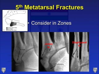
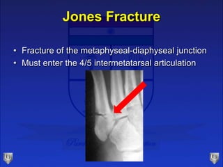
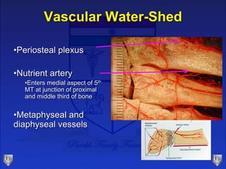
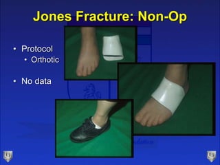
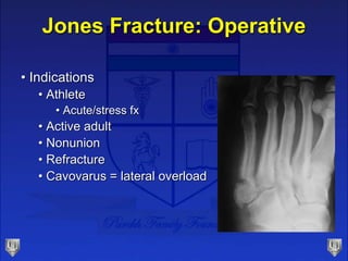
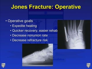
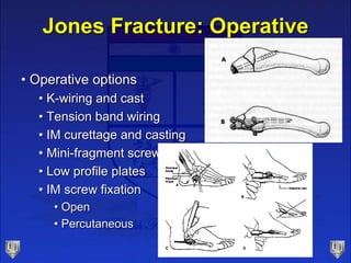
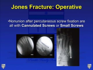
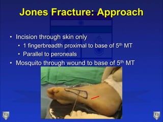
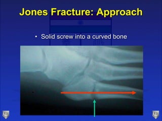
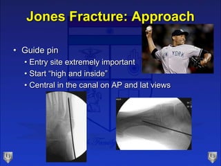
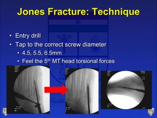
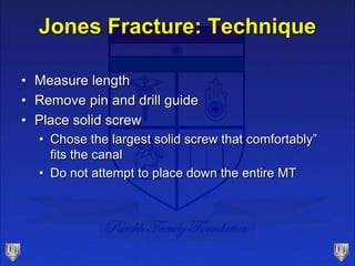
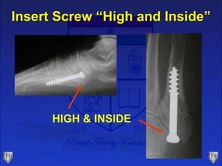
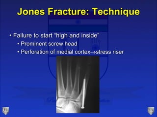
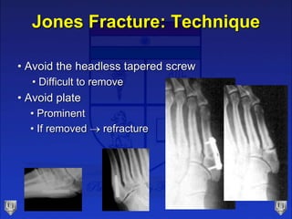
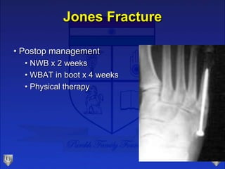
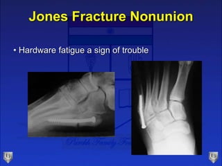
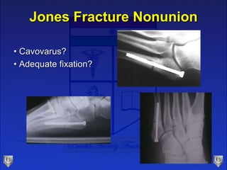
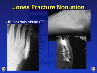
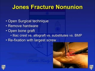
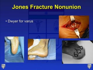
Jones Fractures
Selene G. Parekh, MD, MBA
Associate Professor
North Carolina Orthopaedic Clinic
Department of Orthopaedic Surgery
Adjunct Faculty Fuqua Business School
Duke University
Durham, NC
919.471.9622
www.seleneparekhmd.com
@seleneparekhmd
5th Metatarsal Fractures
• Consider in Zones
Jones
fx
Diaphyseal
fx
Jones Fracture
• Fracture of the metaphyseal-diaphyseal junction
• Must enter the 4/5 intermetatarsal articulation
Mechanism of Injury
• Vertical and mediolateral forces concentrated
over the 5th MT
• Forced adduction
• Can result in a fracture between shaft and
immobile base
• Greater propensity
• Forefoot supination
• Knee/ankle/hindfoot varus
Anatomy
• Peroneus brevis
• Peroneus tertius
• Abductor digiti minimi
• Lateral band of plantar fascia
• Plantar and dorsal interosseous
• Flexor digiti minimi brevis
Vascular Water-Shed
•Periosteal plexus
•Nutrient artery
•Enters medial aspect of 5th
MT at junction of proximal
and middle third of bone
•Metaphyseal and
diaphyseal vessels
Jones Fracture: Non-Op
• Indications
• Elderly
• Non-athletic
• Low-demand
• Protocol
• NWB cast 4-8 weeks
• 100 Fractures of 5th MT: Naval base
• 72% union (avg. time 21.2 wks)Clapper, ClinOrth 1995
Jones Fracture: Non-Op
• Protocol
• Orthotic
• No data
Jones Fracture: Operative
• Indications
• Athlete
• Acute/stress fx
• Active adult
• Nonunion
• Refracture
• Cavovarus = lateral overload
Jones Fracture: Operative
• Operative goals
• Expedite healing
• Quicker recovery; easier rehab
• Decrease nonunion rate
• Decrease refracture risk
Jones Fracture: Operative
• Operative options
• K-wiring and cast
• Tension band wiring
• IM curettage and casting
• Mini-fragment screws
• Low profile plates
• IM screw fixation
• Open
• Percutaneous
Jones Fracture: Operative
•Screw Fixation
• Screw choices
• 4.5 mm malleolar (Synthes)
• 5.0 mm cannulated (ACE/DePuy)
• 6.5 mm short thread (Synthes)
Jones Fracture: Operative
•Nonunion after percutaneous screw fixation are
all with Cannulated Screws or Small Screws
Jones Fracture: Operative
• Percutaneous Screw Fixation
• Solid screw
• Large fragment screws
• Dedicated system
• Cannulated bone preparation
• Solid screw fixation
Jones Fracture: Set-up
• Supine
• Ankle block
• Mini C-arm
• Flex knee and place foot on base
Jones Fracture: Approach
• Incision through skin only
• 1 fingerbreadth proximal to base of 5th MT
• Parallel to peroneals
• Mosquito through wound to base of 5th MT
Jones Fracture: Approach
• Solid screw into a curved bone
Jones Fracture: Approach
• Guide pin
• Entry site extremely important
• Start “high and inside”
• Central in the canal on AP and lat views
Jones Fracture: Technique
• Entry drill
• Tap to the correct screw diameter
• 4.5, 5.5, 6.5mm
• Feel the 5th MT head torsional forces
Jones Fracture: Technique
• Measure length
• Remove pin and drill guide
• Place solid screw
• Chose the largest solid screw that comfortably”
fits the canal
• Do not attempt to place down the entire MT
Insert Screw “High and Inside”
HIGH & INSIDE
Jones Fracture: Technique
• Failure to start “high and inside”
• Prominent screw head
• Perforation of medial cortexstress riser
Jones Fracture: Technique
• Avoid the headless tapered screw
• Difficult to remove
• Avoid plate
• Prominent
• If removed  refracture
Jones Fracture
• Postop management
• NWB x 2 weeks
• WBAT in boot x 4 weeks
• Physical therapy
Jones Fracture
• Begin running in modified shoewear at 6-8 weeks
if clinically nontender
Jones Fracture
• Follow radiographs
• Healing may not be
evident for 12 weeks
12 wks p.o.
Jones Fracture
• Leave screw forever!
Pitfalls of IM Screw
• Infection
• Delayed wound healing
• Distal perforation (stress riser)
• Sural neuritis/neuroma
• Peroneal tendinitis
• Persistent nonunion
Jones Fracture Nonunion
• Hardware fatigue a sign of trouble
Jones Fracture Nonunion
• Cavovarus?
• Adequate fixation?
Jones Fracture Nonunion
• If uncertain obtain CT
Jones Fracture Nonunion
• Open Surgical technique
• Remove hardware
• Open bone graft
• Iliac crest vs. allograft vs. substitutes vs. BMP
• Re-fixation with largest screw
Jones Fracture Nonunion
• Dwyer for varus
RE
ECT
the ankle
the foot

Lecture 16 parekh jones