Downloaded 215 times


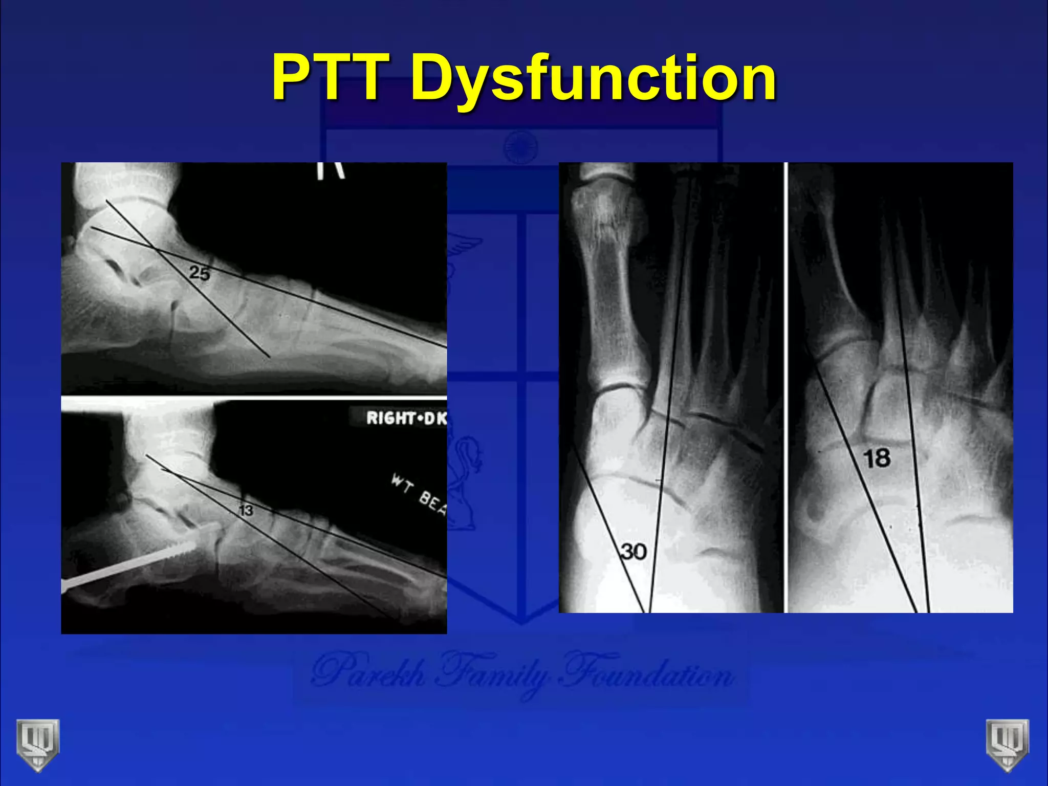


Stage 2 posterior tibial tendon dysfunction is characterized by an elongated and partially torn tendon, resulting in a flexible deformity with increased heel valgus and possible forefoot abduction. Treatment begins conservatively with orthotics but may progress to surgical procedures like flexor digitorum longus transfer, medial displacement calcaneal osteotomy, and lateral column lengthening to restore the arch height and alignment. More advanced cases may require fusion of the talonavicular joint or additional medial column procedures to address deformity in the midfoot. The goal of surgical treatment is to correct all aspects of the flexible deformity through procedures on the hindfoot, midfoot and forefoot.


