SlideShare a Scribd company logo
Embryology of Head and neck
• The mesenchyme for formation of the head region is derived from
mesoderm, neural crest, and thickened regions of ectoderm known as
ectodermal placodes.
G.LUFUKUJA
PHARYNGEAL APPARATUS
(Branchial arch)
• Neural crest cells originate in the neuroectoderm of
forebrain, midbrain, and hindbrain regions and migrate
ventrally into the pharyngeal arches and rostrally around the
forebrain and optic cup into the facial region. In these
locations they form mid-facial and pharyngeal arch skeletal
structures
• There are 5 pairs of branchial arches, that form on either side
of the pharyngeal foregut on day 22 are the embryologic
basis of all the differentiated structures of the head and neck
Pharyngeal Apparatus
• The head and neck region of four week human
embryo somewhat resemble these regions of
a fish embryo of comparable stage
• This explains the former use of designation
branchial apparatus
• Branchial is derived from the Greek word
branchia or gill
Pharyngeal Apparatus
Pharyngeal apparatus consists of:
• Pharyngeal arches
• Pharyngeal pouches
• Pharyngeal grooves/clefts
• Pharyngeal membrane
Pharyngeal Arches
• Pharyngeal arches begin to develop early in the
fourth week as neural crest cells migrate into the
head and neck region
• The first pair of pharyngeal arches (primordium of
jaws) appears as a surface elevations lateral to the
developing pharynx
• Soon other arches appear as obliquely disposed,
rounded ridges on each side of the future head and
neck regions
G.LUFUKUJA 7
G.LUFUKUJA 8
Branchial arches
Pharyngeal Arches
• By the end of the fourth week, four pairs of
pharyngeal arches are visible externally
• The fifth and sixth arches are rudimentary and are
not visible on the surface of the embryo
• The pharyngeal arches are separated from each
other by fissures called pharyngeal grooves
• They are numbered in craniocaudal sequence
Pharyngeal Arches
• The first pharyngeal arch (mandibular arch)
develops maxillary and mandibular
prominences
• The first pair of pharyngeal arches plays a
major role in facial development
• The second pharyngeal arch (hyoid arch)
contributes to the formation of hyoid bone
Pharyngeal Arch Components
• Each pharyngeal arch consists of a core of
mesenchyme
• Is covered externally by ectoderm and internally by
endoderm
• In the third week the original mesenchyme is derived
from mesoderm
• During the fourth week most of the mesenchyme is
derived from neural crest cells that migrate into the
pharyngeal arches
Fate of Pharyngeal Arches
• The pharyngeal arches contribute exclusively to the
formation of the face, nasal cavities, mouth, larynx,
pharynx and neck
• During the fifth week, the second pharyngeal arch
enlarges and overgrows the third and fourth arches,
forming the ectodermal depression called cervical
sinus
• By the end of seventh week the second to fourth
pharyngeal grooves and the cervical sinus have
disappeared, giving the neck a smooth contour
Fate of Pharyngeal Arches
A typical pharyngeal arch contains:
• An aortic arch, an artery that arises from the truncus
arteriosus of the primordial heart
• A cartilaginous rod that forms the skeleton of the arch
• A muscular component that differentiates into muscles in the
head and neck
• A nerve that supplies the mucosa and muscles derived from
the arch
Derivatives of Pharyngeal Arch Cartilages
• The dorsal end of first arch cartilage (Meckel
cartilage) ossifies to form malleus and incus
• The middle part of cartilage forms anterior ligament
of malleus sphenomandibular ligament
• Ventral part of the first arch cartilages form
primordium of the mandible
• The cartilage disappears as mandible develops
around it
Derivatives of Pharyngeal Arch Cartilages
• The dorsal end of second arch cartilage (Reichert
cartilage) ossifies to form the stapes and styloid
process of the temporal bone
• The ventral end of second arch cartilage ossifies to
form the lesser cornu and superior part of the body
of the hyoid bone
• Its perichondrium forms the stylohyoid ligament
Derivatives of Pharyngeal Arch Cartilages
• The third arch cartilage ossifies to form the greater
cornu and the inferior part of the body of the hyoid
bone
• The fourth and sixth arch cartilages fuse to form the
laryngeal cartilages except epiglottis which develops
from hypopharyngeal eminence
• The fifth pharyngeal arch is rudimentary and has no
derivatives
Derivatives of Pharyngeal Arch Muscles
• The musculature of the first pharyngeal arch forms the
muscles of mastication
• The second pharyngeal arch forms the stapedius, stylohyoid,
posterior belly of digastric, auricular and muscles of facial
expression
• The third arch forms the stylopharyngeus
• The fourth arch forms cricothyroid, levator veli palatini and
constrictors of pharynx
• Sixth pharyngeal arch forms the intrinsic muscles of the larynx
Derivatives of Pharyngeal Arch Nerves
• Caudal two branches of Trigeminal nerve (maxillary and
mandibular) supply derivatives of the first pharyngeal arch
• The facial, glossopharyngeal and vagus nerves supply the
second, third and caudal (fourth to sixth) arches respectively
• The fourth arch is supplied by superior laryngeal branch of
vagus nerve
• The sixth arch is supplied by its recurrent laryngeal branch
Pharyngeal Pouches
• The primordial pharynx, derived from the foregut,
widens cranially where it joins the primordial mouth
or stomodeum
• It narrows caudally where it joins the esophagus
• The endoderm of the pharynx lines the internal
aspects of pharyngeal arches and passes into
balloonlike diverticula called pharyngeal pouches
Pharyngeal Pouches
• The pairs of pouches develop in a craniocaudal
sequence between the arches
• The first pair of pouches lies between the first and
second pharyngeal arches
• There are four well defined pairs of pharyngeal
pouches
• The fifth pair is absent or rudimentary
Pharyngeal Pouches
• The endoderm of the pouches contacts the
ectoderm of the pharyngeal grooves and
together they form the double layered
pharyngeal membranes that separate the
pharyngeal pouches from the pharyngeal
grooves
Derivatives of First Pharyngeal Pouch
• The first pharyngeal pouch expands into an elongate
tubotympanic recess
• The expanded distal part of this recess contacts the
first pharyngeal groove, where it contributes to the
formation of the tympanic membrane (eardrum)
• The cavity of the tubotympanic recess gives rise to
the tympanic cavity and mastoid antrum
Derivatives of Second Pharyngeal Pouch
• The second pharyngeal pouch is largely obliterated
as the palatine tonsils develop
• Part of the cavity of this pouch remains as the
tonsillar sinus or fossa
• The endoderm of the pouch proliferates and grows
into the underlying mesenchyme
• The central parts of these buds form crypts
Derivatives of Second Pharyngeal Pouch
• The pouch endoderm forms the surface
epithelium and the lining of the tonsillar
crypts
• At about 20 weeks the mesenchyme around
the crypts differentiates into lymphoid tissue
• These tissues soon organize into the lymphatic
nodules of the palatine tonsil
Derivatives of Third Pharyngeal Pouch
• The third pharyngeal pouch expands and develops a
solid, dorsal bulbar part and a hollow elongate
ventral part
• Its connection with the pharynx is reduced to a
narrow duct that soon degenerates
• By the sixth week the epithelium of each dorsal
bulbar part begins to differentiate into inferior
parathyroid gland
Derivatives of Third Pharyngeal Pouch
• The epithelium of the elongate ventral parts of third
pharyngeal pouch proliferates and their cavities obliterate
• These bilateral primordia of thymus come together in the
median plane to form thymus
• It descends into the superior mediastenum
• The bilobed form of thymus remains throughout life
• Discretely encapsulated and each lobe has its own blood
supply, lymphatic drainage and nerve supply
Derivatives of Third Pharyngeal Pouch
• The primordia of thymus and parathyroid
glands lose their connections with the
pharynx and migrate into the neck
• Later the parathyroid glands separate from
the thymus and lie on the dorsal surface of the
thyroid gland
Derivatives of Fourth Pharyngeal Pouch
• The fourth pharyngeal pouch also expands into
dorsal bulbar and elongate ventral parts
• Its connection with the pharynx is reduced to a
narrow duct that soon degenerates
• By the sixth week, each dorsal part develops into a
superior parathyroid gland
• It lies on the dorsal surface of the thyroid gland
Derivatives of Fourth Pharyngeal Pouch
• The parathyroid glands derived from the third
pouches descend with the thymus and are
carried to a more inferior position than the
parathyroid derived from the fourth pouches
• This explains why the parathyroid glands
derived from the third pair of pouches are
located inferior to those from the fourth
pouches
The Fifth Pharyngeal Pouch
• When this develops, this rudimentary pouch
becomes part of the fourth pharyngeal pouch
and helps to form the ultimopharyngeal body
Pharyngeal Grooves
• During the fourth and fifth weeks, head and neck
region of the human embryo exhibit four pharyngeal
grooves or clefts on each side
• These grooves separate the pharyngeal arches
externally
• Only first pair persists as the external acoustic
meatus
• The other grooves normally obliterated with the
cervical sinus as the neck develops
Development of the Tongue
• The adult tongue is derived from embryonic processes arising
from the first three pharyngeal arches and a small
contribution from the fourth arch.
• The anterior two thirds of the adult tongue (the movable part) is
formed by two lateral swellings and a single one, the tuberculum
impar, all of which are derived from the first pharyngeal arch
44G.LUFUKUJA
Development of the Tongue…
• The posterior third (the fixed part or the root of the tongue) is
formed by an embryonic process, the hypobranchial eminence
or copula. This process is derived mainly from the second and
third pharyngeal arches with small part from the fourth
pharyngeal arch.
• The muscles of the tongue are derived from a different embryonic
source, namely the occipital myotomes.
Development of the Tongue
Development of the Tongue…
• The junction of the anterior 2/3 and the posterior 1/3 of the adult
tongue is marked by a V-shaped structure, the sulcus terminalis
and at its tip is a blind foramen, the foramen caecum. This site
marks the area from which an epithelial structure arise during
embryonic development and extend inferiorly to the anterior part
of the future neck where it forms the thyroid gland.
Thyroid Gland
• The thyroid gland appears as an
epithelial proliferation in the floor of the
pharynx between the tuberculum impar
and the copula at a point later indicated
by the foramen cecum . Subsequently
the thyroid descends in front of the
pharyngeal gut as a bilobed
diverticulum.
• During this migration, the thyroid
remains connected to the tongue by a
narrow canal, the thyroglossal duct.
50
The Face
• The face is formed from 7 primordia surrounding the stomodeum
• Single frontonasal prominence -cranial boundary of the stomodeum
• Paired median nasal process
• Paired maxillary process
• Paired mandibularprocess
G.LUFUKUJA
Development of the Face…
• The Frontonasal prominence forms the forehead. The nasal
prominence is split into lateral and medial nasal processes by
formation of a nasal placode
• Thus a horseshoe-shaped nasal processes surrounds the nasal
placode, which undergoes apoptosis to form a depression; the
nasal pit. Initially, the maxillary and lateral nasal prominences
are separated by a deep furrow, the nasolacrimal groove.
• Ectoderm in the floor of this groove forms a solid epithelial cord
that detaches from the overlying ectoderm. After canalization, the
cord forms the nasolacrimal duct; its upper end widens to form
the lacrimal sac.
Intermaxillary segment
• As a result of medial growth of the maxillary prominences, the
two medial nasal prominences merge not only at the surface but
also at a deeper level. The structure formed by the two merged
prominences is the intermaxillary segment
54G.LUFUKUJA
55
Anomalies
• Craniofacial: These are defects associated with tissue fusion.
Estimated 1/3 of all congenital defects are craniaofacial
• Facial Clefts - Anomalies associated with defective fusion of
Facial prominences
 1. Cleft lip - failure of maxillary prominence to fuse with
intermaxillary process
 2. Cleft palate - failure of the lateral palatine processes to fuse
56G.LUFUKUJA
Thank you
57G.LUFUKUJA

More Related Content

What's hot

Skull Development
Skull DevelopmentSkull Development
Skull Development
Pro Faather
 
Embryology of head and neck
Embryology of head and neckEmbryology of head and neck
Embryology of head and neck
Dr Bhavik Miyani
 
Development of face
Development of faceDevelopment of face
Development of face
Dr Sudeep Madhusudan Chaudhari
 
Development of Palate and Tongue PPT
Development of Palate and Tongue PPTDevelopment of Palate and Tongue PPT
Development of Palate and Tongue PPT
oral and maxillofacial pathology
 
PHARYNGEAL ARCHES,POUCHES
PHARYNGEAL ARCHES,POUCHESPHARYNGEAL ARCHES,POUCHES
PHARYNGEAL ARCHES,POUCHES
Jamil Kifayatullah
 
3. formation of notochord
3. formation of notochord3. formation of notochord
3. formation of notochord
Dr. Mohammad Mahmoud
 
Development of the tongue
Development of the tongueDevelopment of the tongue
Development of the tongueLheanne Tesoro
 
Development of face, palate and jaw
Development of face, palate and jawDevelopment of face, palate and jaw
Development of face, palate and jaw
K BHATTACHARJEE
 
Development of face
Development of faceDevelopment of face
Development of face
dr. supratim tripathi
 
Tongue development, applied anatomy and prosthetic implications
Tongue development, applied anatomy and prosthetic implicationsTongue development, applied anatomy and prosthetic implications
Tongue development, applied anatomy and prosthetic implications
Dr. KRITI TREHAN
 
DEVELOPMENT OF PAROTID GLAND
DEVELOPMENT OF PAROTID GLANDDEVELOPMENT OF PAROTID GLAND
DEVELOPMENT OF PAROTID GLAND
akash chauhan
 
Development of face and oral cavity*
Development of face and oral cavity*Development of face and oral cavity*
Development of face and oral cavity*Meloy Macainag
 
Development of Face and Palate including Anomalies of palate
Development of Face and Palate including Anomalies of palate Development of Face and Palate including Anomalies of palate
Development of Face and Palate including Anomalies of palate
Ragya Bharadwaj
 
The hyoglossal muscle
The hyoglossal muscleThe hyoglossal muscle
The hyoglossal muscle
Idris Siddiqui
 
Development of face
Development of faceDevelopment of face
Development of face
Dr. Deepashree Paul
 
Foramina of the Skull and the Structures that Pass Through
Foramina of the Skull and the Structures that Pass ThroughForamina of the Skull and the Structures that Pass Through
Foramina of the Skull and the Structures that Pass Through
Ahmad Amro Baradee
 
Development of Respiratory System i lung bud & larynx
Development of Respiratory System i lung bud & larynxDevelopment of Respiratory System i lung bud & larynx
Development of Respiratory System i lung bud & larynx
Rohit Paswan
 
Development of Face, Nose and Palate (Special Embryology)
Development of Face, Nose and Palate (Special Embryology)Development of Face, Nose and Palate (Special Embryology)
Development of Face, Nose and Palate (Special Embryology)
Dr. Sherif Fahmy
 
Folding of embryo
Folding of embryoFolding of embryo
Folding of embryo
Farhan Ali
 
Development of Face
Development of FaceDevelopment of Face

What's hot (20)

Skull Development
Skull DevelopmentSkull Development
Skull Development
 
Embryology of head and neck
Embryology of head and neckEmbryology of head and neck
Embryology of head and neck
 
Development of face
Development of faceDevelopment of face
Development of face
 
Development of Palate and Tongue PPT
Development of Palate and Tongue PPTDevelopment of Palate and Tongue PPT
Development of Palate and Tongue PPT
 
PHARYNGEAL ARCHES,POUCHES
PHARYNGEAL ARCHES,POUCHESPHARYNGEAL ARCHES,POUCHES
PHARYNGEAL ARCHES,POUCHES
 
3. formation of notochord
3. formation of notochord3. formation of notochord
3. formation of notochord
 
Development of the tongue
Development of the tongueDevelopment of the tongue
Development of the tongue
 
Development of face, palate and jaw
Development of face, palate and jawDevelopment of face, palate and jaw
Development of face, palate and jaw
 
Development of face
Development of faceDevelopment of face
Development of face
 
Tongue development, applied anatomy and prosthetic implications
Tongue development, applied anatomy and prosthetic implicationsTongue development, applied anatomy and prosthetic implications
Tongue development, applied anatomy and prosthetic implications
 
DEVELOPMENT OF PAROTID GLAND
DEVELOPMENT OF PAROTID GLANDDEVELOPMENT OF PAROTID GLAND
DEVELOPMENT OF PAROTID GLAND
 
Development of face and oral cavity*
Development of face and oral cavity*Development of face and oral cavity*
Development of face and oral cavity*
 
Development of Face and Palate including Anomalies of palate
Development of Face and Palate including Anomalies of palate Development of Face and Palate including Anomalies of palate
Development of Face and Palate including Anomalies of palate
 
The hyoglossal muscle
The hyoglossal muscleThe hyoglossal muscle
The hyoglossal muscle
 
Development of face
Development of faceDevelopment of face
Development of face
 
Foramina of the Skull and the Structures that Pass Through
Foramina of the Skull and the Structures that Pass ThroughForamina of the Skull and the Structures that Pass Through
Foramina of the Skull and the Structures that Pass Through
 
Development of Respiratory System i lung bud & larynx
Development of Respiratory System i lung bud & larynxDevelopment of Respiratory System i lung bud & larynx
Development of Respiratory System i lung bud & larynx
 
Development of Face, Nose and Palate (Special Embryology)
Development of Face, Nose and Palate (Special Embryology)Development of Face, Nose and Palate (Special Embryology)
Development of Face, Nose and Palate (Special Embryology)
 
Folding of embryo
Folding of embryoFolding of embryo
Folding of embryo
 
Development of Face
Development of FaceDevelopment of Face
Development of Face
 

Similar to Embryology of head and neck

Development of Pharyngeal arches and pouches ppt
Development of Pharyngeal arches and pouches pptDevelopment of Pharyngeal arches and pouches ppt
Development of Pharyngeal arches and pouches ppt
BharanidharanS5
 
HEAD AND NECK (2).pptx
HEAD AND NECK (2).pptxHEAD AND NECK (2).pptx
HEAD AND NECK (2).pptx
Divinefavour12
 
Embryology of head&neck es - copy
Embryology of head&neck es - copyEmbryology of head&neck es - copy
Embryology of head&neck es - copy
sallamahmed1
 
phayrangeal apparatus By Dr.Aisha Sadaf IBMS -KMU-peshawar
phayrangeal apparatus By Dr.Aisha Sadaf IBMS -KMU-peshawarphayrangeal apparatus By Dr.Aisha Sadaf IBMS -KMU-peshawar
phayrangeal apparatus By Dr.Aisha Sadaf IBMS -KMU-peshawarAisha Sadaf
 
THE_EMBRYOLOGY_OF_HEAD_&_NECK[1].ppt
THE_EMBRYOLOGY_OF_HEAD_&_NECK[1].pptTHE_EMBRYOLOGY_OF_HEAD_&_NECK[1].ppt
THE_EMBRYOLOGY_OF_HEAD_&_NECK[1].ppt
Dr Ndayisaba Corneille
 
Branchial arches
Branchial archesBranchial arches
Branchial arches
DrpriyonaJohn
 
the_pharyngeal_arches.pdf
the_pharyngeal_arches.pdfthe_pharyngeal_arches.pdf
the_pharyngeal_arches.pdf
Akash875331
 
Embryology of Pharyngeal Arches by Dr Niazur Rahman
Embryology of Pharyngeal Arches by Dr Niazur RahmanEmbryology of Pharyngeal Arches by Dr Niazur Rahman
Embryology of Pharyngeal Arches by Dr Niazur Rahman
S M Niazur Rahman
 
Embryo of Ear and Nose
Embryo of Ear and NoseEmbryo of Ear and Nose
Embryology of branchial arches applied anatomy by osama elhamzawy
Embryology of branchial arches applied anatomy by osama elhamzawyEmbryology of branchial arches applied anatomy by osama elhamzawy
Embryology of branchial arches applied anatomy by osama elhamzawy
Osama Elhamzawy
 
Pharyngeal Arches, Pouches and clefts-1.pptx
Pharyngeal Arches, Pouches and clefts-1.pptxPharyngeal Arches, Pouches and clefts-1.pptx
Pharyngeal Arches, Pouches and clefts-1.pptx
EmmanuelIshioma
 
Embrylogy of head and neck part 2
Embrylogy of head and neck part 2Embrylogy of head and neck part 2
Embrylogy of head and neck part 2
deepika seshagiri
 
Development of respiratory system
Development of respiratory systemDevelopment of respiratory system
Development of respiratory system
Dr. Mohammad Mahmoud
 
Birth defect system according to System wise in that Respiratory System Birth...
Birth defect system according to System wise in that Respiratory System Birth...Birth defect system according to System wise in that Respiratory System Birth...
Birth defect system according to System wise in that Respiratory System Birth...
sonal patel
 
Growth and development of facial structures
Growth and development of facial structures Growth and development of facial structures
Growth and development of facial structures
Kunal Ajay Patankar
 
DEVELOPMENT OF FACE
DEVELOPMENT OF FACEDEVELOPMENT OF FACE
DEVELOPMENT OF FACE
Dr. Swathi Yennemadi
 
Embryology of ear8
Embryology of ear8Embryology of ear8
Embryology of ear8
Dr Zeeshan Ahmad
 
Embryology In Orthodontics
Embryology In OrthodonticsEmbryology In Orthodontics
Embryology In Orthodontics
Miliya Parveen
 
Pharyngeal apparatus.pptx
Pharyngeal apparatus.pptxPharyngeal apparatus.pptx
Pharyngeal apparatus.pptx
Sundip Charmode
 
Embryonic period of development
Embryonic period of developmentEmbryonic period of development
Embryonic period of development
Dr.Nadeesha Abegunasekara
 

Similar to Embryology of head and neck (20)

Development of Pharyngeal arches and pouches ppt
Development of Pharyngeal arches and pouches pptDevelopment of Pharyngeal arches and pouches ppt
Development of Pharyngeal arches and pouches ppt
 
HEAD AND NECK (2).pptx
HEAD AND NECK (2).pptxHEAD AND NECK (2).pptx
HEAD AND NECK (2).pptx
 
Embryology of head&neck es - copy
Embryology of head&neck es - copyEmbryology of head&neck es - copy
Embryology of head&neck es - copy
 
phayrangeal apparatus By Dr.Aisha Sadaf IBMS -KMU-peshawar
phayrangeal apparatus By Dr.Aisha Sadaf IBMS -KMU-peshawarphayrangeal apparatus By Dr.Aisha Sadaf IBMS -KMU-peshawar
phayrangeal apparatus By Dr.Aisha Sadaf IBMS -KMU-peshawar
 
THE_EMBRYOLOGY_OF_HEAD_&_NECK[1].ppt
THE_EMBRYOLOGY_OF_HEAD_&_NECK[1].pptTHE_EMBRYOLOGY_OF_HEAD_&_NECK[1].ppt
THE_EMBRYOLOGY_OF_HEAD_&_NECK[1].ppt
 
Branchial arches
Branchial archesBranchial arches
Branchial arches
 
the_pharyngeal_arches.pdf
the_pharyngeal_arches.pdfthe_pharyngeal_arches.pdf
the_pharyngeal_arches.pdf
 
Embryology of Pharyngeal Arches by Dr Niazur Rahman
Embryology of Pharyngeal Arches by Dr Niazur RahmanEmbryology of Pharyngeal Arches by Dr Niazur Rahman
Embryology of Pharyngeal Arches by Dr Niazur Rahman
 
Embryo of Ear and Nose
Embryo of Ear and NoseEmbryo of Ear and Nose
Embryo of Ear and Nose
 
Embryology of branchial arches applied anatomy by osama elhamzawy
Embryology of branchial arches applied anatomy by osama elhamzawyEmbryology of branchial arches applied anatomy by osama elhamzawy
Embryology of branchial arches applied anatomy by osama elhamzawy
 
Pharyngeal Arches, Pouches and clefts-1.pptx
Pharyngeal Arches, Pouches and clefts-1.pptxPharyngeal Arches, Pouches and clefts-1.pptx
Pharyngeal Arches, Pouches and clefts-1.pptx
 
Embrylogy of head and neck part 2
Embrylogy of head and neck part 2Embrylogy of head and neck part 2
Embrylogy of head and neck part 2
 
Development of respiratory system
Development of respiratory systemDevelopment of respiratory system
Development of respiratory system
 
Birth defect system according to System wise in that Respiratory System Birth...
Birth defect system according to System wise in that Respiratory System Birth...Birth defect system according to System wise in that Respiratory System Birth...
Birth defect system according to System wise in that Respiratory System Birth...
 
Growth and development of facial structures
Growth and development of facial structures Growth and development of facial structures
Growth and development of facial structures
 
DEVELOPMENT OF FACE
DEVELOPMENT OF FACEDEVELOPMENT OF FACE
DEVELOPMENT OF FACE
 
Embryology of ear8
Embryology of ear8Embryology of ear8
Embryology of ear8
 
Embryology In Orthodontics
Embryology In OrthodonticsEmbryology In Orthodontics
Embryology In Orthodontics
 
Pharyngeal apparatus.pptx
Pharyngeal apparatus.pptxPharyngeal apparatus.pptx
Pharyngeal apparatus.pptx
 
Embryonic period of development
Embryonic period of developmentEmbryonic period of development
Embryonic period of development
 

Recently uploaded

Guidance_and_Counselling.pdf B.Ed. 4th Semester
Guidance_and_Counselling.pdf B.Ed. 4th SemesterGuidance_and_Counselling.pdf B.Ed. 4th Semester
Guidance_and_Counselling.pdf B.Ed. 4th Semester
Atul Kumar Singh
 
Azure Interview Questions and Answers PDF By ScholarHat
Azure Interview Questions and Answers PDF By ScholarHatAzure Interview Questions and Answers PDF By ScholarHat
Azure Interview Questions and Answers PDF By ScholarHat
Scholarhat
 
2024.06.01 Introducing a competency framework for languag learning materials ...
2024.06.01 Introducing a competency framework for languag learning materials ...2024.06.01 Introducing a competency framework for languag learning materials ...
2024.06.01 Introducing a competency framework for languag learning materials ...
Sandy Millin
 
1.4 modern child centered education - mahatma gandhi-2.pptx
1.4 modern child centered education - mahatma gandhi-2.pptx1.4 modern child centered education - mahatma gandhi-2.pptx
1.4 modern child centered education - mahatma gandhi-2.pptx
JosvitaDsouza2
 
Digital Tools and AI for Teaching Learning and Research
Digital Tools and AI for Teaching Learning and ResearchDigital Tools and AI for Teaching Learning and Research
Digital Tools and AI for Teaching Learning and Research
Vikramjit Singh
 
Synthetic Fiber Construction in lab .pptx
Synthetic Fiber Construction in lab .pptxSynthetic Fiber Construction in lab .pptx
Synthetic Fiber Construction in lab .pptx
Pavel ( NSTU)
 
Lapbook sobre os Regimes Totalitários.pdf
Lapbook sobre os Regimes Totalitários.pdfLapbook sobre os Regimes Totalitários.pdf
Lapbook sobre os Regimes Totalitários.pdf
Jean Carlos Nunes Paixão
 
How to Make a Field invisible in Odoo 17
How to Make a Field invisible in Odoo 17How to Make a Field invisible in Odoo 17
How to Make a Field invisible in Odoo 17
Celine George
 
The Accursed House by Émile Gaboriau.pptx
The Accursed House by Émile Gaboriau.pptxThe Accursed House by Émile Gaboriau.pptx
The Accursed House by Émile Gaboriau.pptx
DhatriParmar
 
The French Revolution Class 9 Study Material pdf free download
The French Revolution Class 9 Study Material pdf free downloadThe French Revolution Class 9 Study Material pdf free download
The French Revolution Class 9 Study Material pdf free download
Vivekanand Anglo Vedic Academy
 
The Challenger.pdf DNHS Official Publication
The Challenger.pdf DNHS Official PublicationThe Challenger.pdf DNHS Official Publication
The Challenger.pdf DNHS Official Publication
Delapenabediema
 
Honest Reviews of Tim Han LMA Course Program.pptx
Honest Reviews of Tim Han LMA Course Program.pptxHonest Reviews of Tim Han LMA Course Program.pptx
Honest Reviews of Tim Han LMA Course Program.pptx
timhan337
 
Unit 8 - Information and Communication Technology (Paper I).pdf
Unit 8 - Information and Communication Technology (Paper I).pdfUnit 8 - Information and Communication Technology (Paper I).pdf
Unit 8 - Information and Communication Technology (Paper I).pdf
Thiyagu K
 
June 3, 2024 Anti-Semitism Letter Sent to MIT President Kornbluth and MIT Cor...
June 3, 2024 Anti-Semitism Letter Sent to MIT President Kornbluth and MIT Cor...June 3, 2024 Anti-Semitism Letter Sent to MIT President Kornbluth and MIT Cor...
June 3, 2024 Anti-Semitism Letter Sent to MIT President Kornbluth and MIT Cor...
Levi Shapiro
 
The approach at University of Liverpool.pptx
The approach at University of Liverpool.pptxThe approach at University of Liverpool.pptx
The approach at University of Liverpool.pptx
Jisc
 
Overview on Edible Vaccine: Pros & Cons with Mechanism
Overview on Edible Vaccine: Pros & Cons with MechanismOverview on Edible Vaccine: Pros & Cons with Mechanism
Overview on Edible Vaccine: Pros & Cons with Mechanism
DeeptiGupta154
 
A Strategic Approach: GenAI in Education
A Strategic Approach: GenAI in EducationA Strategic Approach: GenAI in Education
A Strategic Approach: GenAI in Education
Peter Windle
 
Multithreading_in_C++ - std::thread, race condition
Multithreading_in_C++ - std::thread, race conditionMultithreading_in_C++ - std::thread, race condition
Multithreading_in_C++ - std::thread, race condition
Mohammed Sikander
 
Best Digital Marketing Institute In NOIDA
Best Digital Marketing Institute In NOIDABest Digital Marketing Institute In NOIDA
Best Digital Marketing Institute In NOIDA
deeptiverma2406
 
How libraries can support authors with open access requirements for UKRI fund...
How libraries can support authors with open access requirements for UKRI fund...How libraries can support authors with open access requirements for UKRI fund...
How libraries can support authors with open access requirements for UKRI fund...
Jisc
 

Recently uploaded (20)

Guidance_and_Counselling.pdf B.Ed. 4th Semester
Guidance_and_Counselling.pdf B.Ed. 4th SemesterGuidance_and_Counselling.pdf B.Ed. 4th Semester
Guidance_and_Counselling.pdf B.Ed. 4th Semester
 
Azure Interview Questions and Answers PDF By ScholarHat
Azure Interview Questions and Answers PDF By ScholarHatAzure Interview Questions and Answers PDF By ScholarHat
Azure Interview Questions and Answers PDF By ScholarHat
 
2024.06.01 Introducing a competency framework for languag learning materials ...
2024.06.01 Introducing a competency framework for languag learning materials ...2024.06.01 Introducing a competency framework for languag learning materials ...
2024.06.01 Introducing a competency framework for languag learning materials ...
 
1.4 modern child centered education - mahatma gandhi-2.pptx
1.4 modern child centered education - mahatma gandhi-2.pptx1.4 modern child centered education - mahatma gandhi-2.pptx
1.4 modern child centered education - mahatma gandhi-2.pptx
 
Digital Tools and AI for Teaching Learning and Research
Digital Tools and AI for Teaching Learning and ResearchDigital Tools and AI for Teaching Learning and Research
Digital Tools and AI for Teaching Learning and Research
 
Synthetic Fiber Construction in lab .pptx
Synthetic Fiber Construction in lab .pptxSynthetic Fiber Construction in lab .pptx
Synthetic Fiber Construction in lab .pptx
 
Lapbook sobre os Regimes Totalitários.pdf
Lapbook sobre os Regimes Totalitários.pdfLapbook sobre os Regimes Totalitários.pdf
Lapbook sobre os Regimes Totalitários.pdf
 
How to Make a Field invisible in Odoo 17
How to Make a Field invisible in Odoo 17How to Make a Field invisible in Odoo 17
How to Make a Field invisible in Odoo 17
 
The Accursed House by Émile Gaboriau.pptx
The Accursed House by Émile Gaboriau.pptxThe Accursed House by Émile Gaboriau.pptx
The Accursed House by Émile Gaboriau.pptx
 
The French Revolution Class 9 Study Material pdf free download
The French Revolution Class 9 Study Material pdf free downloadThe French Revolution Class 9 Study Material pdf free download
The French Revolution Class 9 Study Material pdf free download
 
The Challenger.pdf DNHS Official Publication
The Challenger.pdf DNHS Official PublicationThe Challenger.pdf DNHS Official Publication
The Challenger.pdf DNHS Official Publication
 
Honest Reviews of Tim Han LMA Course Program.pptx
Honest Reviews of Tim Han LMA Course Program.pptxHonest Reviews of Tim Han LMA Course Program.pptx
Honest Reviews of Tim Han LMA Course Program.pptx
 
Unit 8 - Information and Communication Technology (Paper I).pdf
Unit 8 - Information and Communication Technology (Paper I).pdfUnit 8 - Information and Communication Technology (Paper I).pdf
Unit 8 - Information and Communication Technology (Paper I).pdf
 
June 3, 2024 Anti-Semitism Letter Sent to MIT President Kornbluth and MIT Cor...
June 3, 2024 Anti-Semitism Letter Sent to MIT President Kornbluth and MIT Cor...June 3, 2024 Anti-Semitism Letter Sent to MIT President Kornbluth and MIT Cor...
June 3, 2024 Anti-Semitism Letter Sent to MIT President Kornbluth and MIT Cor...
 
The approach at University of Liverpool.pptx
The approach at University of Liverpool.pptxThe approach at University of Liverpool.pptx
The approach at University of Liverpool.pptx
 
Overview on Edible Vaccine: Pros & Cons with Mechanism
Overview on Edible Vaccine: Pros & Cons with MechanismOverview on Edible Vaccine: Pros & Cons with Mechanism
Overview on Edible Vaccine: Pros & Cons with Mechanism
 
A Strategic Approach: GenAI in Education
A Strategic Approach: GenAI in EducationA Strategic Approach: GenAI in Education
A Strategic Approach: GenAI in Education
 
Multithreading_in_C++ - std::thread, race condition
Multithreading_in_C++ - std::thread, race conditionMultithreading_in_C++ - std::thread, race condition
Multithreading_in_C++ - std::thread, race condition
 
Best Digital Marketing Institute In NOIDA
Best Digital Marketing Institute In NOIDABest Digital Marketing Institute In NOIDA
Best Digital Marketing Institute In NOIDA
 
How libraries can support authors with open access requirements for UKRI fund...
How libraries can support authors with open access requirements for UKRI fund...How libraries can support authors with open access requirements for UKRI fund...
How libraries can support authors with open access requirements for UKRI fund...
 

Embryology of head and neck

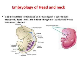
  • 1. Embryology of Head and neck • The mesenchyme for formation of the head region is derived from mesoderm, neural crest, and thickened regions of ectoderm known as ectodermal placodes. G.LUFUKUJA
  • 2. PHARYNGEAL APPARATUS (Branchial arch) • Neural crest cells originate in the neuroectoderm of forebrain, midbrain, and hindbrain regions and migrate ventrally into the pharyngeal arches and rostrally around the forebrain and optic cup into the facial region. In these locations they form mid-facial and pharyngeal arch skeletal structures • There are 5 pairs of branchial arches, that form on either side of the pharyngeal foregut on day 22 are the embryologic basis of all the differentiated structures of the head and neck
  • 3. Pharyngeal Apparatus • The head and neck region of four week human embryo somewhat resemble these regions of a fish embryo of comparable stage • This explains the former use of designation branchial apparatus • Branchial is derived from the Greek word branchia or gill
  • 4. Pharyngeal Apparatus Pharyngeal apparatus consists of: • Pharyngeal arches • Pharyngeal pouches • Pharyngeal grooves/clefts • Pharyngeal membrane
  • 5. Pharyngeal Arches • Pharyngeal arches begin to develop early in the fourth week as neural crest cells migrate into the head and neck region • The first pair of pharyngeal arches (primordium of jaws) appears as a surface elevations lateral to the developing pharynx • Soon other arches appear as obliquely disposed, rounded ridges on each side of the future head and neck regions
  • 6.
  • 9. Pharyngeal Arches • By the end of the fourth week, four pairs of pharyngeal arches are visible externally • The fifth and sixth arches are rudimentary and are not visible on the surface of the embryo • The pharyngeal arches are separated from each other by fissures called pharyngeal grooves • They are numbered in craniocaudal sequence
  • 10. Pharyngeal Arches • The first pharyngeal arch (mandibular arch) develops maxillary and mandibular prominences • The first pair of pharyngeal arches plays a major role in facial development • The second pharyngeal arch (hyoid arch) contributes to the formation of hyoid bone
  • 11.
  • 12. Pharyngeal Arch Components • Each pharyngeal arch consists of a core of mesenchyme • Is covered externally by ectoderm and internally by endoderm • In the third week the original mesenchyme is derived from mesoderm • During the fourth week most of the mesenchyme is derived from neural crest cells that migrate into the pharyngeal arches
  • 13. Fate of Pharyngeal Arches • The pharyngeal arches contribute exclusively to the formation of the face, nasal cavities, mouth, larynx, pharynx and neck • During the fifth week, the second pharyngeal arch enlarges and overgrows the third and fourth arches, forming the ectodermal depression called cervical sinus • By the end of seventh week the second to fourth pharyngeal grooves and the cervical sinus have disappeared, giving the neck a smooth contour
  • 14.
  • 15. Fate of Pharyngeal Arches A typical pharyngeal arch contains: • An aortic arch, an artery that arises from the truncus arteriosus of the primordial heart • A cartilaginous rod that forms the skeleton of the arch • A muscular component that differentiates into muscles in the head and neck • A nerve that supplies the mucosa and muscles derived from the arch
  • 16.
  • 17. Derivatives of Pharyngeal Arch Cartilages • The dorsal end of first arch cartilage (Meckel cartilage) ossifies to form malleus and incus • The middle part of cartilage forms anterior ligament of malleus sphenomandibular ligament • Ventral part of the first arch cartilages form primordium of the mandible • The cartilage disappears as mandible develops around it
  • 18.
  • 19. Derivatives of Pharyngeal Arch Cartilages • The dorsal end of second arch cartilage (Reichert cartilage) ossifies to form the stapes and styloid process of the temporal bone • The ventral end of second arch cartilage ossifies to form the lesser cornu and superior part of the body of the hyoid bone • Its perichondrium forms the stylohyoid ligament
  • 20. Derivatives of Pharyngeal Arch Cartilages • The third arch cartilage ossifies to form the greater cornu and the inferior part of the body of the hyoid bone • The fourth and sixth arch cartilages fuse to form the laryngeal cartilages except epiglottis which develops from hypopharyngeal eminence • The fifth pharyngeal arch is rudimentary and has no derivatives
  • 21.
  • 22. Derivatives of Pharyngeal Arch Muscles • The musculature of the first pharyngeal arch forms the muscles of mastication • The second pharyngeal arch forms the stapedius, stylohyoid, posterior belly of digastric, auricular and muscles of facial expression • The third arch forms the stylopharyngeus • The fourth arch forms cricothyroid, levator veli palatini and constrictors of pharynx • Sixth pharyngeal arch forms the intrinsic muscles of the larynx
  • 23.
  • 24. Derivatives of Pharyngeal Arch Nerves • Caudal two branches of Trigeminal nerve (maxillary and mandibular) supply derivatives of the first pharyngeal arch • The facial, glossopharyngeal and vagus nerves supply the second, third and caudal (fourth to sixth) arches respectively • The fourth arch is supplied by superior laryngeal branch of vagus nerve • The sixth arch is supplied by its recurrent laryngeal branch
  • 25.
  • 26. Pharyngeal Pouches • The primordial pharynx, derived from the foregut, widens cranially where it joins the primordial mouth or stomodeum • It narrows caudally where it joins the esophagus • The endoderm of the pharynx lines the internal aspects of pharyngeal arches and passes into balloonlike diverticula called pharyngeal pouches
  • 27.
  • 28. Pharyngeal Pouches • The pairs of pouches develop in a craniocaudal sequence between the arches • The first pair of pouches lies between the first and second pharyngeal arches • There are four well defined pairs of pharyngeal pouches • The fifth pair is absent or rudimentary
  • 29. Pharyngeal Pouches • The endoderm of the pouches contacts the ectoderm of the pharyngeal grooves and together they form the double layered pharyngeal membranes that separate the pharyngeal pouches from the pharyngeal grooves
  • 30. Derivatives of First Pharyngeal Pouch • The first pharyngeal pouch expands into an elongate tubotympanic recess • The expanded distal part of this recess contacts the first pharyngeal groove, where it contributes to the formation of the tympanic membrane (eardrum) • The cavity of the tubotympanic recess gives rise to the tympanic cavity and mastoid antrum
  • 31.
  • 32. Derivatives of Second Pharyngeal Pouch • The second pharyngeal pouch is largely obliterated as the palatine tonsils develop • Part of the cavity of this pouch remains as the tonsillar sinus or fossa • The endoderm of the pouch proliferates and grows into the underlying mesenchyme • The central parts of these buds form crypts
  • 33.
  • 34. Derivatives of Second Pharyngeal Pouch • The pouch endoderm forms the surface epithelium and the lining of the tonsillar crypts • At about 20 weeks the mesenchyme around the crypts differentiates into lymphoid tissue • These tissues soon organize into the lymphatic nodules of the palatine tonsil
  • 35. Derivatives of Third Pharyngeal Pouch • The third pharyngeal pouch expands and develops a solid, dorsal bulbar part and a hollow elongate ventral part • Its connection with the pharynx is reduced to a narrow duct that soon degenerates • By the sixth week the epithelium of each dorsal bulbar part begins to differentiate into inferior parathyroid gland
  • 36. Derivatives of Third Pharyngeal Pouch • The epithelium of the elongate ventral parts of third pharyngeal pouch proliferates and their cavities obliterate • These bilateral primordia of thymus come together in the median plane to form thymus • It descends into the superior mediastenum • The bilobed form of thymus remains throughout life • Discretely encapsulated and each lobe has its own blood supply, lymphatic drainage and nerve supply
  • 37.
  • 38. Derivatives of Third Pharyngeal Pouch • The primordia of thymus and parathyroid glands lose their connections with the pharynx and migrate into the neck • Later the parathyroid glands separate from the thymus and lie on the dorsal surface of the thyroid gland
  • 39. Derivatives of Fourth Pharyngeal Pouch • The fourth pharyngeal pouch also expands into dorsal bulbar and elongate ventral parts • Its connection with the pharynx is reduced to a narrow duct that soon degenerates • By the sixth week, each dorsal part develops into a superior parathyroid gland • It lies on the dorsal surface of the thyroid gland
  • 40.
  • 41. Derivatives of Fourth Pharyngeal Pouch • The parathyroid glands derived from the third pouches descend with the thymus and are carried to a more inferior position than the parathyroid derived from the fourth pouches • This explains why the parathyroid glands derived from the third pair of pouches are located inferior to those from the fourth pouches
  • 42. The Fifth Pharyngeal Pouch • When this develops, this rudimentary pouch becomes part of the fourth pharyngeal pouch and helps to form the ultimopharyngeal body
  • 43. Pharyngeal Grooves • During the fourth and fifth weeks, head and neck region of the human embryo exhibit four pharyngeal grooves or clefts on each side • These grooves separate the pharyngeal arches externally • Only first pair persists as the external acoustic meatus • The other grooves normally obliterated with the cervical sinus as the neck develops
  • 44. Development of the Tongue • The adult tongue is derived from embryonic processes arising from the first three pharyngeal arches and a small contribution from the fourth arch. • The anterior two thirds of the adult tongue (the movable part) is formed by two lateral swellings and a single one, the tuberculum impar, all of which are derived from the first pharyngeal arch 44G.LUFUKUJA
  • 45. Development of the Tongue… • The posterior third (the fixed part or the root of the tongue) is formed by an embryonic process, the hypobranchial eminence or copula. This process is derived mainly from the second and third pharyngeal arches with small part from the fourth pharyngeal arch. • The muscles of the tongue are derived from a different embryonic source, namely the occipital myotomes.
  • 47. Development of the Tongue… • The junction of the anterior 2/3 and the posterior 1/3 of the adult tongue is marked by a V-shaped structure, the sulcus terminalis and at its tip is a blind foramen, the foramen caecum. This site marks the area from which an epithelial structure arise during embryonic development and extend inferiorly to the anterior part of the future neck where it forms the thyroid gland.
  • 48.
  • 49. Thyroid Gland • The thyroid gland appears as an epithelial proliferation in the floor of the pharynx between the tuberculum impar and the copula at a point later indicated by the foramen cecum . Subsequently the thyroid descends in front of the pharyngeal gut as a bilobed diverticulum. • During this migration, the thyroid remains connected to the tongue by a narrow canal, the thyroglossal duct.
  • 50. 50 The Face • The face is formed from 7 primordia surrounding the stomodeum • Single frontonasal prominence -cranial boundary of the stomodeum • Paired median nasal process • Paired maxillary process • Paired mandibularprocess G.LUFUKUJA
  • 51. Development of the Face… • The Frontonasal prominence forms the forehead. The nasal prominence is split into lateral and medial nasal processes by formation of a nasal placode • Thus a horseshoe-shaped nasal processes surrounds the nasal placode, which undergoes apoptosis to form a depression; the nasal pit. Initially, the maxillary and lateral nasal prominences are separated by a deep furrow, the nasolacrimal groove. • Ectoderm in the floor of this groove forms a solid epithelial cord that detaches from the overlying ectoderm. After canalization, the cord forms the nasolacrimal duct; its upper end widens to form the lacrimal sac.
  • 52.
  • 53. Intermaxillary segment • As a result of medial growth of the maxillary prominences, the two medial nasal prominences merge not only at the surface but also at a deeper level. The structure formed by the two merged prominences is the intermaxillary segment
  • 55. 55 Anomalies • Craniofacial: These are defects associated with tissue fusion. Estimated 1/3 of all congenital defects are craniaofacial • Facial Clefts - Anomalies associated with defective fusion of Facial prominences  1. Cleft lip - failure of maxillary prominence to fuse with intermaxillary process  2. Cleft palate - failure of the lateral palatine processes to fuse