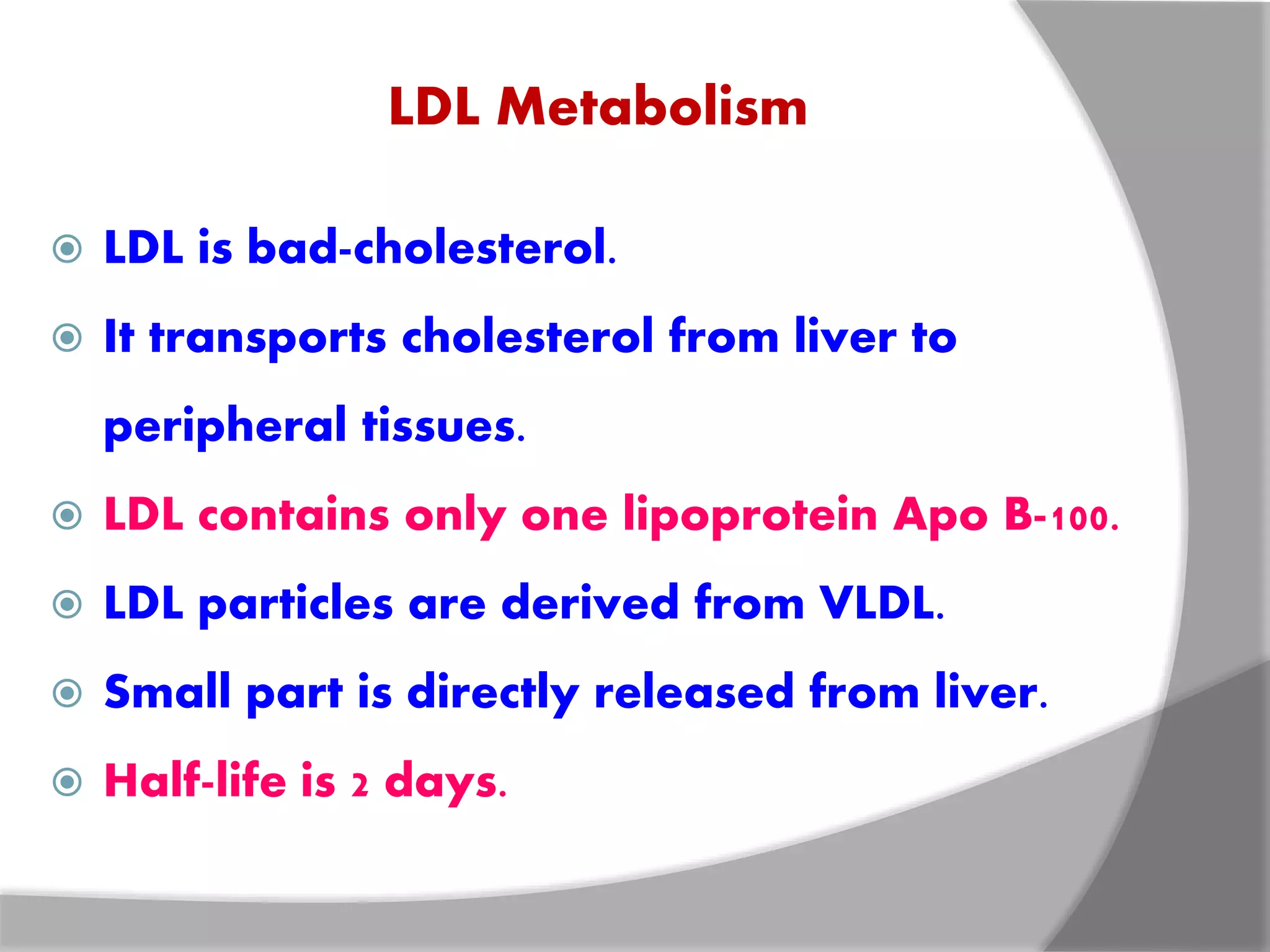
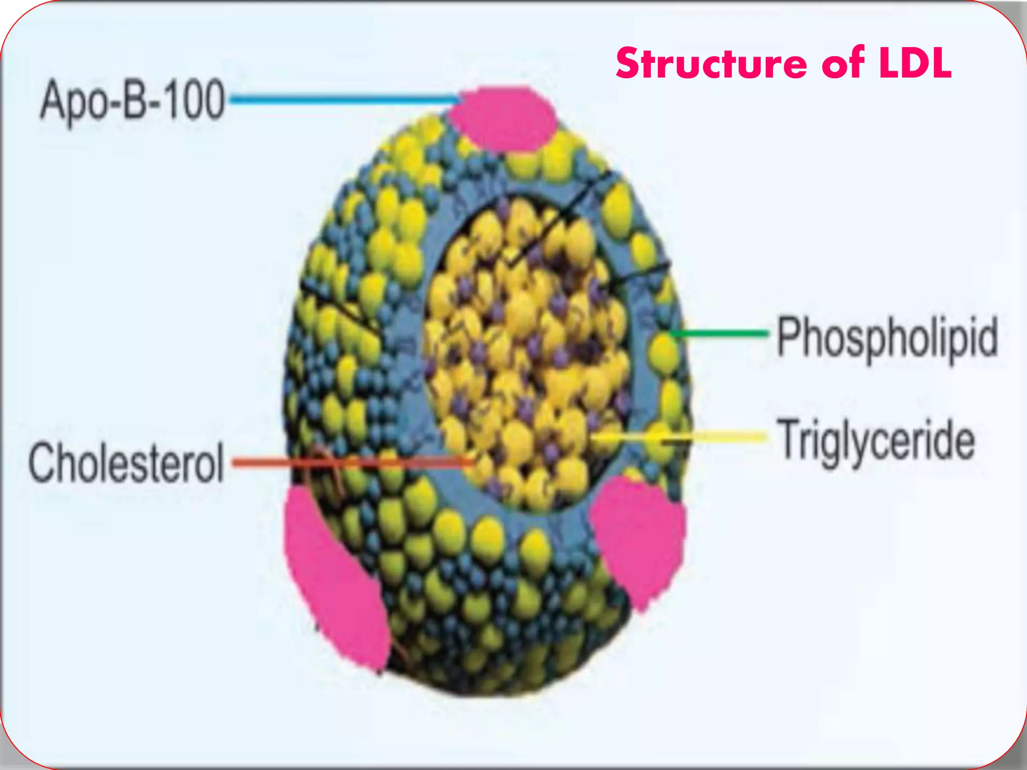
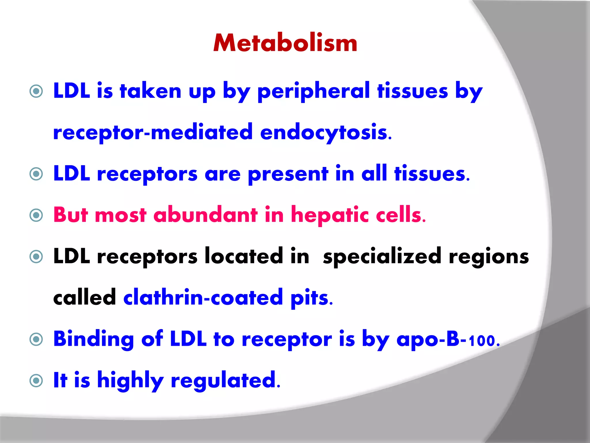
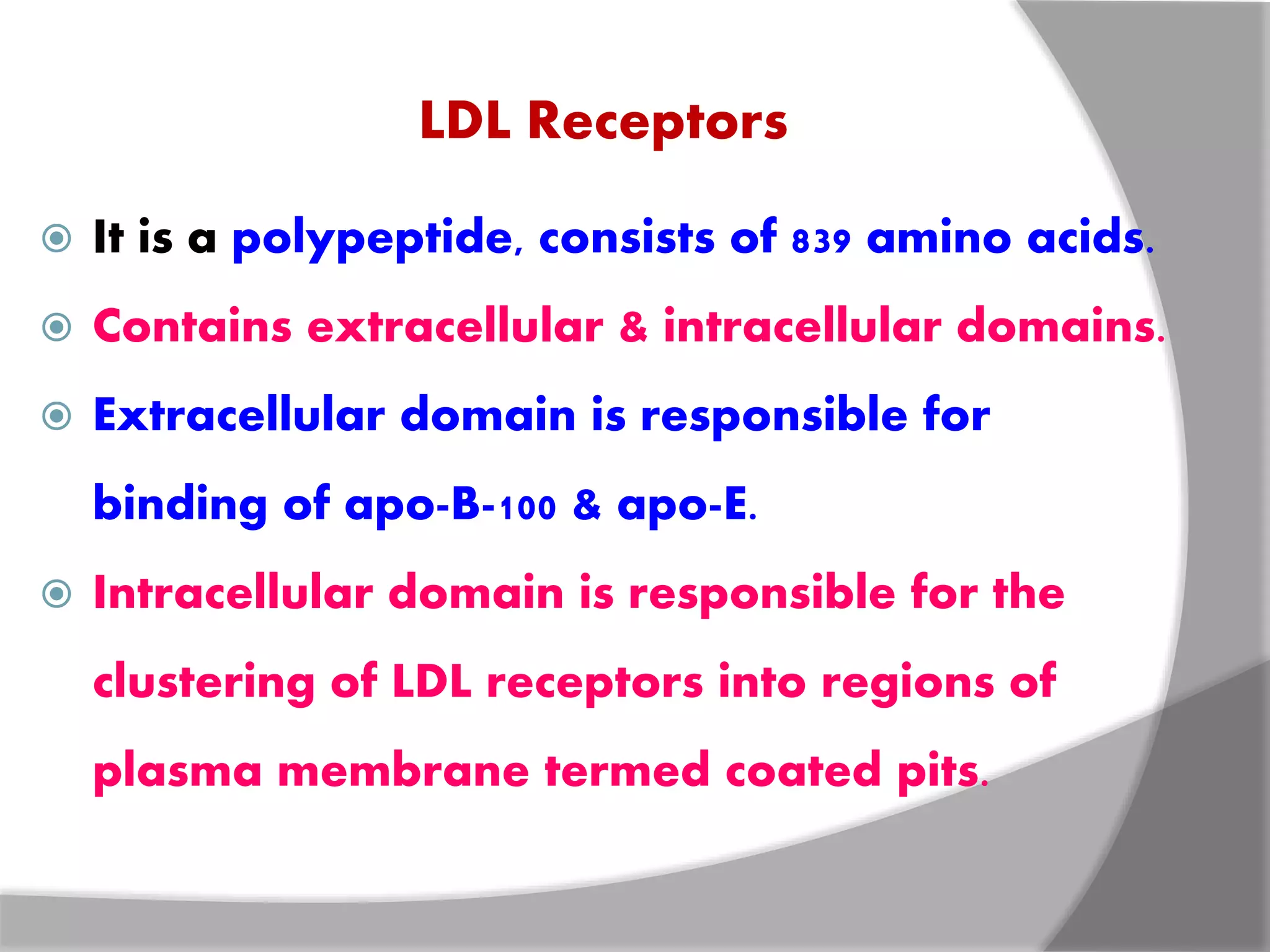
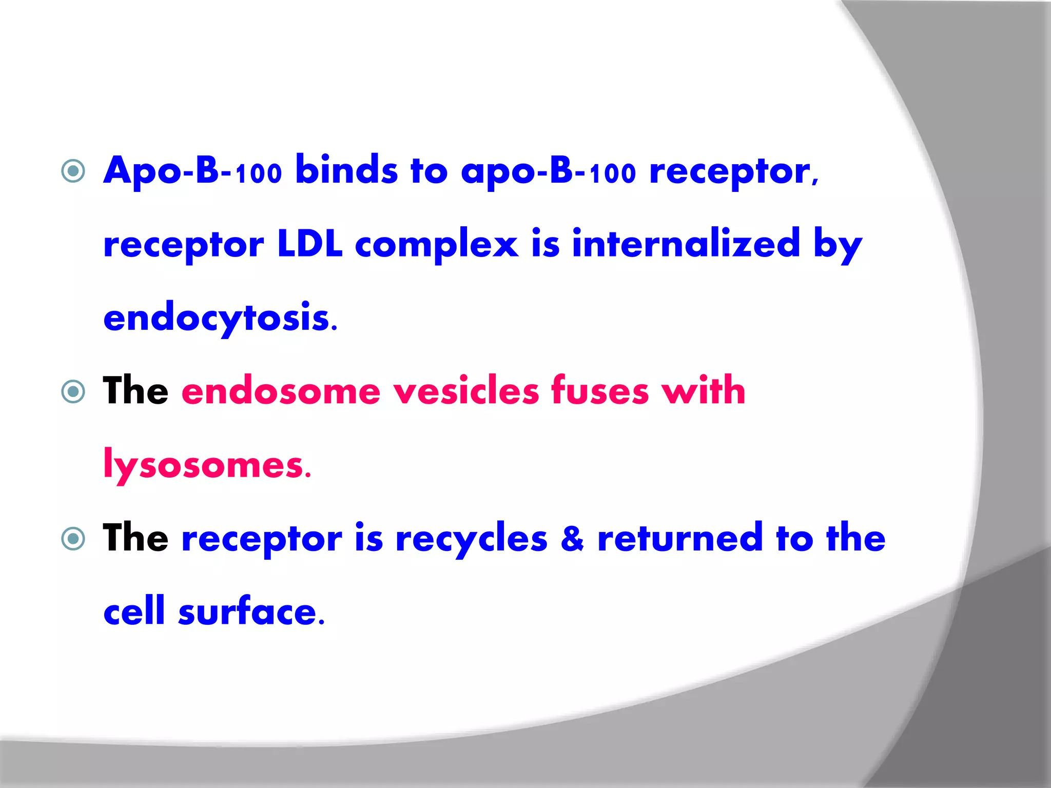
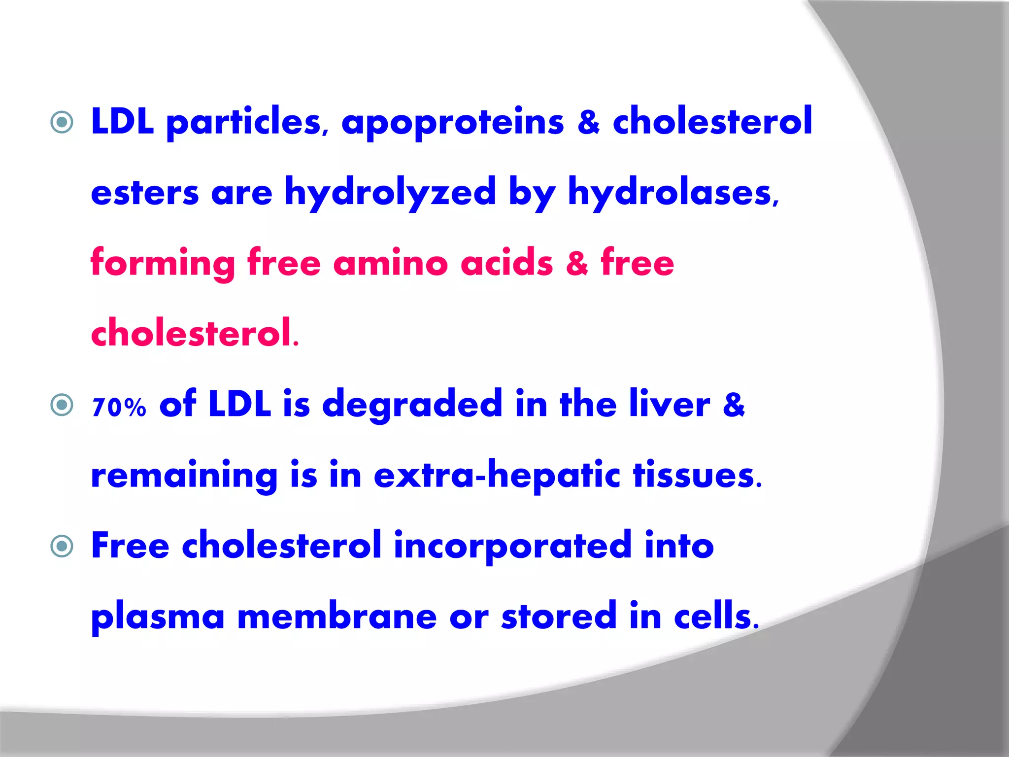
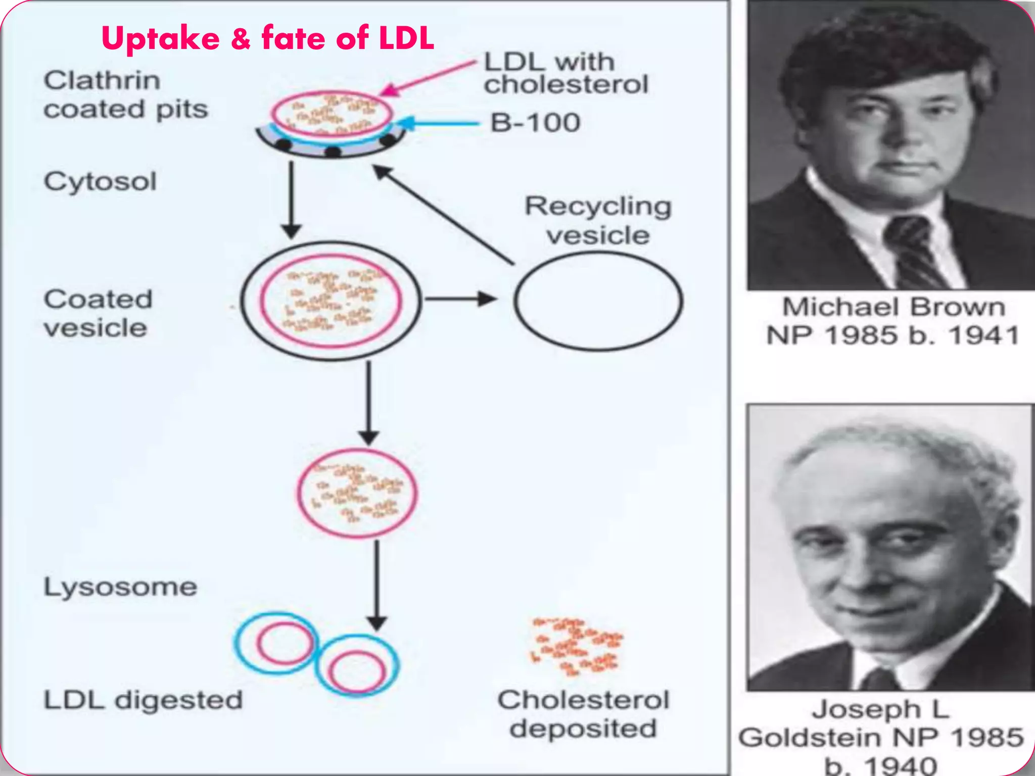
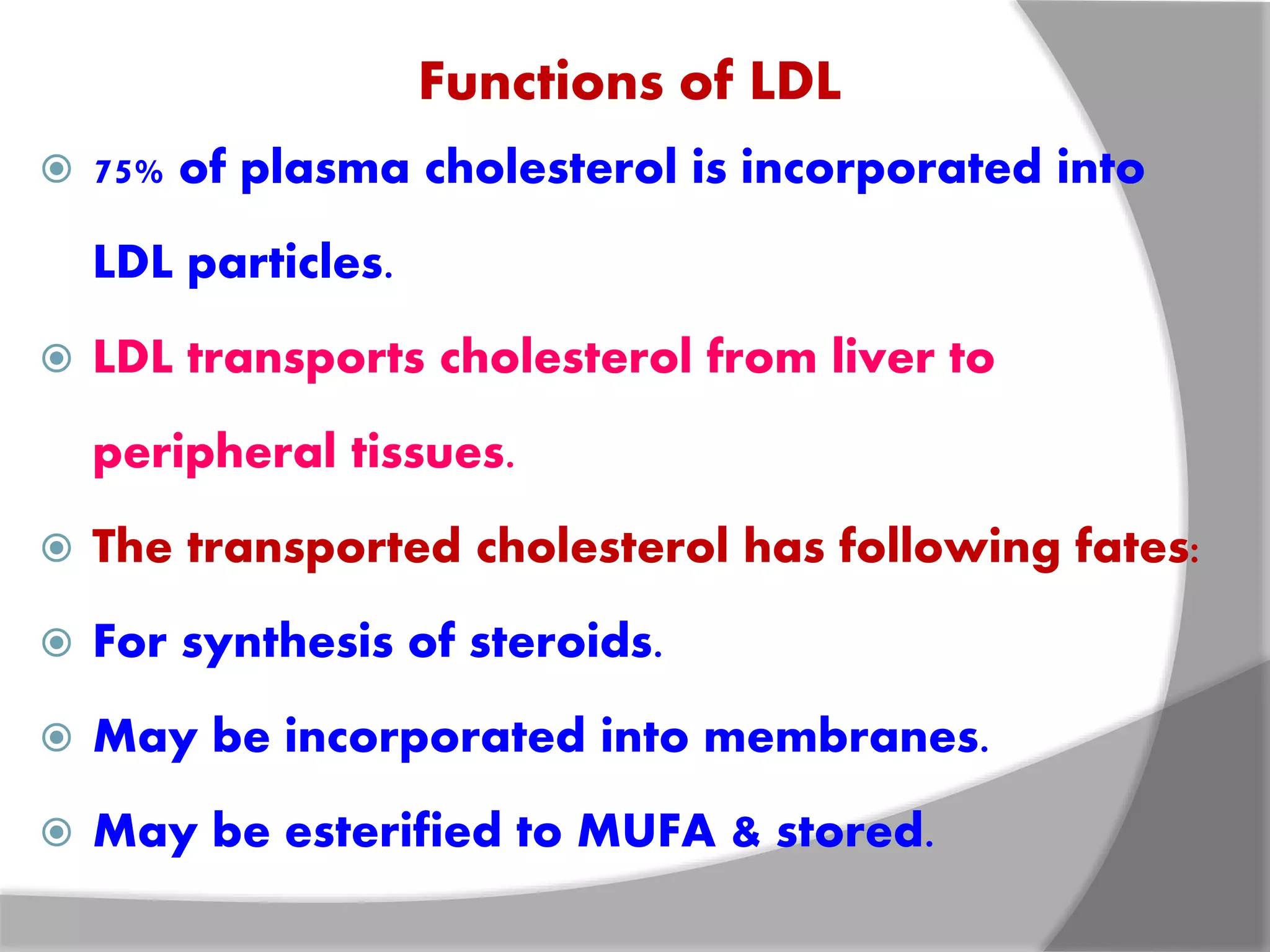
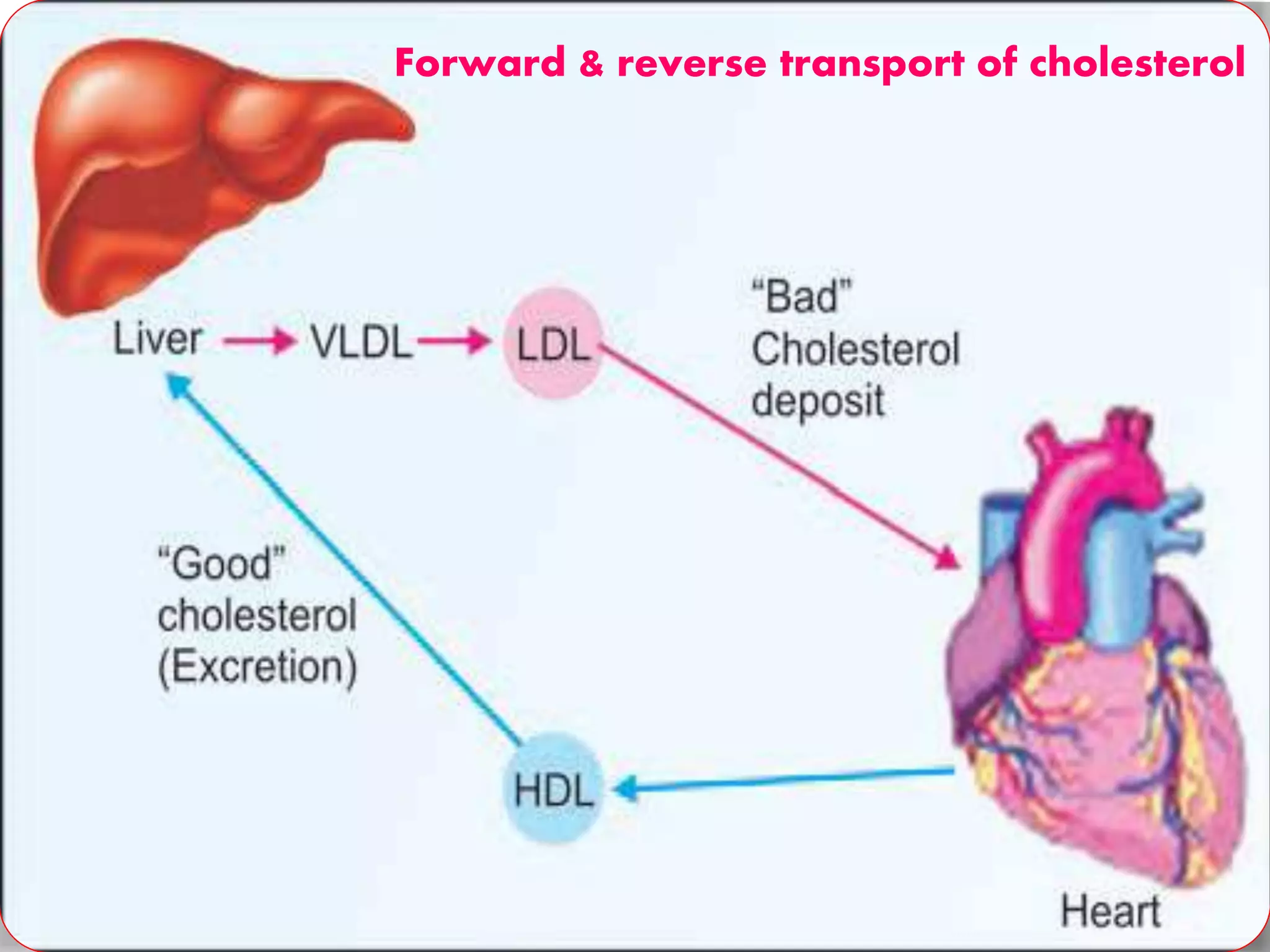
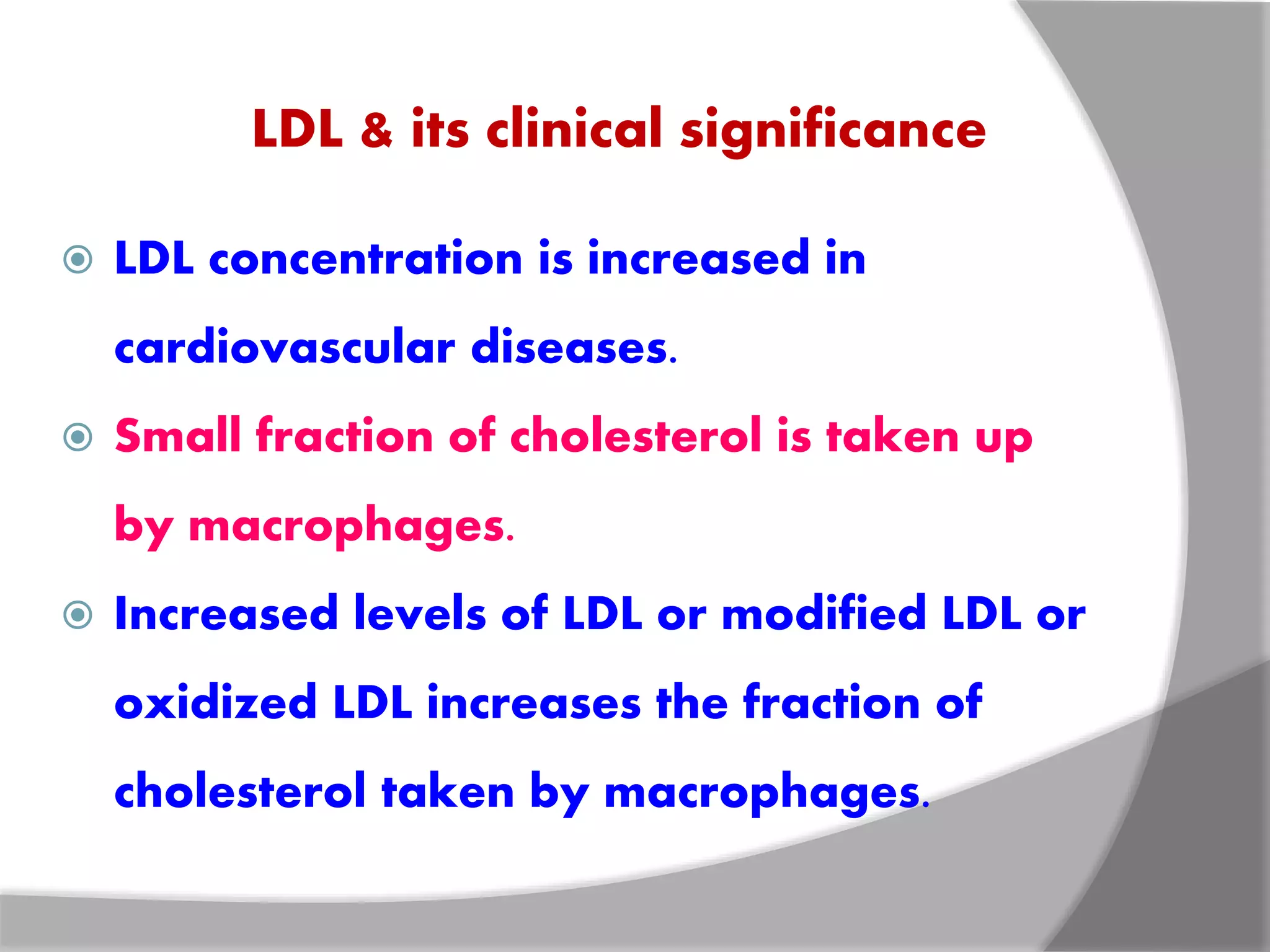
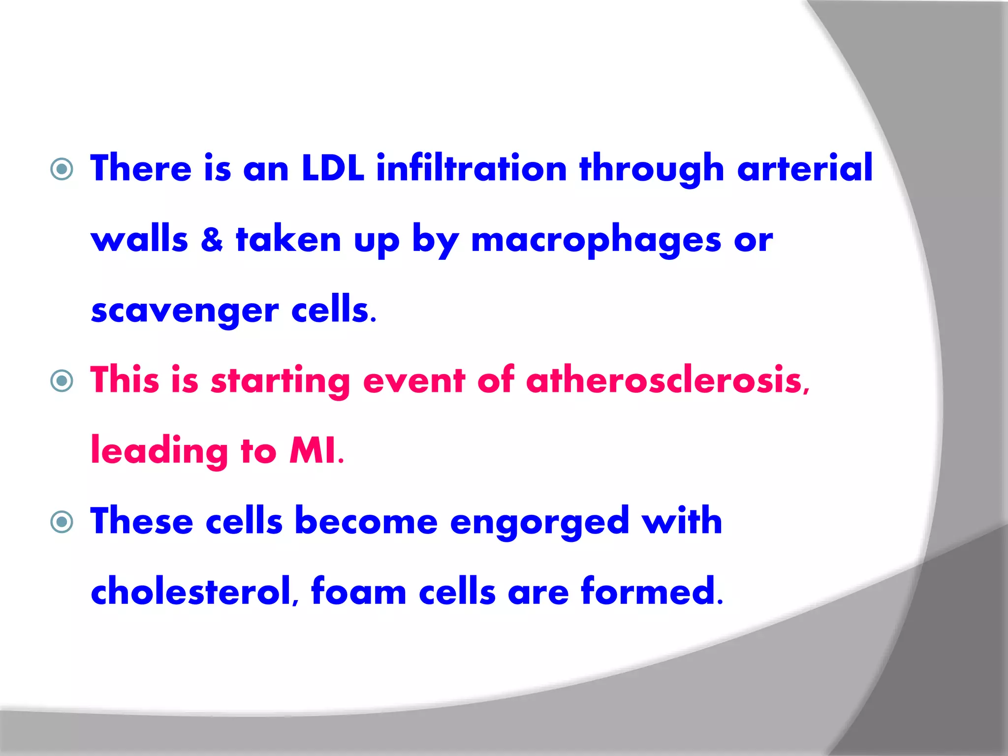
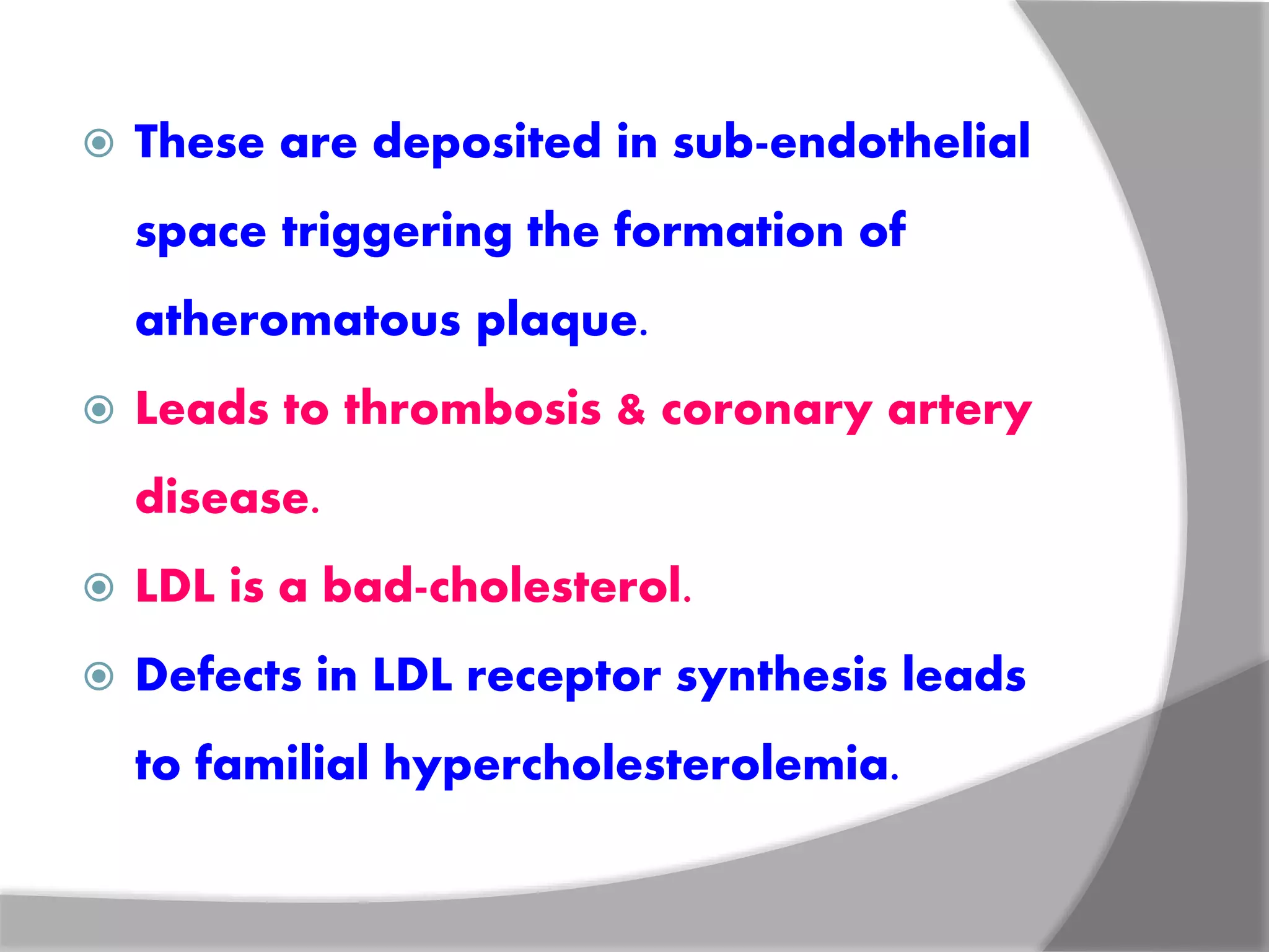
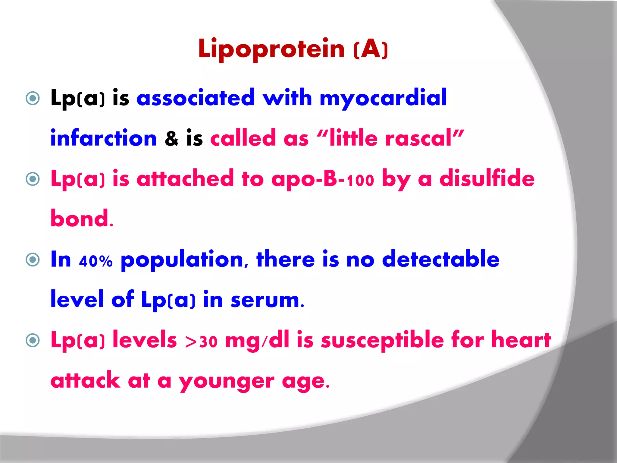
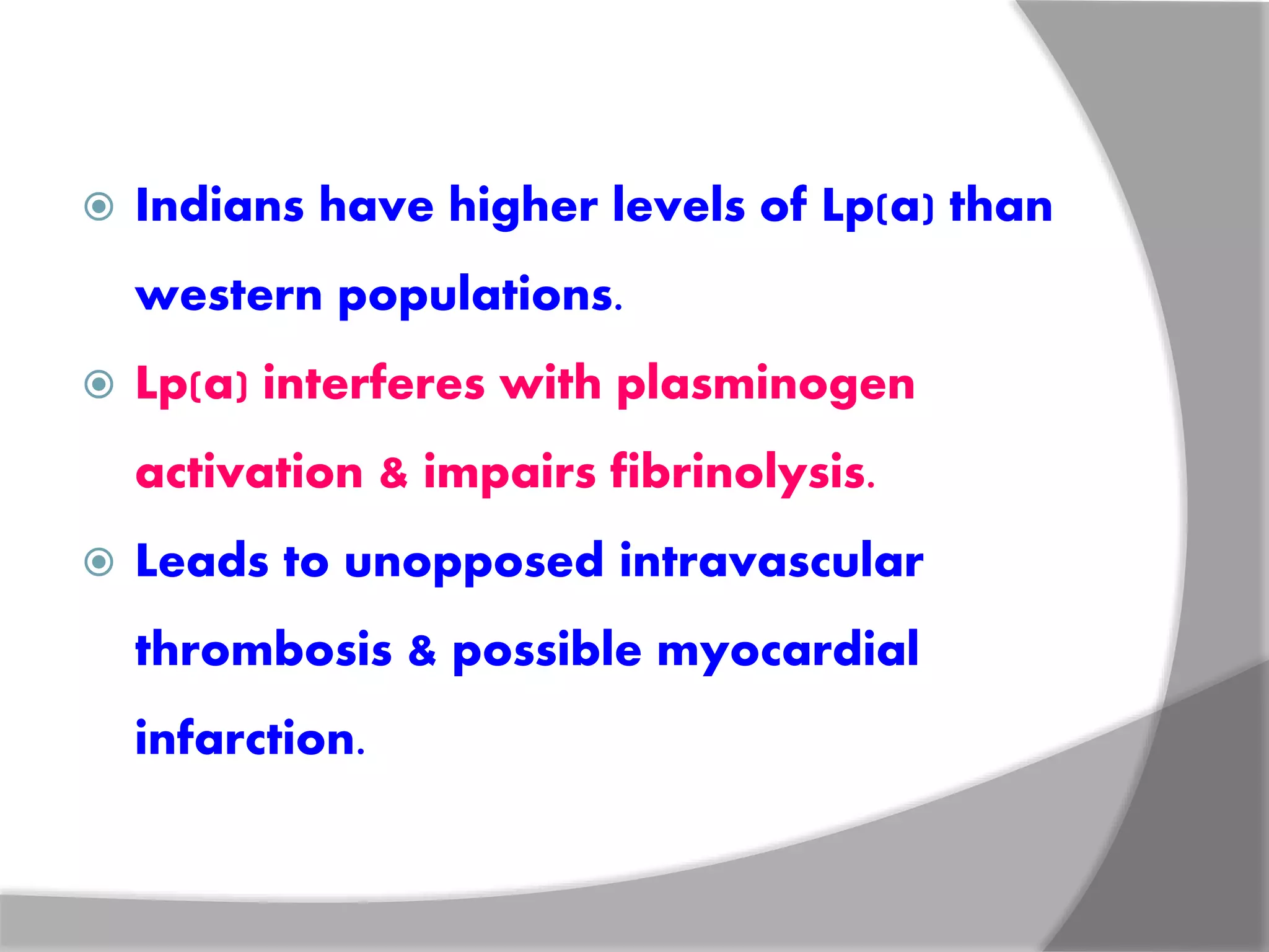
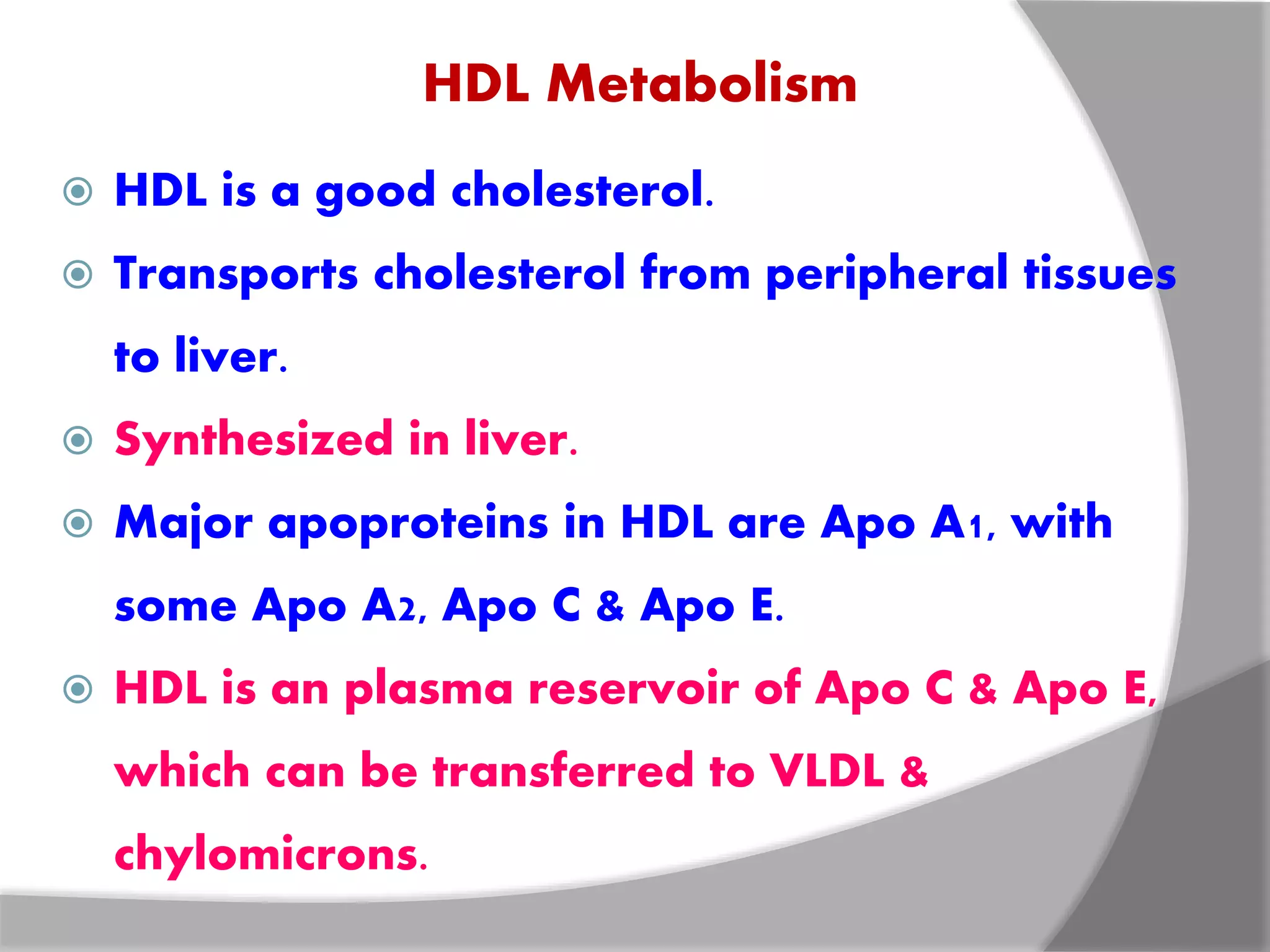
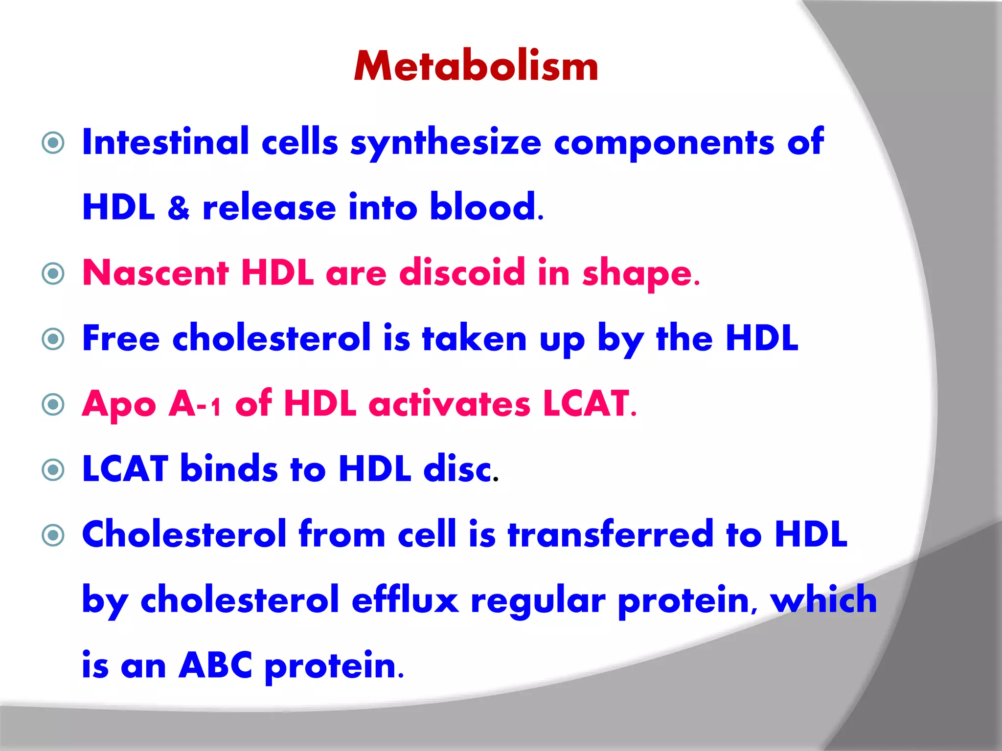
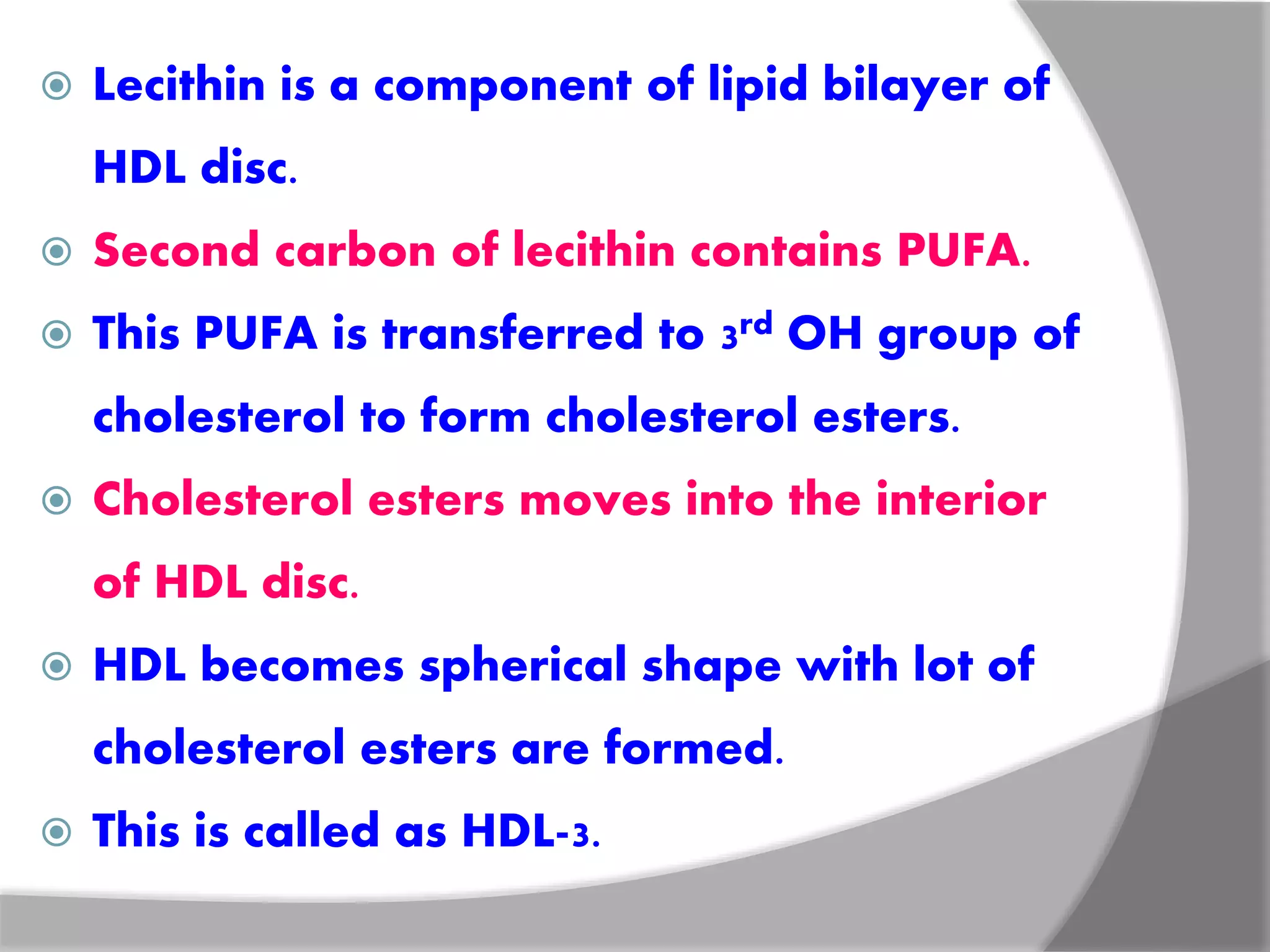
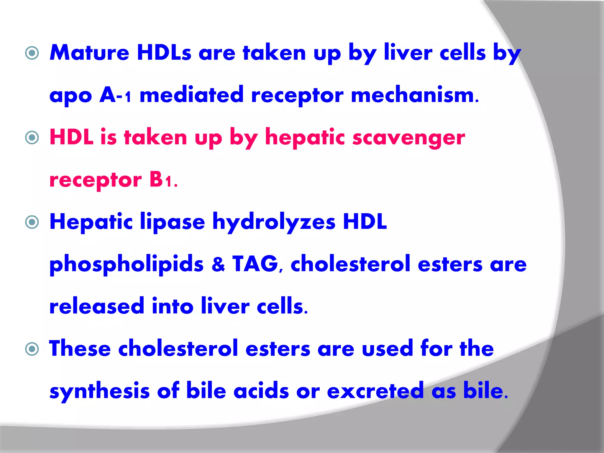
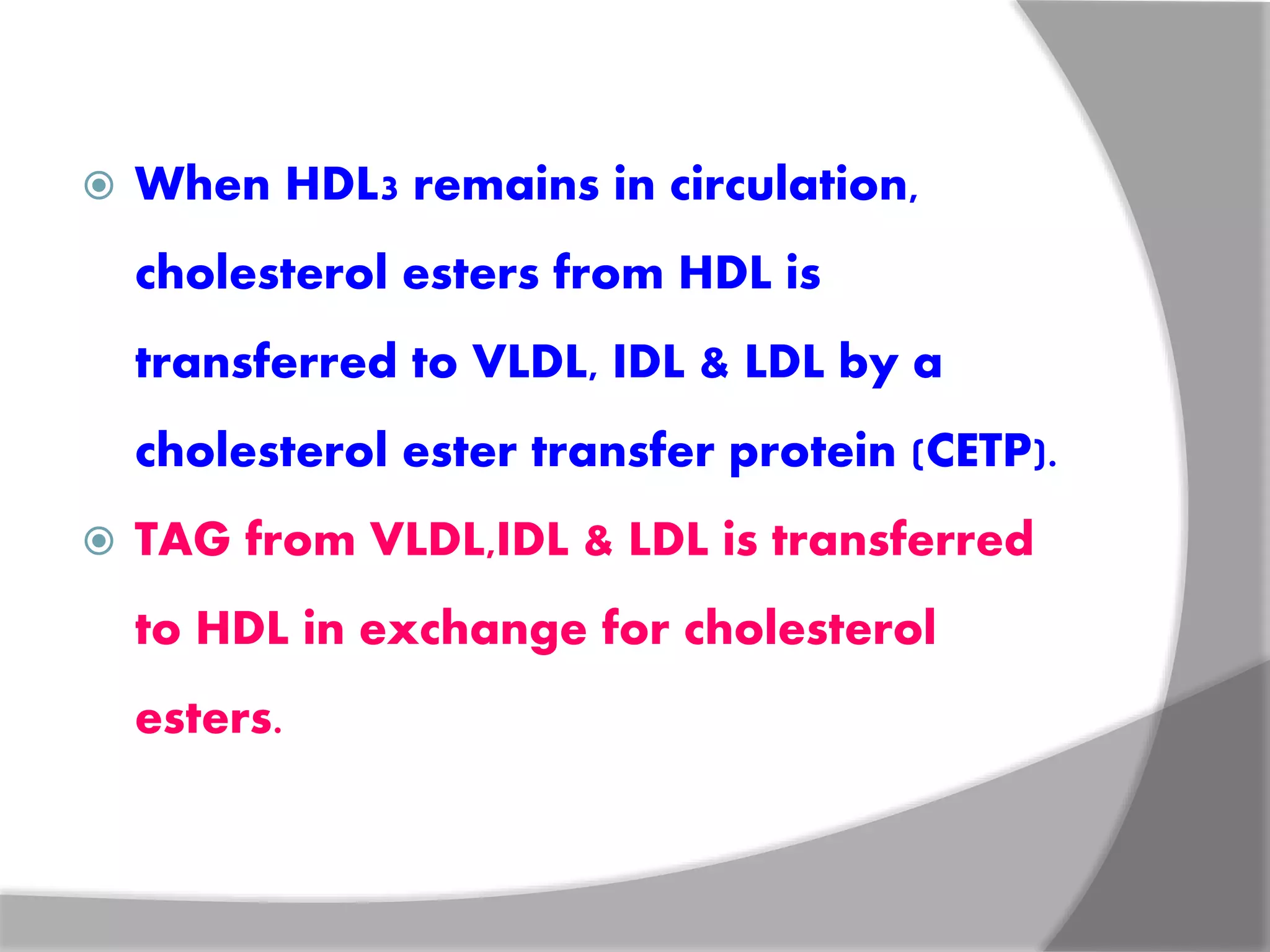
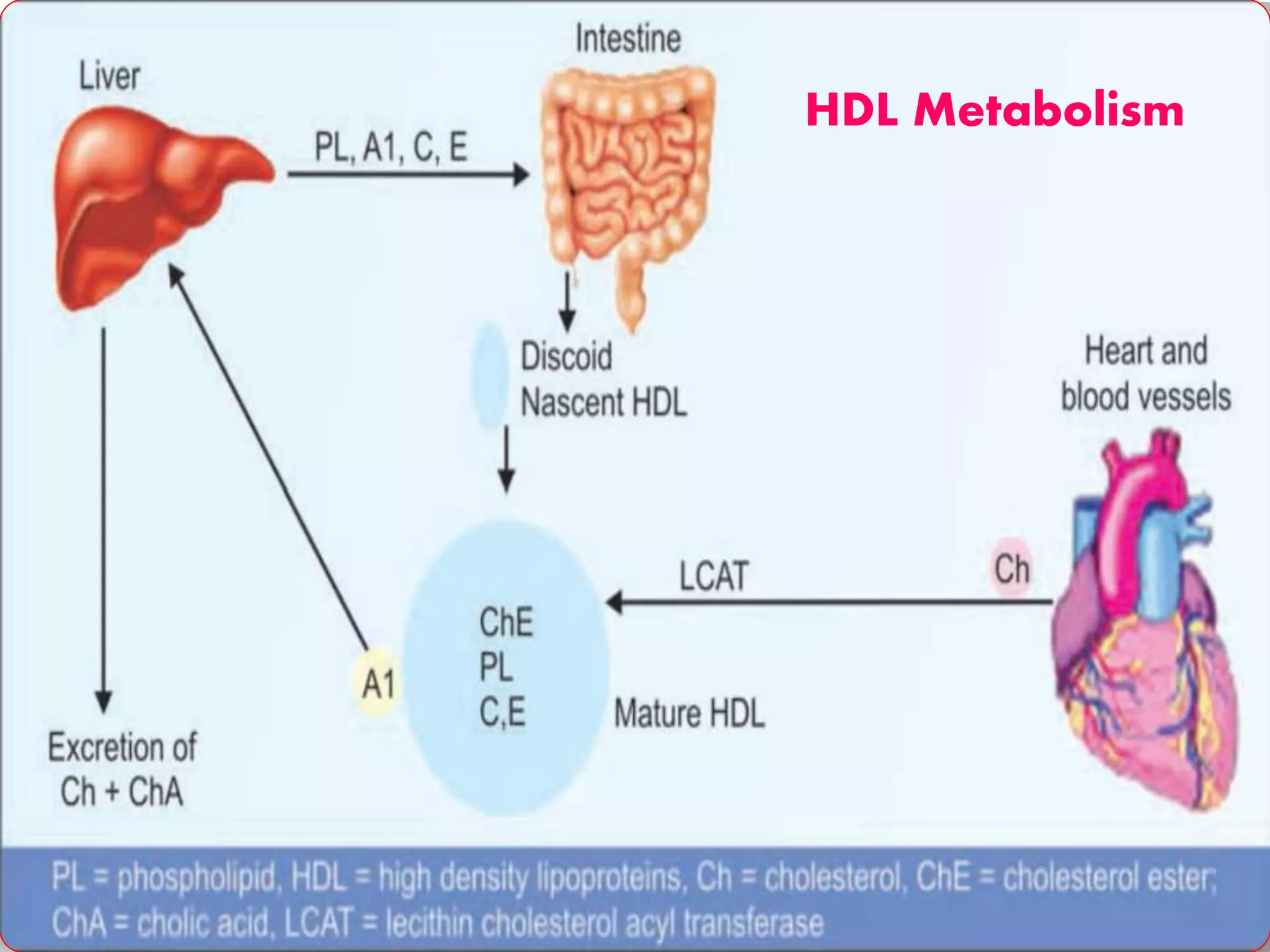
Gandham.Rajeev
Email:gandhamrajeev33@gmail.com
LDL Metabolism
 LDL is bad-cholesterol.
 It transports cholesterol from liver to
peripheral tissues.
 LDL contains only one lipoprotein Apo B-100.
 LDL particles are derived from VLDL.
 Small part is directly released from liver.
 Half-life is 2 days.
Structure of LDL
Metabolism
 LDL is taken up by peripheral tissues by
receptor-mediated endocytosis.
 LDL receptors are present in all tissues.
 But most abundant in hepatic cells.
 LDL receptors located in specialized regions
called clathrin-coated pits.
 Binding of LDL to receptor is by apo-B-100.
 It is highly regulated.
LDL Receptors
 It is a polypeptide, consists of 839 amino acids.
 Contains extracellular & intracellular domains.
 Extracellular domain is responsible for
binding of apo-B-100 & apo-E.
 Intracellular domain is responsible for the
clustering of LDL receptors into regions of
plasma membrane termed coated pits.
 Apo-B-100 binds to apo-B-100 receptor,
receptor LDL complex is internalized by
endocytosis.
 The endosome vesicles fuses with
lysosomes.
 The receptor is recycles & returned to the
cell surface.
 LDL particles, apoproteins & cholesterol
esters are hydrolyzed by hydrolases,
forming free amino acids & free
cholesterol.
 70% of LDL is degraded in the liver &
remaining is in extra-hepatic tissues.
 Free cholesterol incorporated into
plasma membrane or stored in cells.
Uptake & fate of LDL
Functions of LDL
 75% of plasma cholesterol is incorporated into
LDL particles.
 LDL transports cholesterol from liver to
peripheral tissues.
 The transported cholesterol has following fates:
 For synthesis of steroids.
 May be incorporated into membranes.
 May be esterified to MUFA & stored.
Forward & reverse transport of cholesterol
LDL & its clinical significance
 LDL concentration is increased in
cardiovascular diseases.
 Small fraction of cholesterol is taken up
by macrophages.
 Increased levels of LDL or modified LDL or
oxidized LDL increases the fraction of
cholesterol taken by macrophages.
 There is an LDL infiltration through arterial
walls & taken up by macrophages or
scavenger cells.
 This is starting event of atherosclerosis,
leading to MI.
 These cells become engorged with
cholesterol, foam cells are formed.
 These are deposited in sub-endothelial
space triggering the formation of
atheromatous plaque.
 Leads to thrombosis & coronary artery
disease.
 LDL is a bad-cholesterol.
 Defects in LDL receptor synthesis leads
to familial hypercholesterolemia.
Lipoprotein (A)
 Lp(a) is associated with myocardial
infarction & is called as “little rascal”
 Lp(a) is attached to apo-B-100 by a disulfide
bond.
 In 40% population, there is no detectable
level of Lp(a) in serum.
 Lp(a) levels >30 mg/dl is susceptible for heart
attack at a younger age.
 Indians have higher levels of Lp(a) than
western populations.
 Lp(a) interferes with plasminogen
activation & impairs fibrinolysis.
 Leads to unopposed intravascular
thrombosis & possible myocardial
infarction.
HDL Metabolism
 HDL is a good cholesterol.
 Transports cholesterol from peripheral tissues
to liver.
 Synthesized in liver.
 Major apoproteins in HDL are Apo A1, with
some Apo A2, Apo C & Apo E.
 HDL is an plasma reservoir of Apo C & Apo E,
which can be transferred to VLDL &
chylomicrons.
Metabolism
 Intestinal cells synthesize components of
HDL & release into blood.
 Nascent HDL are discoid in shape.
 Free cholesterol is taken up by the HDL
 Apo A-1 of HDL activates LCAT.
 LCAT binds to HDL disc.
 Cholesterol from cell is transferred to HDL
by cholesterol efflux regular protein, which
is an ABC protein.
 Lecithin is a component of lipid bilayer of
HDL disc.
 Second carbon of lecithin contains PUFA.
 This PUFA is transferred to 3rd OH group of
cholesterol to form cholesterol esters.
 Cholesterol esters moves into the interior
of HDL disc.
 HDL becomes spherical shape with lot of
cholesterol esters are formed.
 This is called as HDL-3.
 Mature HDLs are taken up by liver cells by
apo A-1 mediated receptor mechanism.
 HDL is taken up by hepatic scavenger
receptor B1.
 Hepatic lipase hydrolyzes HDL
phospholipids & TAG, cholesterol esters are
released into liver cells.
 These cholesterol esters are used for the
synthesis of bile acids or excreted as bile.
 When HDL3 remains in circulation,
cholesterol esters from HDL is
transferred to VLDL, IDL & LDL by a
cholesterol ester transfer protein (CETP).
 TAG from VLDL,IDL & LDL is transferred
to HDL in exchange for cholesterol
esters.
 HDL particles rich in TAG & spherical are
called as HDl-2
 These particles are first acted upon by
hepatic triglyceride lipase (HTGL)
 Efflux of cholesterol from peripheral cells
to HDL is mediated by ABC transporter
protein.
HDL Metabolism
Functions of HDL
 HDL is the transports cholesterol from
peripheral tissues to liver, called as reverse
cholesterol transport.
 Cholesterol is excreted through bile.
 Cholesterol excretion needs prior
esterification with PUFA.
 PUFA reduces serum cholesterol levels.
 PUFA is anti-atherogenic.
HDL & its clinical significance
 Serum HDL levels are inversely related to
the incidence of MI.
 HDL is “anti-atherogenic” or protective in
nature.
 It is a good cholesterol.
 HDL levels <35mg/dl increases risk,
>65mg/dl reduces the risk of CAD.
Free fatty acids
 Complexed with albumin in plasma.
 FFA is derived from lipolysis of TAG stored in
adipose tissue by hormone sensitive lipase.
 FFA may be long chain saturated or
unsaturated FA. & are transported to heart,
skeletal muscle, liver & other tissues.
 FFA are either oxidized or incorporated into
tissue lipids.
 In tissue cells, FFA-albumin complex is
dissociated, FFA binds with fatty acid
transport protein.
 It is a co-transport with sodium.
 Half-life of FFA is 1-2 minutes.
 During starvation, 40-50% of energy is met
by oxidation of FFA.
References
 Textbook of Biochemistry-U Satyanarayana
 Textbook of Biochemistry-DM Vasudevan
Thank You

LDL & HDL METABOLISM

  • 1.
  • 2.
    LDL Metabolism  LDLis bad-cholesterol.  It transports cholesterol from liver to peripheral tissues.  LDL contains only one lipoprotein Apo B-100.  LDL particles are derived from VLDL.  Small part is directly released from liver.  Half-life is 2 days.
  • 3.
  • 4.
    Metabolism  LDL istaken up by peripheral tissues by receptor-mediated endocytosis.  LDL receptors are present in all tissues.  But most abundant in hepatic cells.  LDL receptors located in specialized regions called clathrin-coated pits.  Binding of LDL to receptor is by apo-B-100.  It is highly regulated.
  • 5.
    LDL Receptors  Itis a polypeptide, consists of 839 amino acids.  Contains extracellular & intracellular domains.  Extracellular domain is responsible for binding of apo-B-100 & apo-E.  Intracellular domain is responsible for the clustering of LDL receptors into regions of plasma membrane termed coated pits.
  • 6.
     Apo-B-100 bindsto apo-B-100 receptor, receptor LDL complex is internalized by endocytosis.  The endosome vesicles fuses with lysosomes.  The receptor is recycles & returned to the cell surface.
  • 7.
     LDL particles,apoproteins & cholesterol esters are hydrolyzed by hydrolases, forming free amino acids & free cholesterol.  70% of LDL is degraded in the liver & remaining is in extra-hepatic tissues.  Free cholesterol incorporated into plasma membrane or stored in cells.
  • 8.
  • 9.
    Functions of LDL 75% of plasma cholesterol is incorporated into LDL particles.  LDL transports cholesterol from liver to peripheral tissues.  The transported cholesterol has following fates:  For synthesis of steroids.  May be incorporated into membranes.  May be esterified to MUFA & stored.
  • 10.
    Forward & reversetransport of cholesterol
  • 11.
    LDL & itsclinical significance  LDL concentration is increased in cardiovascular diseases.  Small fraction of cholesterol is taken up by macrophages.  Increased levels of LDL or modified LDL or oxidized LDL increases the fraction of cholesterol taken by macrophages.
  • 12.
     There isan LDL infiltration through arterial walls & taken up by macrophages or scavenger cells.  This is starting event of atherosclerosis, leading to MI.  These cells become engorged with cholesterol, foam cells are formed.
  • 13.
     These aredeposited in sub-endothelial space triggering the formation of atheromatous plaque.  Leads to thrombosis & coronary artery disease.  LDL is a bad-cholesterol.  Defects in LDL receptor synthesis leads to familial hypercholesterolemia.
  • 14.
    Lipoprotein (A)  Lp(a)is associated with myocardial infarction & is called as “little rascal”  Lp(a) is attached to apo-B-100 by a disulfide bond.  In 40% population, there is no detectable level of Lp(a) in serum.  Lp(a) levels >30 mg/dl is susceptible for heart attack at a younger age.
  • 15.
     Indians havehigher levels of Lp(a) than western populations.  Lp(a) interferes with plasminogen activation & impairs fibrinolysis.  Leads to unopposed intravascular thrombosis & possible myocardial infarction.
  • 16.
    HDL Metabolism  HDLis a good cholesterol.  Transports cholesterol from peripheral tissues to liver.  Synthesized in liver.  Major apoproteins in HDL are Apo A1, with some Apo A2, Apo C & Apo E.  HDL is an plasma reservoir of Apo C & Apo E, which can be transferred to VLDL & chylomicrons.
  • 17.
    Metabolism  Intestinal cellssynthesize components of HDL & release into blood.  Nascent HDL are discoid in shape.  Free cholesterol is taken up by the HDL  Apo A-1 of HDL activates LCAT.  LCAT binds to HDL disc.  Cholesterol from cell is transferred to HDL by cholesterol efflux regular protein, which is an ABC protein.
  • 18.
     Lecithin isa component of lipid bilayer of HDL disc.  Second carbon of lecithin contains PUFA.  This PUFA is transferred to 3rd OH group of cholesterol to form cholesterol esters.  Cholesterol esters moves into the interior of HDL disc.  HDL becomes spherical shape with lot of cholesterol esters are formed.  This is called as HDL-3.
  • 19.
     Mature HDLsare taken up by liver cells by apo A-1 mediated receptor mechanism.  HDL is taken up by hepatic scavenger receptor B1.  Hepatic lipase hydrolyzes HDL phospholipids & TAG, cholesterol esters are released into liver cells.  These cholesterol esters are used for the synthesis of bile acids or excreted as bile.
  • 20.
     When HDL3remains in circulation, cholesterol esters from HDL is transferred to VLDL, IDL & LDL by a cholesterol ester transfer protein (CETP).  TAG from VLDL,IDL & LDL is transferred to HDL in exchange for cholesterol esters.
  • 21.
     HDL particlesrich in TAG & spherical are called as HDl-2  These particles are first acted upon by hepatic triglyceride lipase (HTGL)  Efflux of cholesterol from peripheral cells to HDL is mediated by ABC transporter protein.
  • 22.
  • 23.
    Functions of HDL HDL is the transports cholesterol from peripheral tissues to liver, called as reverse cholesterol transport.  Cholesterol is excreted through bile.  Cholesterol excretion needs prior esterification with PUFA.  PUFA reduces serum cholesterol levels.  PUFA is anti-atherogenic.
  • 24.
    HDL & itsclinical significance  Serum HDL levels are inversely related to the incidence of MI.  HDL is “anti-atherogenic” or protective in nature.  It is a good cholesterol.  HDL levels <35mg/dl increases risk, >65mg/dl reduces the risk of CAD.
  • 25.
    Free fatty acids Complexed with albumin in plasma.  FFA is derived from lipolysis of TAG stored in adipose tissue by hormone sensitive lipase.  FFA may be long chain saturated or unsaturated FA. & are transported to heart, skeletal muscle, liver & other tissues.  FFA are either oxidized or incorporated into tissue lipids.
  • 26.
     In tissuecells, FFA-albumin complex is dissociated, FFA binds with fatty acid transport protein.  It is a co-transport with sodium.  Half-life of FFA is 1-2 minutes.  During starvation, 40-50% of energy is met by oxidation of FFA.
  • 27.
    References  Textbook ofBiochemistry-U Satyanarayana  Textbook of Biochemistry-DM Vasudevan
  • 28.