LC-MS combines liquid chromatography separation with mass spectrometry detection. It is useful for bioactivity screening, proteomics, and other applications. Key aspects of LC-MS include the HPLC separation, ionization sources like ESI and APCI, mass analyzers like quadrupole and time-of-flight, and the ability to identify proteins and peptides from complex mixtures. Proteomics uses these techniques to study protein expression, structure, function, and interactions on a large scale.
LIQUID CHROMATOGRAPHY-MASS
SPECTROMETRY
Liquidchromatography – mass spectrometry (LC/MS) is a
technique that uses liquid chromatography (or HPLC) with
the mass spectrometry.
It is an analytical chemistry technique that combines the
physical separation capabilities of liquid chromatography
with the mass analysis capabilities of mass spectrometry.
3.
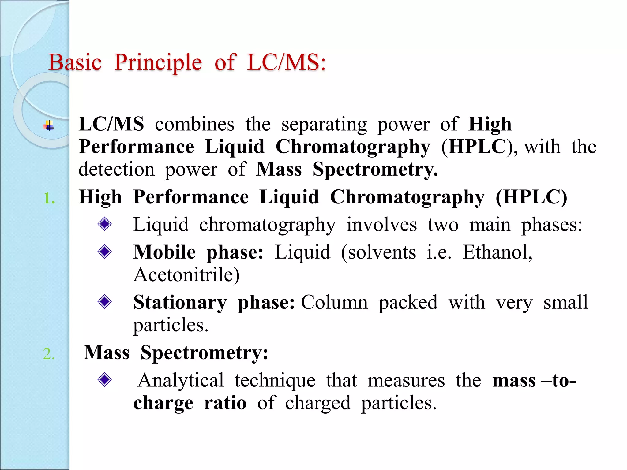
Basic Principle ofLC/MS:
LC/MS combines the separating power of High
Performance Liquid Chromatography (HPLC), with the
detection power of Mass Spectrometry.
1. High Performance Liquid Chromatography (HPLC)
Liquid chromatography involves two main phases:
Mobile phase: Liquid (solvents i.e. Ethanol,
Acetonitrile)
Stationary phase: Column packed with very small
particles.
2. Mass Spectrometry:
Analytical technique that measures the mass –to-
charge ratio of charged particles.
4.
Theory of LC/MS
HPLC is a method for separating a complex mixture in
to its components.
High sensitivity of mass spectroscopy provides the
information for identification of compounds or structural
elucidation of compounds.
Combination of these two techniques in LC-MS.
As the metabolites appear from the end of the column
they enter the mass detector, where the solvent is
removed and the metabolites are ionized.
5.
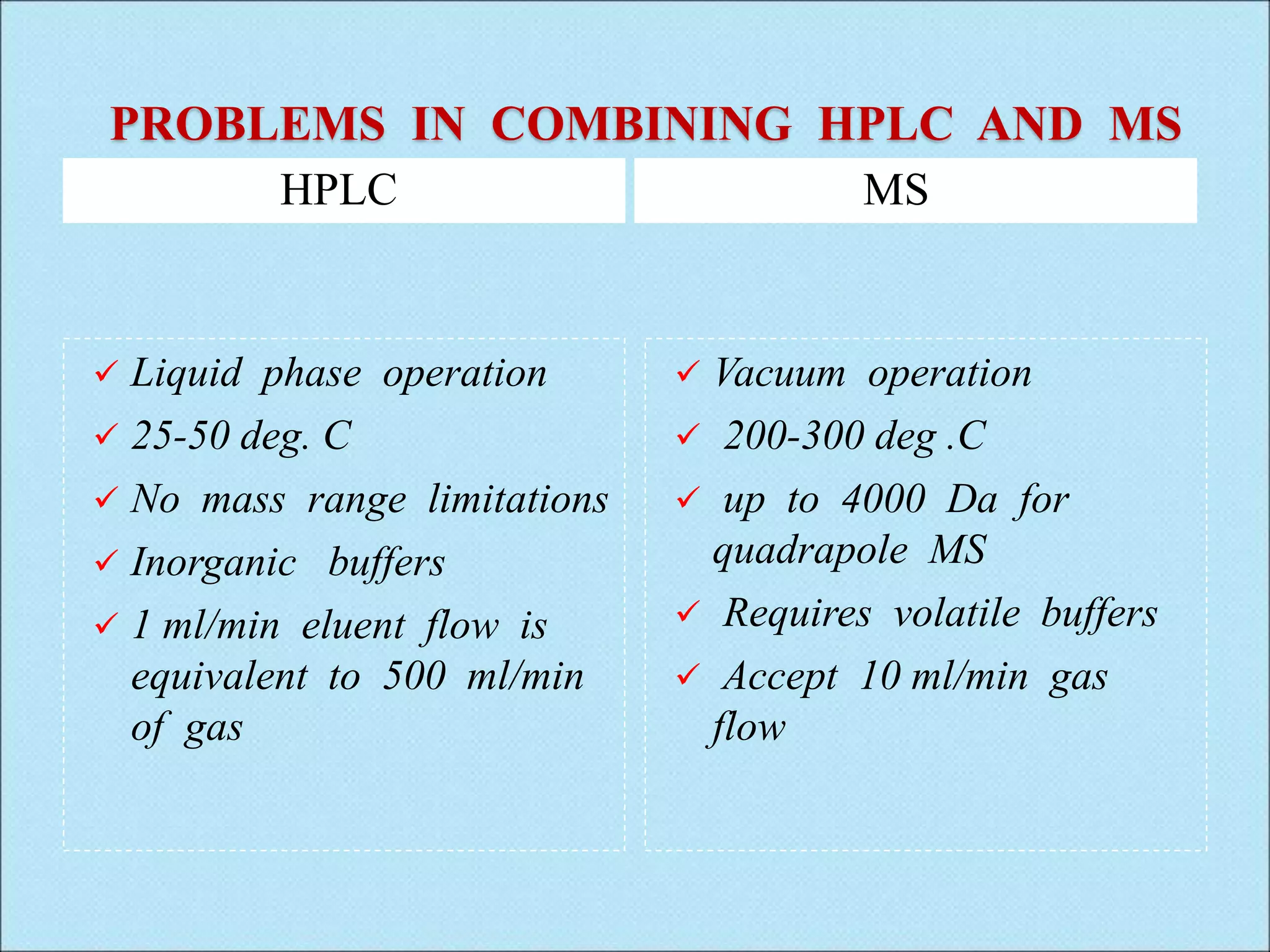
PROBLEMS IN COMBININGHPLC AND MS
HPLC MS
Liquid phase operation
25-50 deg. C
No mass range limitations
Inorganic buffers
1 ml/min eluent flow is
equivalent to 500 ml/min
of gas
Vacuum operation
200-300 deg .C
up to 4000 Da for
quadrapole MS
Requires volatile buffers
Accept 10 ml/min gas
flow
6.
High Pressure LiquidChromatography
Liquid chromatography generally utilizes very small
particles packed and operating at relatively high
pressure, and is referred to as high performance liquid
chromatography (HPLC).
Modern LC/MS methods use HPLC instrumentation,
essentially exclusively, for sample introduction.
In HPLC, the sample is forced by a liquid at high
pressure ( the mobile phase) through a column that is
packed with a stationary phase generally composed of
irregularly or spherically shaped particles chosen or
derivatized to accomplish particular types of separation.
RP-LC is most often used as the means to introduce
samples into the MS, in LC-MS instrumentation.
7.
MASS SPECTROMETRY
Massspectrometry (MS) is an analytical technique that
measures the mass-to-charge ratio of charged particles.
MS works by ionizing chemical compounds to generate
charged molecules or molecule fragments and measuring
their mass-to-charge ratios.
In a typical MS procedure, a sample is loaded onto the
instrument and undergoes vaporization.
The components of the sample are ionized by one of a
variety of methods ( e.g., by impacting them with an
electron beam), Which results in the formation of
charged particles (ions).
The ions are separated according to their mass-to-charge
ratio in an analyzer by electromagnetic fields.
8.
INSTRUMENTATION OF LC/MS
1) HPLC constitutes the lc part:
Solvent system (mobile phase)
Pumps
Mixer
Injector
Column
2) Mass spectrometer
A) Ion sources
Electrospray ionization
Atmospheric pressure chemical ionization
Atmospheric pressure photoionization
B) Mass Analyzer
Quadrupole
Time of flight
Ion trap
Fourier transform-ion cyclotron resonance
10.
MOBILE PHASE
The mobilephase is the solvent that moves the solute
through out column.
General requirements:-
1) Low cost, UV transparency, high purity.
2) Low viscosity, low toxicity, non flammability.
3) Non corrosive to LC system component.
Solvent strength and selectivity:-
It is the ability of solvent to elute solutes from a
column.
11.
COLUMN
The use ofdi-functional or tri-functional silanes to
create bonded groups with two or three attachment
points leading to phases with higher stability in low or
higher PH and lower bleed for LCMS
Most widely used columns for LC-MS are:-
1) Fast LC column.
the use of short column. (15-50mm)
2) Micro LC column.
the use of large column. (20-150mm)
12.
SAMPLE PREPARATION
Sample preparationgenerally consists of
concentrating the analyte and removing compounds that
can cause background ions or suppress ionization.
Examples of sample preparation include:
On-column concentration to increase analyte
concentration.
Desalting to reduce the sodium and potassium adduct
formation that commonly occurs in electrospray
Filtration to separate a low molecular-weight drug from
proteins in plasma, milk, or tissue.
13.
ION SOURCES
Ion sources:
Electrospray ionization (ESI)
Atmospheric pressure chemical ionization (APCI)
Atmospheric pressure photoionization (APPI)
Electrospray ionization (ESI)
Process
API-ES is a process of ionization followed by
evaporation. It occurs in three basic steps:
1. Nebulization and charging
2. Desolvation and;
3. Ion evaporation
14.
NEBULIZATION:
The Hplceffluent is pumbed through a nebulizing needle
which is at ground potential.
The spray goes through a semi-cylindrical electrode Which
is at a high potential.
The potential difference between the needle and the
electrode produces a strong electrical field.
This field charges the surface of the liquid and forms a
spray of charged droplets.
There is a concentric flow of gas which assists in the
nebulization process.
16.
DESOLVATION:
The chargeddroplets are attracted toward the capillary
sampling orifice.
There is counterflow of heated nitrogen gas which shrinks
the droplets and carries away the uncharged material.
IONIZATION:
As the droplets shrink, they approach a point where the
electrostatic (coulombic) forces exceed the cohesive forces.
This process continues until the analyte ions are ultimately
desorbed into the gas phase.
These gas-phase ions pass through the capillary sampling
orifice into the low pressure region of the ion source and
the mass analyzer.
17.
Atmospheric Pressure ChemicalIonization:
Process:
APCL, a process of evaporation followd by ionization,
is complementary to API-ES.
NEBULIZATION AND DESOLVATION:
APCI nebulization is similar to that in API-ES.
However, APCI nebulization occurs in a hot (typically
250ºC-400ºC) vaporizer chamber.
The heat rapidly evaporates the spray droplets, resulting in
gas-phase HPLC solvent and anlytes molecules
18.
IONIZATION:
The gas-phasesolvent molecules are ionized by
the discharge from a corona needle.
In APCI there is a charge transfer from the
ionized solvent reagent ions to the analyte
molecules in a way that is similar to chemical
ionization in GC/MS.
These analyte ions then are transported through the
ion optics to the filter and detector.
19.
ATMOSPHERIC PRESSURE PHOTOIONIZATION
Atmospheric pressure photoionization (APPI) for LC/MS is
a relatively new technique.
As in APCI, a vaporizer converts the LC eluent to the
gas phase.
A discharge lamp generates photons in a narrow range of
ionization energies.
The range of energies is carefully chosen to ionize as
many analyte molecules as possible while minimizing the
ionization of solvent molecules.
The resulting ions pass through a capillary sampling
orifice into the mass analyzer.
21.
MASS ANALYZERS
Mass analyzeris also called ion separator.
Mass analyzer is the heart of the mass spectrophotometer
that takes ionized masses and separates them based on
charge to mass ratios.
TYPES OF MASS ANALYZERS
Quadrupole mass analyzer
Time of flight analyzer (TOF)
Quadrupole ion trap mass analyzer
22.
QUADRUPOLE:
A quadrupole massanalyzer consists of four parallel rods
arranged in a square.
The analyte ions are directed down the center of the
square.
Voltages applied to the rods generate electromagnetic
fields.
These fields determine which mass-to-charge ratio of ions
can pass through the filter at a given time.
Quadrupole tend to be the simplest and least expensive
mass analyzers.
24.
Quadrupole massanalyzers can operate in two modes:
Scanning (scan) mode.
Selected ion monitering (SIM) mode
Scan mode:-
The mass analyzer monitors a range of mass-to-charge ratios.
In mass analyzer monitors only a few mass-to-charge ratios.
SIM mode:-
Significantly more sensitive than scan mode but provides
information about fewer ions.
Scan mode is typically used for qualitative analyzes or for
quantification when all analyte masses are not known in advance.
SIM mode is used for quantification and monitering of target
compounds.
25.
TIME OF FLIGHT
In a time of flight (TOF) mass analyzer, a uniform
electromagnetic force is applied to all ions at the same
time, causing them to accelerate down a flight tube.
Lighter ions travel faster and arrive at the detector first,
so the mass-to-charge ratios of the ions are determined
by their arrived times.
Time-of-flight mass analyzers have a wide mass range
and can be very accurate in their mass measurements.
27.
ION TRAP
Anion trap mass analyzer consists of a circular ring
electrode plus two end caps that together from a chamber.
Ions entering the chamber are “trapped” there by
electromagnetic fields.
Another field can be applied to selectivity eject ions from
the trap.
Ion traps have the advantage of being able to perform
multiple stages of mass spectrometry without additional
mass analyzers
29.
DETECTORS
Once theions have passed the mass analyzer they have
to be detected and transformed into a usable signal.
The detector is an important element generating secondary
electrons, which are further amplified, or by inducing a
current (generated by moving charges).
Ion detector fall into two main classes:
Point detectors
Array detectors
30.
POINT DETECTOR:
Ionsare not spatially resolved and sequentially
impinge upon a detector situated at a single point
within the spectrometer geometry
ARRAY DETECTOR:
Ions are spatially resolved and all ions arrive
simultaneously (or near simultaneously) and are recorded
along a plane using a bank of detector.
31.
APPLICATION OF LC/MS
Pharmaceutical Applications:
Rapid chromatography of benzodiazepines
Identification of bile acid metabolite
Biochemical applications:
Rapid protein identification using capillary
LC/MS/MS and database searching.
Clinical Applications:
High-senstivity detection of trimipramine and
thioridazine
32.
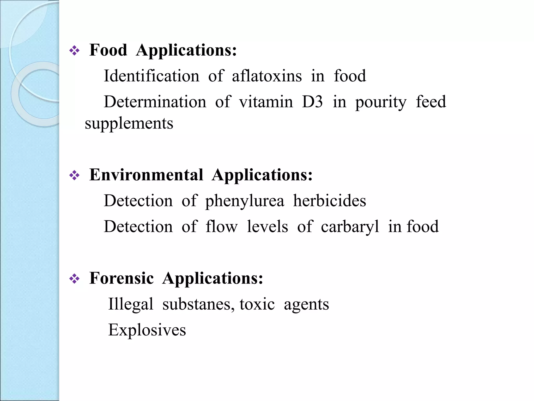
Food Applications:
Identificationof aflatoxins in food
Determination of vitamin D3 in pourity feed
supplements
Environmental Applications:
Detection of phenylurea herbicides
Detection of flow levels of carbaryl in food
Forensic Applications:
Illegal substanes, toxic agents
Explosives
33.
PROTEOMICS
Proteome isa defines the complete set
of proteins expressed during a cell’s entire
lifetime.
Proteomics is the study of the proteome;
it uses technologies ranging from genetic
analysis to mass spectrometry.
Proteomics assesses
Activities
Modifications
Localization
Interactions of proteins in
complexes
34.
ROLE OF PROTEOMICS
To study the structure and function of protein
To study the 3D structure of protein
Study of qualitative and quantitative analysis of
proteins
INTRODUCTION
Proteome indicatesthe total proteins expressed
by a genome in a cell or tissue.
With the development of proteomic techniques,
proteome analysis provides a fast, non – invasive
diagnostic tool for patients with various diseases.
The advent of highly sensitive proteomic
technologies can identify proteins associated with
development of diseases well before any
clinically identifiable alteration.
MS has a high resolving power and identifies
proteins with more accuracy.
37.
TYPES OF PROTEOMICS
1.Structural Proteomics:- The ultimate aim of this
proteomics is to build a body of structural
information that will help predict the probable
structure and potential function for almost any
protein from knowledge of its coding sequence.
2. Functional proteomics:- It refers to the use of
proteomics techniques to analyze the characterization
of molecular protein-networks involved in a living
cell.
3. Expression proteomics:- It refers to the quantitative
study of protein expression between sample differing
by some variable.
38.
PROTEIN STRUCTURE
Primarystructure:- is sequence
of specific amino acid in
polypeptide chain.
Secondary structure:- the
primary polypeptide chain gets
properly folded in the form of
alpha-helix, beta pleated sheet,
random coils and turns.
39.
Tertiary structure:-Secondary
structure interact with each
other chemically to form the 3
dimensional shape of the
proteins.
Quardinary structure:-
interaction between different
polypeptide unit.
40.
EXPERIMENTAL WORK FLOW
Propersample selection and sample storage
Sample pre- processing
Immuno depletion/protein concentration etc
Protein separation by 1D SDS-PAGE/ 2DE
or
Fractionation by LC
In gel trptic digestion/ in-solution digestion
Protein in identification LC-MS/MS
Protein identification (sequest & Mascot
Bioinformatic Analysis
41.
PROCEDURE OF PROTEOMICS
oSeparation of proteins
one dimensional electrophoresis
2D electrophoresis (modern)
Multi-dimensional HPLC (modern)
o Analysis of proteins
Mass Spectrometry (modern)
o Database utilization
42.
FIVE STEPS OFPROTEOME
ANALYSIS
1. Sample collection, handling and storage.
2. Separation of individual proteins by 2-D electrophoresis.
3. Protein characterization.
4. Identification by mass spectrometry or other methods.
5. Storage, manipulation, and comparison of the data using
bioinformatics.
43.
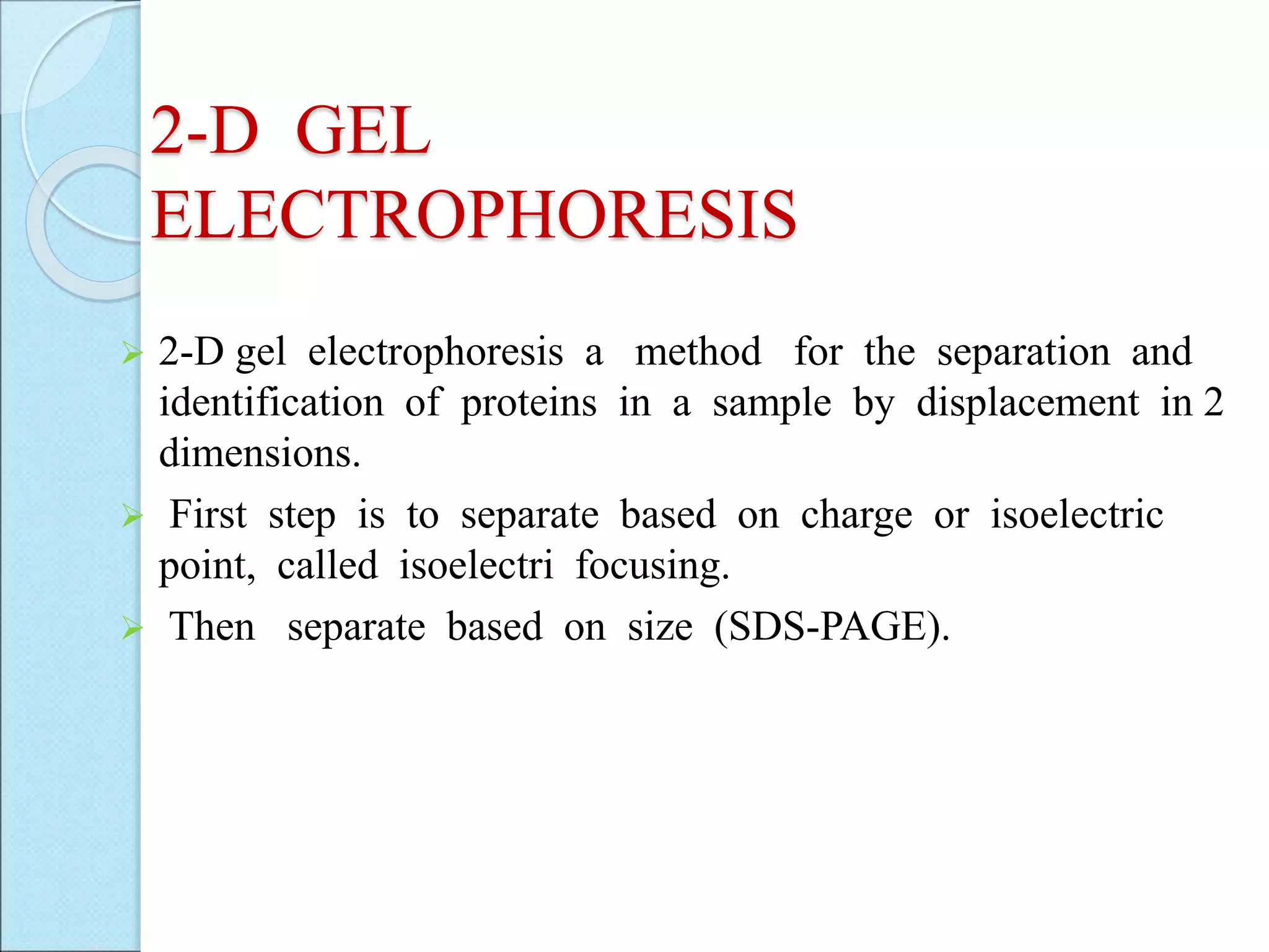
2-D GEL
ELECTROPHORESIS
2-Dgel electrophoresis a method for the separation and
identification of proteins in a sample by displacement in 2
dimensions.
First step is to separate based on charge or isoelectric
point, called isoelectri focusing.
Then separate based on size (SDS-PAGE).
44.
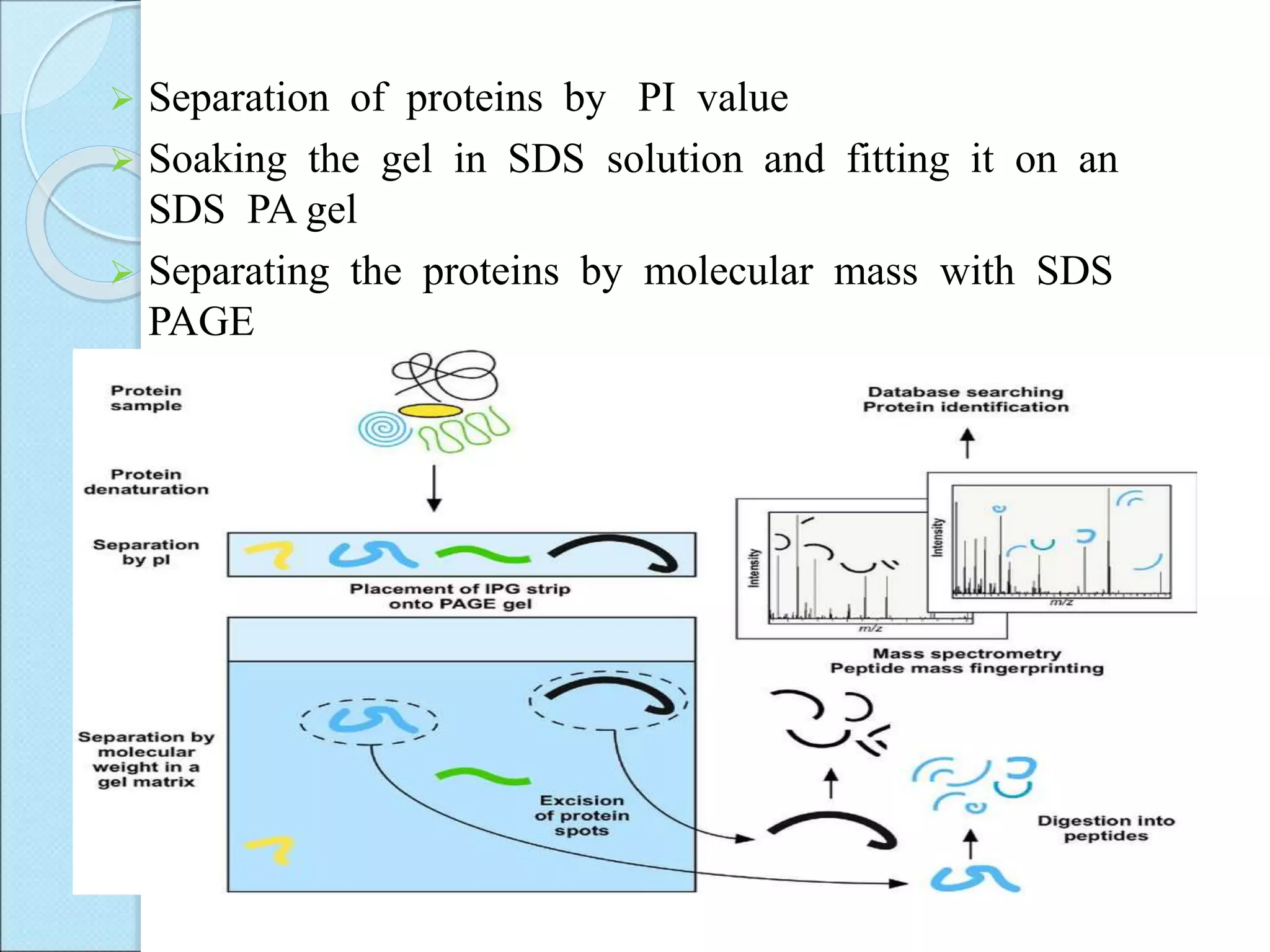
Separation ofproteins by PI value
Soaking the gel in SDS solution and fitting it on an
SDS PA gel
Separating the proteins by molecular mass with SDS
PAGE
45.
MASS SPECTROMETRY
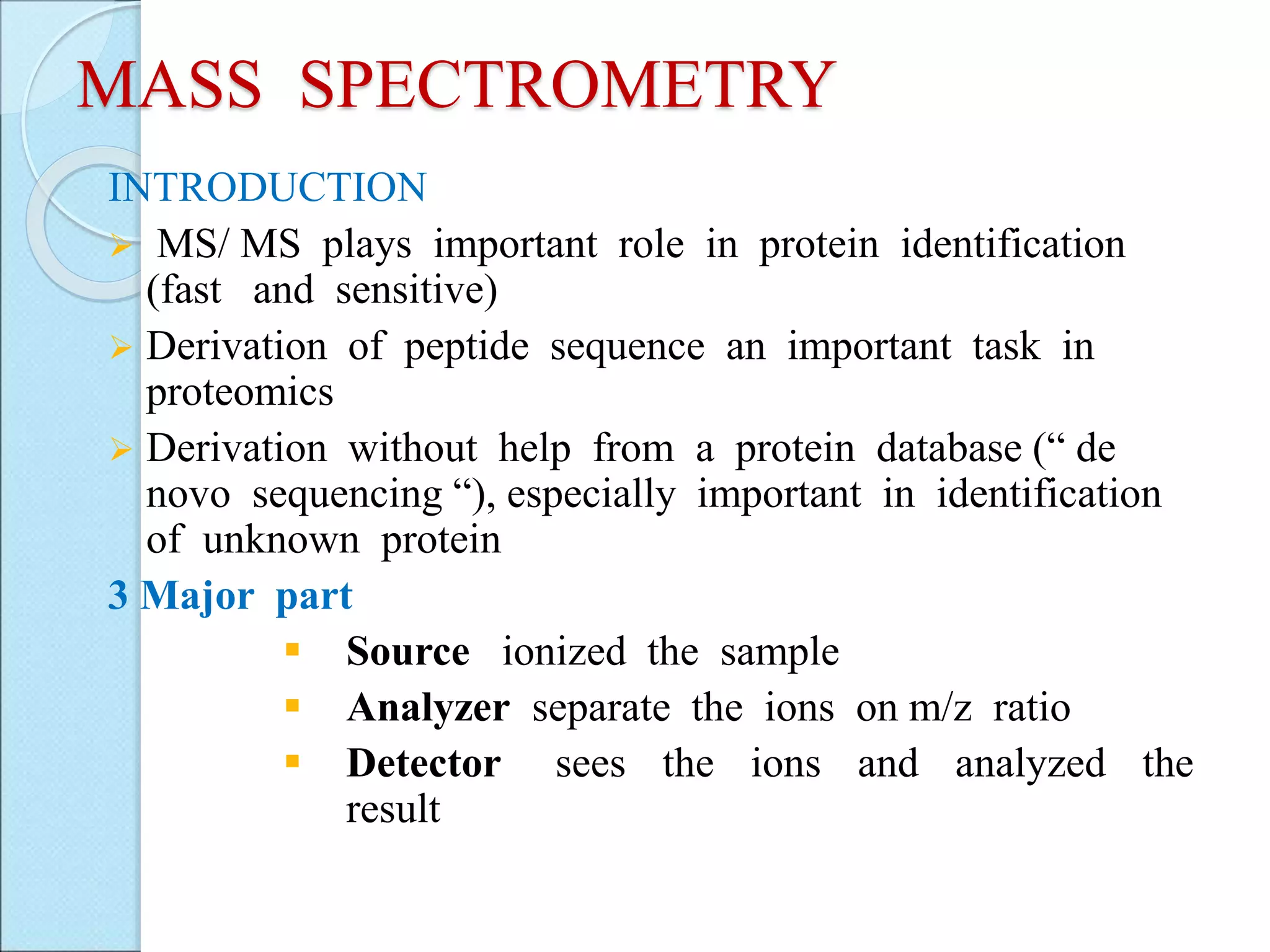
INTRODUCTION
MS/MS plays important role in protein identification
(fast and sensitive)
Derivation of peptide sequence an important task in
proteomics
Derivation without help from a protein database (“ de
novo sequencing “), especially important in identification
of unknown protein
3 Major part
Source ionized the sample
Analyzer separate the ions on m/z ratio
Detector sees the ions and analyzed the
result
47.
Basic steps
Isolatecell or other protein source
Lyses cells and isolate proteins
Break up protein into smaller ( but still relatively large)
amino acid chains
Separate chains (2D Gel, gas or liquid chromatography)
Analyze separated protein parts by mass spectrometry
Mass Spectrum
Proteins consist of 20 different types of a. a. With
different masses (except for one pair leu and lle)
Different peptides produce different spectra
Use the spectrum of a peptide to determine its
sequence
48.
Protein Identification
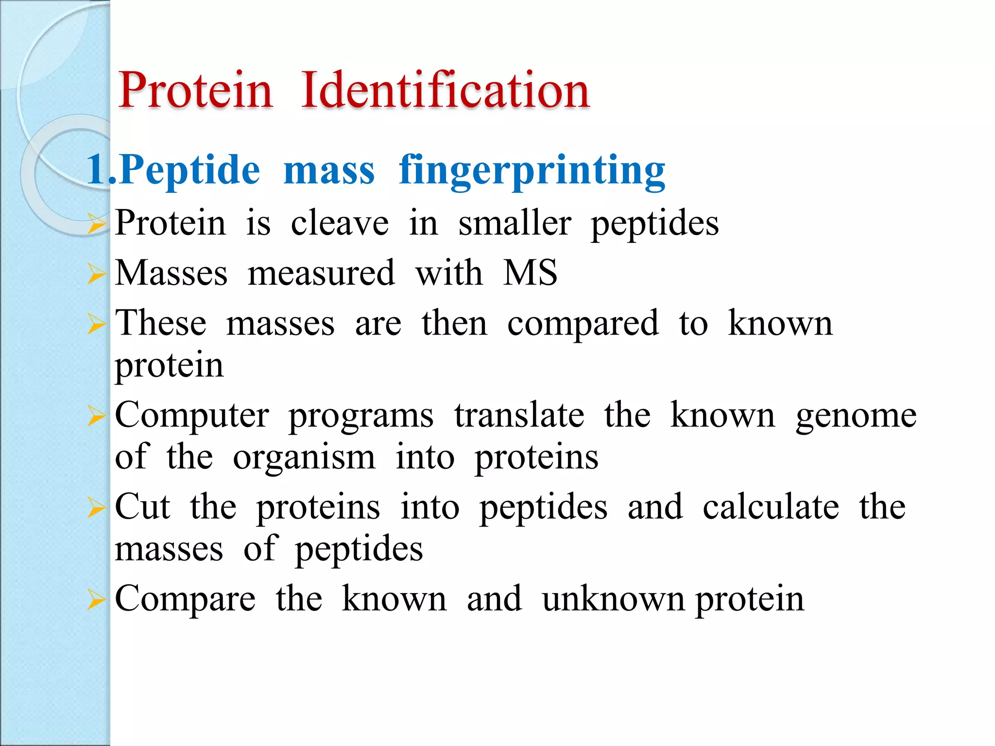
1.Peptide massfingerprinting
Protein is cleave in smaller peptides
Masses measured with MS
These masses are then compared to known
protein
Computer programs translate the known genome
of the organism into proteins
Cut the proteins into peptides and calculate the
masses of peptides
Compare the known and unknown protein
49.
2.Tandem MS
TwoMass Specs, (MS1, MS2)
A specific peak corresponding to a specific peptide
chain is identified and fragmented to from ions
The ions are analyzed by MS2, and identified as amino
acids
This way each selected piece of the whole protein can
be broken up and analyzed
50.
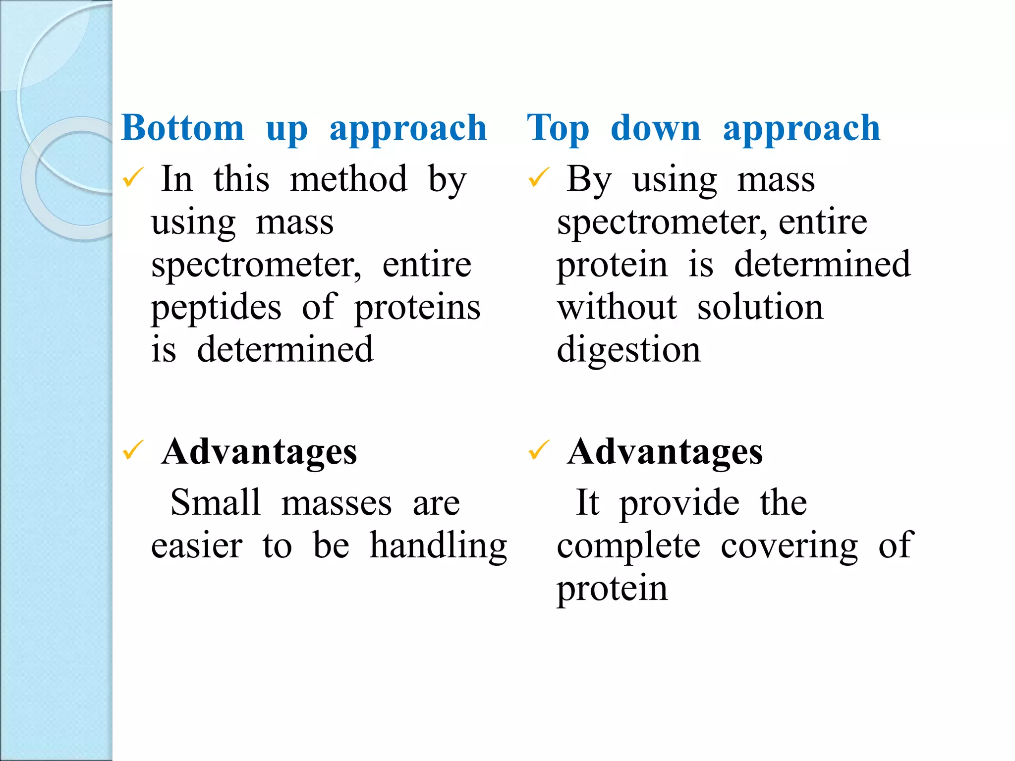
Bottom up approach
In this method by
using mass
spectrometer, entire
peptides of proteins
is determined
Advantages
Small masses are
easier to be handling
Top down approach
By using mass
spectrometer, entire
protein is determined
without solution
digestion
Advantages
It provide the
complete covering of
protein
51.
Electro sprayionization (ESI)
• In this method high electric field is applied to
the tip of capillary, from which solution will
pass through and get the ions of interest.
• Ions said to be multiple ions
• Multiple charged ions measure the high mass
biopolymer
52.
Matrix assistedlaser desorption/ ionization
(MALDI)
By pulses of laser light on the sample, ions of
interest formed.
large bio molecules can be determined and
synthetic polymer greater than 200,000 Dalton.
Advantages
• High speed
• Relative immunity to contaminants
53.
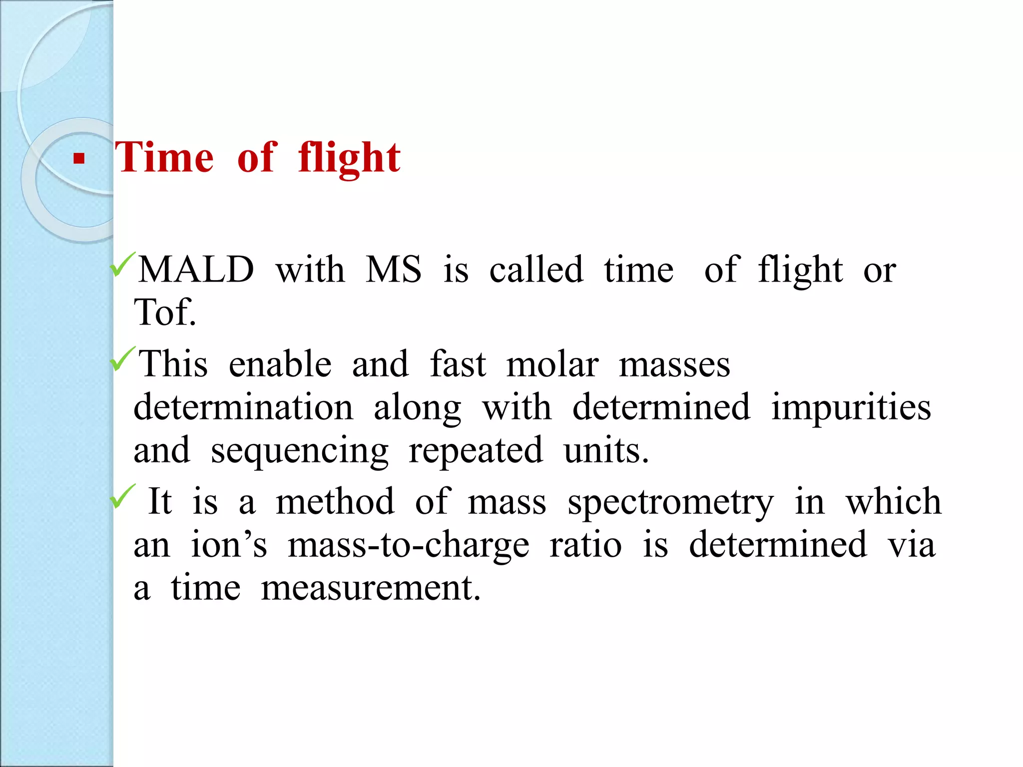
Time offlight
MALD with MS is called time of flight or
Tof.
This enable and fast molar masses
determination along with determined impurities
and sequencing repeated units.
It is a method of mass spectrometry in which
an ion’s mass-to-charge ratio is determined via
a time measurement.
54.
Application of proteomics
Protein sample identification/confirmation
Proteins sample purity determination
Detection of post-translation modification
Detection of amino acids substitution
Mass fingerprint identification of proteins
Nutrition Research
To identify unknown protein of interest
Quantify protein and peptide
Protein Biomarker


![LIQUID CHROMATOGRAPHY- MASS SPECTROSCOPY[LC-MS]](https://cdn.slidesharecdn.com/ss_thumbnails/63-191219160156-thumbnail.jpg?width=640&height=640&fit=bounds)


