Latest guidelines covid_2020.html
•
3 likes•1,394 views

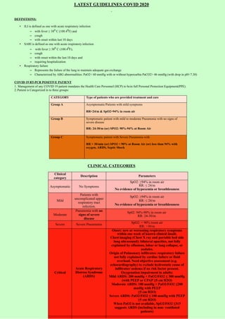
DEFINITIONS: • ILI is defined as one with acute respiratory infection – with fever ≥ 380 C (100.40F) and – cough – with onset within last 10 days • SARI is defined as one with acute respiratory infection – with fever ≥ 380 C (100.40F), – cough – with onset within the last 10 days and – requiring hospitalization • Respiratory failure – Represents the failure of the lung to maintain adequate gas exchange – Characterized by ABG abnormalities: PaO2< 60 mmHg with or without hypercarbia PaCO2> 46 mmHg (with drop in pH<7.30)

More Related Content
What's hot
What's hot (20)
Similar to Latest guidelines covid_2020.html
Similar to Latest guidelines covid_2020.html (20)
More from Ramachandra Barik
More from Ramachandra Barik (20)
Recently uploaded
Recently uploaded (20)
Latest guidelines covid_2020.html
- 1. LATEST GUIDELINES COVID 2020 DEFINITIONS: • ILI is defined as one with acute respiratory infection – with fever ≥ 380 C (100.40F) and – cough – with onset within last 10 days • SARI is defined as one with acute respiratory infection – with fever ≥ 380 C (100.40F), – cough – with onset within the last 10 days and – requiring hospitalization • Respiratory failure – Represents the failure of the lung to maintain adequate gas exchange – Characterized by ABG abnormalities: PaO2< 60 mmHg with or without hypercarbia PaCO2> 46 mmHg (with drop in pH<7.30) COVID 19 RT-PCR POSITIVE PATIENT 1. Management of any COVID 19 patient mandates the Health Care Personnel (HCP) to bein full Personal Protection Equipment(PPE). 2. Patient is Categorized in to three groups: CATEGORY Type of patients who are provided treatment and care Group A Asymptomatic/Patients with mild symptoms RR<24/m & SpO2>94% in room air Group B Symptomatic patient with mild to moderate Pneumonia with no signs of severe disease RR: 24-30/m (or) SPO2: 90%-94% at Room Air Group C Symptomatic patient with Severe Pneumonia with RR > 30/min (or) SPO2 < 90% at Room Air (or) less than 94% with oxygen, ARDS, Septic Shock CLINICAL CATEGORIES Clinical category Description Parameters Asymptomatic No Symptoms SpO2: ≥94% in room air RR: ≤ 24/m No evidence of hypoxemia or breathlessness Mild Patients with uncomplicated upper respiratory tract infection. SpO2: ≥94% in room air RR: ≤ 24/m No evidence of hypoxemia or breathlessness Moderate Pneumonia with no signs of severe disease Sp02: 94%-90% in room air RR: 24-30/m Severe Severe Pneumonia SpO2: < 90% room air RR: >30/m Critical Acute Respiratory Distress Syndrome (ARDS) Onset: new or worsening respiratory symptoms within one week of known clinical insult. Chest imaging (Chest X ray and portable bed side lung ultrasound): bilateral opacities, not fully explained by effusions, lobar or lung collapse, or nodules. Origin of Pulmonary infiltrates: respiratory failure not fully explained by cardiac failure or fluid overload. Need objective assessment (e.g. echocardiography) to exclude hydrostatic cause of infiltrates/ oedema if no risk factor present. Oxygenation impairment in adults: Mild ARDS: 200 mmHg < PaO2/FiO2 ≤ 300 mmHg (with PEEP or CPAP ≥5 cm H2O) Moderate ARDS: 100 mmHg < PaO2/FiO2 ≤200 mmHg with PEEP ≥5 cm H2O) Severe ARDS: PaO2/FiO2 ≤ 100 mmHg with PEEP ≥5 cm H2O) When PaO2 is not available, SpO2/FiO2 ≤315 suggests ARDS (including in non- ventilated patients)
- 2. Critical Septic Shock Adults: persisting hypotension despite volume resuscitation, requiring vasopressors to maintain MAP ≥65 mmHg and serum lactate level > 2 mmol/L INVESTIGATIONS Timing Mild Moderate Severe/Critical At admission CBC RBS ECG HbA1C (if Diabetic) D-Dimer (If starting on Tab Favipiravir) RFT S.Electrolyte S. Uric Acid • Complete Blood Count (with N/L RATIO) • LFT, RFT, RBS • S.Electrolytes • 12 lead ECG • CHEST X Ray –PA view • CRP, D-DIMER • S. FERRITIN, S.LDH • PROCALCITONIN • TROP – I & T • PT/INR • ABG • CT Thorax (if Available) • Blood culture (if total count is high) • IL – 6 • S. Cortisol • 2D ECHOCARDIOGRAP HY • COVID Antibody IgM/IgGTests •Complete Blood Count (with N/L RATIO) •LFT, RFT, RBS •S. Electrolytes •12 lead ECG •CHEST X Ray –PA view •CRP, D-DIMER •S. FERRITIN, S.LDH •PROCALCITONIN •TROP – I & T •PT/INR •ABG •CT Thorax (if Available) •Blood culture (if total count is high) •IL – 6 •S. Cortisol •S.Mg2+, S.Ca2+ •2D ECHOCARDIOGRA PHY •NTproBNP •HsCRP •S. Lactate •COVID Antibody IgM/IgGTests Repeat Daily _ Complete Blood Count, LFT, RFT ABG Complete Blood Count, LFT, RFT ABG Repeat Every 72hrs If initial D-Dimer is high CRP, D-DIMER S. FERRITIN, S.LDH Chest X ray CRP, D-DIMER S. FERRITIN, S.LDH Chest X ray At the time of discharge _ CRP, D-DIMER S. FERRITIN, S.LDH Chest X ray CRP, D-DIMER S. FERRITIN, S.LDH Chest X ray RT-PCR – Nasal & Throat swab Other Investigations should be done based on patient’s Co-morbid status IDENTIFICATION OF HIGH-RISK PATIENT CO MORBIDITIES CLINICALLY LABORATORY VALUE Age>50 yrs Hypoxia- SPO2<94% Lymphopenia (<20) with Neutrophil/Lymphocyte ratio >17 Ischecmic Heart Disease Tachycardia>100/min CRP>100 mg/L Diabetes Respiratory Distress RR>30/min Serum Ferritin >300 microg/L Hypertension Hypotension Systolic BP < 90mmHg LDH >450 Lung Disease (COPD/Asthma/Post TB Sequele) Altered Sensorium D-Dimer > 1000ng/ml Chronic Kidney Disease/ Chronic Liver Disease Immunosuppression / HIV / Malignancy Obesity Note: Calculation tool for predicting critically ill COVID-19 at admission can be used as reference tool. (Development of Validation of Clinical risk score to predict the occurrence of critical illness in hospitalised patient with COVID19. JAMA internal Medicine –published online, May 12/05/2020) GENERAL MEASURES AND GUIDELINES 1) Categorize in to A, B, C based on Symptoms, SpO2 & Respiratory Rate 2) Supportive Care:
- 3. • Finger Pulse Oximeter for continuous monitoring of Heart rate and Oxygen saturation • Start oxygen with Mask at saturation of 94% or lower • HFNC to be used if there is failed oxygen therapy and Non-invasive ventilation (NIV) to be used appropriately with two limb circuit expiratory filters • Counselling of COVID19 patients ( By Counsellor/psychologist/psychiatrist) • Normal feeding, no dietary restrictions, good oral hydration • Maintenance IV fluids (If indicated) • Maintain blood glucose levels <180 mg/dl. • If Patient is on ACE inhibitors/ARBs, should be continued • Avoid using NSAIDs other than Paracetamol Unless Absolutely Necessary • Avoid using Nebulized drugs to avoid aerosolization of virus. PREFER MDI with SPACER • Antibiotic selection in case of superadded bacterial pneumonia should be according to institution antibiogram. GROUP A - MILD CASES TREATMENT PRECAUTIONS ANTIVIRAL THERAPY • TAB HYDROXYCHLOROQUININE(HCQ) 400MG BD FOR 1 DAY Followed by 200MG 1-0-1 X 4 DAY for patients in COVID CARE CENTER/HOME ISOLATION (OR) Tab FAVIPIRAVIR 1800mg 1-0-1 on Day 1 f/b 800mg 1-0-1 for 6 days (total 7 days) for PATIENTS IN DCHC (OR) If Tab HCQ/Tab FAVIPIRAVIR is contraindicated, then combination of Cap DOXYCYCLIN 100mg 1-0-1 for 5 days + Tab IVERMECTIN 12mg 1-0-0 for 3 days · Cap Oseltamavir 75mg 1-0-1 for 5 days ANTICOAGULATION • Inj ENOXAPARIN 40mg S/C 1-0-0 X 7 DAYS (IF D-DIMER IS MORE THAN 1000NG/ML (OR) X-RAY/CT THORAX SHOWING GROUND GLASS OPACITIES) SUPPORTIVE THERAPY- • TAB ZINC 50 MG 0-1-0 X 7 DAYS • TAB VITAMIN C 500 MG 1-1-1 X 7 DAYS • Tab N Acetylcysteine 600mg 1-1-1 If Patients Having Cough • CATEGORIZATION SHOULD BE REASSESSED REGULARLY • CONTRAINDICATION FOR HCQ- • QT INTERVAL > 480ms • Pre-existing cardiomyopathy and cardiac rhythm disorders • History of Unexplained Syncope • Retinopathy, • Hypersensitivity to HCQ or 4- aminoquinoline compounds • G6PD deficiency • Epilepsy • Hypokalemia (K+ < 3 Meq) • Contraindications for Tab FAVIPIRAVIR: Hyperuricaemia, severe hepatic & renal impairment, Pregnant women and lactating mothers • PREGNANCY IS NOT A CONTRAINDICATION FOR HCQ • # Cap OSELTAMAVIR is advised due to possibility of H1N1 co infection along with COVID19 disease with present weather condition. Its usage will be reviewed at a later date. GROUP B - MODERATE CASES TREATMENT PRECAUTIONS ANTIVIRAL THERAPY · Inj REMDESIVIR 200 mg IV on day 1 followed by 100 mg IV daily for 4 days (total 5 days) IF REMEDESVIR IS NOT AVAILABLE TO START TAB HYDROXYCHLOROQUININE(HCQ) 400MG BD FOR 1 DAY followed by 200MG 1-0-1 X 4 DAY Co-administration of Inj REMDESIVIR with HCQ or chloroquine should be avoided · Cap Oseltamavir 75mg 1-0-1 for 5 days STEROIDS • Inj. Methyl Prednisolone 0.5 -1 mg/kg (or) Inj. Dexamethasone 0.1 – 0.2 mg/kg for 3-5 Days ANTICOAGULATION • Inj ENOXAPARIN 40MG S/C 1-0-0 x 7 DAYS Contraindications for Inj REMDESIVIR: · AST/ALT > 5 times Upper limit of normal (ULN) · Severe renal impairment (i.e., eGFR < 30ml/min/m2 or need for hemodialysis) · Pregnancy or lactating females · Children (< 12 years of age) · No dose adjustment for Inj REMDESIVIR if eGFR >30ml/min · Formula to calculate eGFR in Adults • eGFR, Male: (140 – age in years) × (weight in kg) / 72 × (serum creatinine in mg/dL); • eGFR, Female: (140 – age in years) × (weight in kg) × 0.85 / 72 × (serum creatinine in mg/dL) STEROIDS · to be started preferably within 48 hours of admission (or) if oxygen requirement is increasing and if inflammatory markers are increased. #
- 4. IV ANTIBIOTICS ACCORDING TO LOCAL ANTIBIOGRAM AWAKE PRONING • CONVALASCENT PLASMA THERAPY: 4 to 13 ml/kg (usually 200 ml single dose given slowly over not less than 2 hours) SUPPORTIVE THERAPY · TAB ZINC 50 MG 0-1-0 X 7 DAYS · TAB VITAMIN C 500 MG 1-1-1 X 7 DAYS · TAB N-ACETYL CYSTEINE 1-1-1 IN PATIENTS WITH COUGH · PATIENT SHOULD BE REASSESSED EVERY 12 HRLY AND CONTINOUS MONITORING OF SATURATION. · START ON OXYGEN-NASAL PRONGS 2- 5 L/MIN or FACE MASK 5L/MIN GROUP C - SEVERE/CRITICAL CASES TREATMENT PRECAUTIONS ANTIVIRAL THERAPY · If the patient has not received Inj REMDESIVIR, such patients can be started on Inj REMDESIVIR. Inj REMDESIVIR 200 mg IV on day 1 followed by 100 mg IV daily for 4 days (total 5 days) · Inj. TOCILUZUMAB 8mg/kg (maximum 800 mg at one time) given slowly in 100 ml NS over 1 hour; dose can be repeated once after 12 to 24 hours if needed (Or) Inj ITOLIZUMAB: 1st dose – 1.6mg/kg dose iv infusion. Subsequent dose: weekly 0.8mg/kg dose infusion over 4hours if required · Cap Oseltamavir 75mg 1-0-1 for 5 days STEROIDS · Inj. Methyl Prednisolone 1-2 mg/kg (or) Inj. Dexamethasone 0.2 – 0.4 mg /kg for 5-7 Days ANTICOAGULATION · Inj ENOXAPARIN 1mg/kg body wt s/c 1-0-1 X 7 DAYS PRONE VENTILLATION Inj CEFTRIAXONE 1gm IV 1-0-1 AND CAN BE ESCALATED ACCORDING TO LOCAL ANTIBIOGRAM OR TREATING PHYSICIAN CONSIDER SEPSIVAC (IF AVAILABLE) 0.3ml INTRADERMAL ONCE A DAY FOR 3 DAYS IN CASE OF SEPTIC SHOCK IV Diuretics in case of evidence of Heart Failure secondary to Myocarditis SUPPORTIVE THERAPY · TAB ZINC 50 MG 0-1-0X 7 DAYS · INJ. VITAMIN C 1.5GM IV 6 HOURLY X 5DAYS · TAB N-ACETYL CYSTEINE 1-1-1 Indication for TOCILUZUMAB/ITOLIZUMAB:- 1. IL-6 levels 50-100 fold higher thannormal (Normal range 0 - 9.5pg/ml 2. Worsening trend of the inflammatory markers (Ferritin, LDH, CRP) 3. Deteriorating clinical condition with worsening of PaO2/Fio2 ratio (more than 25% deterioration from the immediate previous value). Contraindications for Inj TOCILIZUMAB/ITOLIZUMAB PLHIV, those with active infections (systemic bacterial/fungal), High Serum. Procalcitonin, Tuberculosis, active hepatitis, Absolute Neutrophil Count < 2000/mm3 and Platelet count < 1,00,000/mm3, hepatic and renal impairment; patients on chronic steroid therapy, Paediatric patients <18 years old; Pregnancy and, Nursing mothers · PATIENT SHOULD BE CONTINOUSLY MONITORED · TO START ON OXYGEN WITH FACE MASK WITH NON REBREATHING BAG @ 8-10 lt/m · BASED ON PaO2/FiO2 ratio, HIGH FLOW NASAL OXYGEN (HFNC)/NIV SHOULD BE GIVEN AND IF PATIENT DETERIORATES INTUBATION SHOULD BE CONSIDERED AND LUNG PROTECTIVE VENTILATION TO BE FOLLOWED AS PER ARDSnet PROTOCOL · ABG TO BE DONE REGULARLY FOR MONITORING OF ACIDOSIS AND HYPOXEMIA · INOTROPHIC SUPPORT (NORADRENALINE – TITRATE ACCORDING TO THE MEAN ARTERIAL PRESSURE) · CORRECTION OF ACIDOSIS · MAINTAIN Hb% GREATER THAN 8gm% SUMMARY OF TREATMENT OF COVID-19PATIENTS BASED on CLINICALCATEGORIES MILD MODERATE SEVERE/CRITICAL An viral Therapy* Tab Hydroxychloroquinine(HCQ) 400mg Bd For 1 Day F/B 200mg 1-0- 1 X 4 Day for pa ents in COVID CARE CENTER/HOME ISOLATION (OR) Tab FAVIPIRAVIR 1800mg 1-0-1 on Day 1 f/b 800mg 1-0-1 for 6 days for PATIENTS IN DCHC (OR) An viral Therapy* Inj REMDESVIR 200 mg IV on day 1 followed by 100 mg IV daily for4 days (Or) IF REMEDESVIR IS NOT AVAILABLE TO START Tab Hydroxychloroquinine(HCQ) 400mg BD For 1 Day F/B 200mg 1-0-1 X 4 Day Co-administra on of Inj REMDESVIR with HCQ or chloroquine should be avoided An viral Therapy* Inj. TOCILUZUMAB 8mg/kg (maximum 800 mg at one me) given slowly in 100 ml NS over 1 hour; dose can be repeated once a er 12 to 24 hours if needed (Or) Inj ITOLIZUMAB: 1 dose – 1.6mg/kg dose iv infusion. Subsequent dose: weekly 0.8mg/kg dose infusion over 4hours if required st
- 5. If Tab HCQ/Tab FAVIPIRAVIR is contraindicated, then combina on of Cap DOXYCYCLIN 100mg 1-0-1 for 5 days + Tab IVERMECTIN 12mg 1-0-0 for 3 Days An coagula on Inj Enoxaparin 40mg S/C 1-0-0 x 7 days (If D- dimer Is More Than 1000ng/Ml or X-ray/CT Thorax Showing Ground glass opacity) Suppor ve Therapy Tab Zinc 50 Mg 0-1-0x 7 Days Tab Vitamin C 500 Mg 1-1-1 X 7 Days Tab N Acetylcysteine 1-1-1 If Pa ents Having Cough STEROIDS Inj. Methyl Prednisolone 0.5 -1 mg/kg (or) Inj. Dexamethasone – 0.2 mg /kg for 3-5 Days ANTICOAGULATION Inj Enoxaparin 40mg S/C 1-0-0 x7 days (if Wt >65kg, 60md 1-0-1 for 7days) Iv An bio cs According to Local An biogram Awake Proning Start on oxygen –Nasal Prongs 2- 5l/min or face mask 5l/min CONVALASCENT PLASMA THERAPY: 4 to 13 ml/kg (usually 200 ml single dose given slowly over not less than 2 hours Suppor ve Therapy Tab Zinc 50 Mg 0-1-0x 7 Days Tab Vitamin C 500 Mg 1-1-1 X 7 Days Tab N Acetylcysteine 1-1-1 If Pa ents Having Cough STEROIDS Inj. Methyl Prednisolone 1-2 mg/kg for 5-7 Days (or) Inj. Dexamethasone 0.2 – 0.4 mg /kg for 5- 7 Days ANTICOAGULATION Inj Enoxaparin 1 Mg/Kg Body Weight S/C 1- 0-1 X 7days Inj Ce riaxone 1 Gm Iv 1-0-1 And Can Be Escalated According To Local An biogram Or Trea ng Physician Start on oxygen with face mask+NRM and change over to HFNC/NIV (based on PaO2/FiO2) IF PATIENT DETERIORATES with HFNC/NIV trial (repeat ABG a er 6hrs suggests worsening of oxygena on) then EARLY INTUBATION SHOULD BE CONSIDERED AND LUNG PROTECTIVE VENTILATION TO BE FOLLOWED AS PER ARDSnet PROTOCOL Prone Ven lla on SEPSIVAC 0.3ml INTRADERMAL ONCE A DAY FOR 3 DAYS Suppor ve Therapy Inj. Vitamin C 1.5gm Iv 6 Hourly X 5 days Tab Zinc 50 Mg 0-1-0x 7 Days Tab N Acetylcysteine 1-1-1 IfPa ents Having Cough 1. Con nous monitoring of oxygen satura on by pulse oximeter and early diagnosis of hypoxemia is essen al in all group of pa ents 2. Indica ons and contraindica ons of the drugs are to be considered before use which is men oned in detail below 3. Transi on of pa ents between the clinical categories is based on SpO2, RR & PaO2/FiO2 ra o 4. Treatment of all co morbid illness to con nue Special Note: · *Cap Oseltamavir 75mg 1-0-1 for 5 days to be added to patients of all categories · All the investigational therapies and drugs approved recently by DGCI should be used with caution and after informed consent from the patient 1. Hydroxychloroquine (HCQ) Dose: Tab HCQ 400MG BD FOR 1 DAY Followed by 200MG 1-0-1 X 4 Days CONTRAINDICATION FOR HCQ • QT INTERVAL > 480ms • Pre-existing cardiomyopathy and cardiac rhythm disorders • History of Unexplained Syncope • Retinopathy, • Hypersensitivity to HCQ or 4-aminoquinoline compounds • G6PD deficiency • Epilepsy • Hypokalemia (K+ < 3 Meq) 2. Anticoagulant Agents Pro Coagulant factors are increased in COVID-19 infection and associated with increased risk of thrombosis Pneumonia and sepsis are complicated by DIC, but although COVID-19 patients do have abnormalities of coagulation and are not atypical of DIC. The most marked abnormality is an elevation of D-Dimer (if D-dimer is more than 1000ng/ml) but without a parallel fall in platelet or prolongation of clotting time, this suggests that local rather disseminated thrombin generation and fibrinolysis is taking place Dose: Inj ENOXAPARIN 40MG S/C Once daily for mild and moderate. Twice daily in severe cases. Other options: • Inj Fondaparinux 2.5mg OD SC • Unfractioned Heparin 5000 Units BD SC Contraindications: ESRD, active bleeding, emergency surgery, platelets < 20,000/mm3, BP >200/120 mmHg) INVESTIGATIONAL THERAPIES (as per MOHFW)
- 6. 1. Remdesivir (under Emergency Use Authorization) may be considered in patients with moderate disease (those on oxygen) with none of the following contraindications: · AST/ALT > 5 times Upper limit of normal (ULN) · Severe renal impairment (i.e., eGFR < 30ml/min/m2 or need for hemodialysis) · Pregnancy or lactating females · Children (< 12 years of age) Dose: 200 mg IV on day 1 followed by 100 mg IV daily for 4 days (total 5 days) 2. Convalescent plasma (Off Label) may be considered in patients with moderate diseasewho are not improving (oxygen requirement is progressively increasing) despite use of steroids. Special prerequisites while considering convalescent plasma include: · ABO compatibility and cross matching of the donor plasma · Neutralizing titer of donor plasma should be above the specific threshold (if the latter is not available, plasma IgG titer (against S- protein RBD) above 1:640 should beused) · Recipient should be closely monitored for several hours post transfusion forany transfusion related adverse events · Use should be avoided in patients with IgA deficiency or immunoglobulin allergy Dose: Dose is variable ranging from 4 to 13 ml/kg (usually 200 ml single dose given slowly over not less than 2 hours 3. Tocilizumab (Off Label) may be considered in patients with severe disease with progressively increasing oxygen requirements and in mechanically ventilated patients not improving despite use of steroids. Long term safety data in COVID 19 remains largely unknown. Special considerations before its use include: o IL-6 levels 50-100 fold higher than normal (Normal range 0 - 9.5pg/ml o Worsening trend of the inflammatory markers (Ferritin, LDH, CRP) o Deteriorating clinical condition with worsening of PaO2/Fio2 ratio(more than 25% deterioration from the immediate previous value) The drug is contraindicated in PLHIV, those with active infections (systemic bacterial/fungal), High Serum. Procalcitonin, Tuberculosis, active hepatitis, Absolute Neutrophil Count < 2000/mm3 and Platelet count < 1,00,000/mm3, hepatic and renal impairment; patients on chronic steroid therapy, Paediatric patients <18 years old; Pregnancy and, Nursing mothers Dose: 8mg/kg (maximum 800 mg at one time) given slowly in 100 ml NS over 1 hour; dose can be repeated once after 12 to 24 hours if needed Drugs Recently approved by DGCI 1. ITOLIZUMAB (An anti-CD6 IgG1 monoclonalantibody) Indication: 1. IL-6 levels 50-100 fold higher than normal (Normal range 0 - 9.5pg/ml 2. Worsening trend of the inflammatory markers (Ferritin, LDH, CRP) 3. Deteriorating clinical condition with worsening of PaO2/Fio2 ratio (more than 25% deterioration from the immediate previous value). Dose: 1st dose – 1.6mg/kg dose iv infusion • Subsequent dose: weekly 0.8mg/kg dose infusion over 4hours if required based onlung function parameters Contraindication: PLHIV, those with active infections (systemic bacterial/fungal), High Serum. Procalcitonin, Tuberculosis, active hepatitis, Absolute Neutrophil Count < 2000/mm3 and Platelet count < 1,00,000/mm3, hepatic and renal impairment; patients on chronic steroid therapy, Paediatric patients <18 years old; Pregnancy and, Nursing mothers Side effects: • In trial Infusion reactions have been reported in 15% of the patients • In clinical practice also infusion reaction ranged from 12% to15% • Other adverse events include Diahorea, Pruritus in 7 – 12 % of cases 2. Tab. FAVIPIRAVIR Mechanism of action: It is considered that favipiravir is metabolized in cells to a ribosyl triphosphate form (favipiravir RTP) and that favipiravir RTP selectively inhibits RNA polymerase involved in influenza viral replication Indications: mild to moderate cases of COVID19 in adults >18yrs old Dose: 1800mg bid followed by 800mg bid upto maximum of 14days Contraindications: Hyperuricaemia, severe hepatic & renal impairment, Pregnant women and lactating mothers Side Effects: increased Uric Acid levels, diarrhea, decreased neutrophil counts, increase in AST/ALT levels Drug Interactions: metabolised partly by Aldehyde Oxidase(AO) and partly by Xanthine Oxidase(XO). Precauitons for co-administration with Pyrazinamide, Repaglinide, Theophyline, Famciclovir PRONE VENTILATION Early self-proning in awake, non-intubated patients – Moderate cases · Any COVID-19 patient with respiratory embarrassment severe enough to be admitted tothe hospital may be considered for rotation and early self-proning. · Care must be taken to not disrupt the flow of oxygen during patient rotation Criteria to be fulfilled Avoid proning
- 7. · Patients with oxygen requirement of >4L · Normal mental status · Able to self-prone or change position with minimal assistance · Hemodynamic instability · Close monitoring not possible · Typical protocols include 30–120 minutes in prone position, followed by30–120 minutes in left lateral decubitus, right lateral decubitus, and upright sitting position (Caputo ND, Strayer RJ, Levitan R. Academic Emergency Medicine 2020;27:375–378) Requirements for safe prone positioning in ARDS · Pre-oxygenate the patient with FiO2 1.0 · Secure the endotracheal tube and arterial and central venous catheters · Adequate number of staff to assist in the turn and to monitor theturn · Supplies to turn (pads for bed, sheet, protection for the patient) · Knowledge of how to perform the turn as well as how to supine the patient in case ofan emergency Contraindications to prone ventilation · Spinal instability requires special care · Intra cranial pressure may increase on turning · Rapidly return to supine in case of CPR or defibrillation When to start proning in SEVERE CASES? · P/F ratio <150 while being ventilated with FiO2 >0.6 and PEEP >5 cm H2O When to stop proning? · When P/F exceeds 150 on FiO2 > 0.6 and > 6 PEEP What portion of the day should patients be keptprone? · As much as possible (16-18 hours a day) · Adult patients with severe ARDS receive prone positioning for more than 12 hours perday (strong recommendation, moderate-high confidence in effect estimates) (ATS-ERS Guideline. Am J RespirCrit Care Med;2017;195(9):1253-1263) Oxygen delivery protocol • SpO2 < 94% ~ Supplement with nasal prongs or simple face mask at 2-5L/min • Monitor continuous SpO2 with finger pulse oximetry • If SpO2 < 94% on simple face mask or nasal prongs, change to non-rebreather mask oxygen (NRB) at 10-15L/min • Oxygen Delivery Devices & approximate FiO2%
- 8. HFNO (High Frequency Nasal Oxygen) and NIV (Non-invasive Ventilation) • When oxygen requirement increases to needing NRB, options of High Frequency Nasal Oxygen (HFNO) or NIV should be considered. • HFNC flow rates to be set from 30 -60 L/min titrating to maintain SpO2 ≥ 92% • HFNC provides PEEP up to 5-6 cm H20 and can deliver FiO2 up to 100% • If HFNC non-available or patient not maintaining SpO2 on flow rates up to 60L/min, initiate on non-invasive ventilation (NIV) only with an ICU ventilator with two limbed circuit and expiratory HME filter with a NIV mode available. Caution is to be exercised to not use potable home BiPAP or CPAP machines with single circuit for these patients. · Appropriate mask with good seal to be ensured when initiated on NIV. Helmetmasks/hoods if available, to be preferred to minimize aerosol contamination. · Once initiated on NIV, close monitoring of respiratory variables hourly is important. · Reassess clinical condition hourly, monitor and observe ABG’s 4-6hrly · Look for signs of clinical improvement in the form of settling tachycardia, improvingSpO2, reduced tachypnea and reduced work of breathing. · On NIV when there are signs of clinical deterioration in the form of worsening sensorium, increased accessory muscles of breathing, raising Pco2, worsening pH on ABG ~ failure of NIV has to be considered and patient has to be planned for intubation and mechanical ventilation after consent from the family. Intubation and Mechanical Ventilation · Indication for intubation: ARDS with PaO2/FiO2 < 200 · Worsening respiratory distress even on NIV · Patient in Shock Initial Settings: Controlled Mode ventilation: VCV (volume-controlled ventilation) or PCV (pressure-controlled ventilation) · Tidal Volume (Vt) 6-8ml/PBW (predicted body weight) · PEEP 8 – 18 cmH2O (follow FiO2-PEEP table) to titrate to target SpO2 90-92% · FiO2 ~ target SpO2 90-92% with lowest FiO2 possible · Respiratory rate 14-18/min (maximum up to35/min) · Plateau pressure < 30 cmH2O and driving pressure < 16cmH2O · ABG targets: PaO2 55-80 mmHg, pH > 7.3 · Measure compliance 6hrly ~ Vt in ml /Pplat – PEEP Notes: Additional steps: If Pplat > 30cmH2O, reduce Vt upto 4ml/PBW · If SpO2 < 88% despite ARDSnet protocol: increase depth of sedation · Optimize secretions clearance/bronchodilation
- 9. Indication for prone ventilation: · Intubation and mechanical ventilation < 36hrs · PaO2/FiO2 < 150, FiO2 > 60%, PEEP > 5, Vt 6ml/PBW · Duration of proning: 12-16 hrs. · Multiple sessions until favorable trends are achieved. · Initiate early muscle relaxant infusion (cis-atracurium or vecuronium) · Early prone ventilation Adjunctive measures when intubated and mechanically ventilated: Ø Antibiotics guided by protocols Ø Steps to reduce VAP (ventilator associated pneumonia) by following VAP bundles Ø Head-end elevation Ø Thrombo-prophylaxis Ø Adequate analgesia and sedation In absence of ABG facility at the hospitals, use SpO2/FiO2 ratio as described in the below table REFERENCES: 1. CLINICAL MANAGEMENT PROTOCOL: COVID-19 Government of India Ministry of Health and Family Welfare Directorate General of Health Services (EMR Division) Version 5. 03.07.20 2. Development of Validation of Clinical risk score to predict the occurrence of critical illness in hospitalised patient with COVID19. JAMA internal Medicine –published online, May 12/05/2020 3. FACT SHEET FOR HEALTH CARE PROVIDERS EMERGENCY USEAUTHORIZATION (EUA) OF REMDESIVIR (GS-5734™ 4. TOCILIZUMAB Drug Monograph 5. ITOLIZUMAB Drug Monograph 6. FAVIPIRAVIR Drug Monograph 7. Matthew Wemple / Joshua O. Benditt. Fishman’s Pulmonary Diseases and Disorders.Fifth Edition. 8. Pratik P. Pandharipande et al. Derivation and validation of SpO2/FiO2 ratio to impute for PaO2/FiO2 ratio in the respiratory component of the Sequential Organ FailureAssessment (SOFA) Score. Crit Care Med. 2009 April ; 37(4): 1317–1321. doi:10.1097/CCM.0b013e31819cefa9