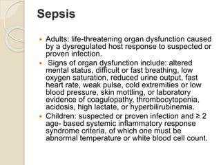
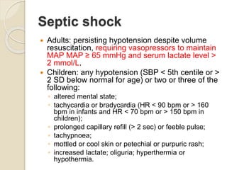
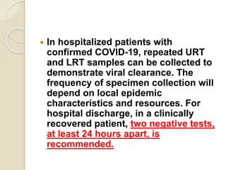
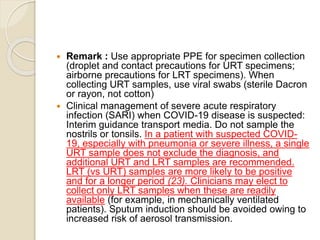
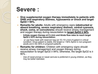
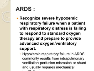
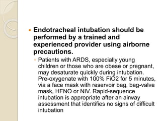
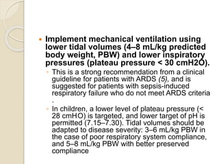
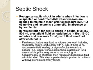
2. Introduction
While most people with COVID-19 develop only
mild or uncomplicated illness,
approximately 14% develop severe disease that
requires hospitalization and oxygen support,
and 5% require admission to an intensive care
unit. In severe cases, COVID-19 can be
complicated by the acute respiratory distress
syndrome (ARDS), sepsis and septic shock,
multiorgan failure, including acute kidney injury
and cardiac injury .
Older age and co-morbid disease have been
reported as risk factors for death, and 2 recent
multivariable analysis confirmed older age,
3. Higher Sequential Organ Failure Assessment
(SOFA) score and d-dimer > 1 μg/L on admission
were associated with higher mortality.
COVID-19 virus was detectable until death in
non-survivors.
The longest observed duration of viral shedding
in survivors was 37 days (3, 4).
There is currently no known difference between
the clinical manifestations of COVID-19 pregnant
and non-pregnant women or adults of
reproductive age.
Pregnant and recently pregnant women with
suspected or confirmed COVID-19 should be
treated with supportive and management
therapies
4. The most common diagnosis in severe COVID-
19 patients is severe pneumonia.
For those with mild illness, hospitalization may
not be required unless there is concern about
rapid deterioration or an inability to promptly
return to hospital, but isolation to contain/mitigate
virus transmission should be prioritized.
All patients cared for outside hospital (i.e. at
home or non-traditional settings) should be
instructed to manage themselves appropriately
according to local/regional public health protocols
for home isolation and return to a designated
COVID-19 hospital if they get worse .
6. Mild illness
Patients uncomplicated upper respiratory
tract viral infection may have non-specific
symptoms such as fever, fatigue, cough (with
or without sputum production), anorexia,
malaise, muscle pain, sore throat, dyspnea,
nasal congestion, or headache. Rarely,
patients may also present with diarrhoea,
nausea, and vomiting .
The elderly and immunosuppressed may
present with atypical symptoms. Symptoms
due to physiologic adaptations of pregnancy
or adverse pregnancy events, such as
dyspnea, fever, GI-symptoms or fatigue, may
overlap with COVID-19 symptoms.
7. Pneumonia :
Adult with pneumonia but no signs of
severe pneumonia and no need for
supplemental oxygen.
Child with non-severe pneumonia
who has cough or difficulty breathing +
fast breathing: fast breathing (in
breaths/min):
◦ < 2 months: ≥ 60; 2–11 months: ≥ 50; 1–5
years: ≥ 40, and no signs of severe
pneumonia.
8. Severe pneumonia
Adolescent or adult: fever or suspected respiratory
infection, plus one of the following: respiratory rate > 30
breaths/min; severe respiratory distress; or SpO2 ≤ 93% on
room air .
Child with cough or difficulty in breathing, plus at least one of
the following:
◦ central cyanosis or SpO2 < 90%;
◦ severe respiratory distress (e.g. grunting, very severe chest
indrawing);
◦ signs of pneumonia with a general danger sign: inability to
breastfeed or drink, lethargy or unconsciousness, or convulsions
◦ Other signs of pneumonia may be present: chest indrawing, fast
breathing (in breaths/min):
< 2 months: ≥ 60;
2–11 months: ≥ 50;
1–5 years: ≥ 40 (16).
◦ While the diagnosis is made on clinical grounds; chest imaging
may identify or exclude some pulmonary complications.
9. Acute respiratory distress
syndrome (ARDS)
Onset: within 1 week of a known clinical
insult or new or worsening respiratory
symptoms.
Chest imaging (radiograph, CT scan, or lung
ultrasound): bilateral opacities, not fully
explained by volume overload, lobar or lung
collapse, or nodules.
Origin of pulmonary infiltrates: respiratory
failure not fully explained by cardiac failure or
fluid overload. Need objective assessment
(e.g. echocardiography) to exclude
hydrostatic cause of infiltrates/oedema if no
risk factor present.
10. Sepsis
Adults: life-threatening organ dysfunction caused
by a dysregulated host response to suspected or
proven infection.
Signs of organ dysfunction include: altered
mental status, difficult or fast breathing, low
oxygen saturation, reduced urine output, fast
heart rate, weak pulse, cold extremities or low
blood pressure, skin mottling, or laboratory
evidence of coagulopathy, thrombocytopenia,
acidosis, high lactate, or hyperbilirubinemia.
Children: suspected or proven infection and ≥ 2
age- based systemic inflammatory response
syndrome criteria, of which one must be
abnormal temperature or white blood cell count.
11. Septic shock
Adults: persisting hypotension despite volume
resuscitation, requiring vasopressors to maintain
MAP MAP ≥ 65 mmHg and serum lactate level >
2 mmol/L.
Children: any hypotension (SBP < 5th centile or >
2 SD below normal for age) or two or three of the
following:
◦ altered mental state;
◦ tachycardia or bradycardia (HR < 90 bpm or > 160
bpm in infants and HR < 70 bpm or > 150 bpm in
children);
◦ prolonged capillary refill (> 2 sec) or feeble pulse;
◦ tachypnoea;
◦ mottled or cool skin or petechial or purpuric rash;
◦ increased lactate; oliguria; hyperthermia or
hypothermia.
12. Investigations :
Collect blood cultures for bacteria that cause
pneumonia and sepsis, ideally before
antimicrobial therapy. DO NOT delay
antimicrobial therapy to collect blood
cultures.
Collect specimens from the upper respiratory
tract (URT; nasopharyngeal and
oropharyngeal) AND, where clinical suspicion
remains and URT specimens are negative,
collect specimens from the lower respiratory
tract when readily available (LRT;
expectorated sputum, endotracheal aspirate,
or bronchoalveolar lavage in ventilated
patient) for COVID-19 virus testing by RT-PCR
and bacterial stains/cultures.
13. In hospitalized patients with
confirmed COVID-19, repeated URT
and LRT samples can be collected to
demonstrate viral clearance. The
frequency of specimen collection will
depend on local epidemic
characteristics and resources. For
hospital discharge, in a clinically
recovered patient, two negative tests,
at least 24 hours apart, is
recommended.
14. Remark : Use appropriate PPE for specimen collection
(droplet and contact precautions for URT specimens;
airborne precautions for LRT specimens). When
collecting URT samples, use viral swabs (sterile Dacron
or rayon, not cotton)
Clinical management of severe acute respiratory
infection (SARI) when COVID-19 disease is suspected:
Interim guidance transport media. Do not sample the
nostrils or tonsils. In a patient with suspected COVID-
19, especially with pneumonia or severe illness, a single
URT sample does not exclude the diagnosis, and
additional URT and LRT samples are recommended.
LRT (vs URT) samples are more likely to be positive
and for a longer period (23). Clinicians may elect to
collect only LRT samples when these are readily
available (for example, in mechanically ventilated
patients). Sputum induction should be avoided owing to
increased risk of aerosol transmission.
15. Remark : Dual infections with other respiratory
viral and bacterial infections have been found in
SARS, MERS and COVID-19 patients . As a
result, a positive test for a non-COVID-19
pathogen does not rule out COVID-19. At this
stage, detailed microbiologic studies are needed
in all suspected cases. Both URT and LRT
specimens can be tested for other respiratory
viruses. In endemic settings arbovirus infection
(dengue/chikungunya) should also be considered
in the differential diagnosis of undifferentiated
febrile illness, particularly when
thrombocytopenia is present. Co-infection with
COVID-19 virus may also occur and a positive
diagnostic test for dengue (e.g. dengue RDTs)
does not exclude the testing for COVID-19 .
16. Lab Interpretation
COVID-19 pneumonia manifests with
chest CT imaging abnormalities, even
in asymptomatic patients, with rapid
evolution from focal unilateral to
diffuse bilateral ground-glass opacities
that progressed to or co-existed with
consolidations within 1–3 weeks.
17. The complete blood count results
indicated that the number of
lymphocytes and platelets were
significantly decreased in both severe
and non-severe COVID-19 patients.
Severe COVID-19 patients had
significantly higher levels of C-reactive
protein (CRP)
19. Mild :
Patients with mild disease do not require hospital
interventions, but isolation is necessary to contain
virus transmission and will depend on national
strategy and resources.
Remark: Although most patients with mild disease may
not have indications for hospitalization, it is necessary
to implement appropriate IPC to contain and mitigate
transmission. This can be done either in hospital, if
there are only sporadic cases or small clusters, or in
repurposed, non-traditional settings; or at home.
Provide patients with mild COVID-19 with
symptomatic treatment such as antipyretics for
fever.
Counsel patients with mild COVID-19 about signs
and symptoms of complicated disease. If they
develop any of these symptoms, they should seek
urgent care through national referral systems.
20. Severe :
Give supplemental oxygen therapy immediately to patients with
SARI and respiratory distress, hypoxaemia or shock and target
SpO2 > 94%.
Remarks for adults: Adults with emergency signs (obstructed or
absent breathing, severe respiratory distress, central cyanosis,
shock, coma, or convulsions) should receive airway management
and oxygen therapy during resuscitation to target SpO2 ≥ 94%.
◦ Initiate oxygen therapy at 5 L/min and titrate flow rates to reach target
SpO2 ≥ 93% during resuscitation;
◦ or use face mask with reservoir bag (at 10–15 L/min) if patient in critical
condition. Once patient is stable, the target is > 90% SpO2 in non-pregnant
adults and ≥ 92–95% in pregnant patients .
Remarks for children: Children with emergency signs should
receive airway management and oxygen therapy during
resuscitation to target SpO2 ≥ 94%; otherwise, the target SpO2 is ≥
90% .
◦ Use of nasal prongs or nasal cannula is preferred in young children, as they
may be better tolerated.
21. Haematology and biochemistry
laboratory testing and ECG should be
performed at admission and as clinically
indicated to monitor for complications,
such as acute liver injury, acute kidney
injury, acute cardiac injury, or shock.
Determine which chronic
therapies(eg.HTN,DM etc) should be
continued and which therapies should be
stopped temporarily. Monitor for drug-
drug interactions.
22. Use conservative fluid management in
patients with SARI when there is no
evidence of shock.
Patients with SARI should be treated
cautiously with intravenous fluids,
because aggressive fluid
resuscitation may worsen
oxygenation, especially in settings
where there is limited availability of
mechanical ventilation This applies for
care of children and adults
23. Give empiric antimicrobials to treat
all likely pathogens causing SARI
and sepsis as soon as possible,
within 1 hour of initial assessment
for patients with sepsis.
24. ARDS :
Recognize severe hypoxemic
respiratory failure when a patient
with respiratory distress is failing
to respond to standard oxygen
therapy and prepare to provide
advanced oxygen/ventilatory
support.
◦ Hypoxemic respiratory failure in ARDS
commonly results from intrapulmonary
ventilation-perfusion mismatch or shunt
and usually requires mechanical
25. Endotracheal intubation should be
performed by a trained and
experienced provider using airborne
precautions.
◦ Patients with ARDS, especially young
children or those who are obese or pregnant,
may desaturate quickly during intubation.
Pre-oxygenate with 100% FiO2 for 5 minutes,
via a face mask with reservoir bag, bag-valve
mask, HFNO or NIV. Rapid-sequence
intubation is appropriate after an airway
assessment that identifies no signs of difficult
intubation
26. Implement mechanical ventilation using
lower tidal volumes (4–8 mL/kg predicted
body weight, PBW) and lower inspiratory
pressures (plateau pressure < 30 cmH2O).
◦ This is a strong recommendation from a clinical
guideline for patients with ARDS (5), and is
suggested for patients with sepsis-induced
respiratory failure who do not meet ARDS criteria
.
◦ In children, a lower level of plateau pressure (<
28 cmH2O) is targeted, and lower target of pH is
permitted (7.15–7.30). Tidal volumes should be
adapted to disease severity: 3–6 mL/kg PBW in
the case of poor respiratory system compliance,
and 5–8 mL/kg PBW with better preserved
compliance
27. In adult patients with severe ARDS, prone
ventilation for 12–16 hours per day is
recommended.
Use a conservative fluid management strategy for
ARDS patients without tissue hypoperfusion
In patients with moderate or severe ARDS, higher
PEEP instead of lower PEEP is suggested.
In patients with moderate-severe ARDS (PaO2/FiO2
< 150), neuromuscular blockade by continuous
infusion should not be routinely used.
Use in-line catheters for airway suctioning and
clamp endotracheal tube when disconnection is
required (for example, transfer to a transport
ventilator)
28. Septic Shock
Recognize septic shock in adults when infection is
suspected or confirmed AND vasopressors are
needed to maintain mean arterial pressure (MAP) ≥
65 mmHg and lactate is ≥ 2 mmol/L, in absence of
hypovolemia .
In resuscitation for septic shock in adults, give 250–
500 mL crystalloid fluid as rapid bolus in first 15–30
minutes and reassess for signs of fluid overload
after each bolus
◦ Fluid resuscitation may lead to volume overload, including
respiratory failure, particularly with ARDS. If there is no
response to fluid loading or signs of volume overload
appear (e.g. jugular venous distension, crackles on lung
auscultation, pulmonary oedema on imaging, or
hepatomegaly in children), then reduce or discontinue fluid
administration. This step is particularly important in patients
with hypoxemic respiratory failure
29. Do not use hypotonic crystalloids,
starches, or gelatins for resuscitation.
In adults, administer vasopressors when
shock persists during or after fluid
resuscitation. The initial blood pressure
target is MAP ≥ 65 mmHg in adults and
improvement of markers of perfusion.
◦ If central venous catheters are not available,
vasopressors can be given through a peripheral
IV, but use a large vein and closely monitor for
signs of extravasation and local tissue necrosis.
If extravasation occurs, stop infusion.
Vasopressors can also be administered through
intraosseous needles.
30. Do not routinely give systemic
corticosteroids for treatment of viral
pneumonia outside clinical trials.
◦ A systematic review of observational
studies of corticosteroids administered
to patients with SARS reported no
survival benefit and possible harms
(avascular necrosis, psychosis,
diabetes, and delayed viral clearance)