Vip Call Girls Anna Salai Chennai 👉 8250192130 ❣️💯 Top Class Girls Available
Ent signs by yassin hasan
1. ENT Signs:
By Dr. Yassin Hasan
Otolaryngologist
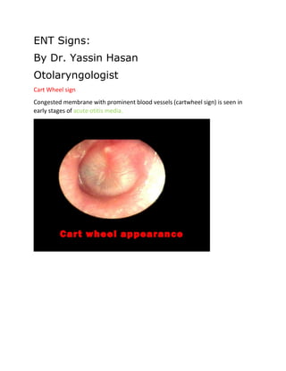
Cart Wheel sign
Congested membrane with prominent blood vessels (cartwheel sign) is seen in
early stages of acute otitis media.
2. LIGHT HOUSE SIGN:
Acute suppurativeotitis media
small pin hole perforation with a pulsatile ear discharge
There is collection of pus behind the tympanic membrane. Thus
pus comes out under pressure and synchronizing with each
arterial pulse, called as pulsatile otorrhea or light‑house sign.
3. Schwartz sign or Flamingo flush sign:
German otologist Hermann Schwartze
This sign of active otosclerosis (such as during pregnancy) is seen
as a pink reflex (reddish hue seen
over the promontory) through intact tympanic membrane in the area of oval
window.
4. Rising sun sign :
Tip of iceberg
red vascular hue seen behind the intact tympanic membrane
glomus tumour
high jugular bulb
aberrant carotid artery in the floor of middle ear
5. “Pulsation sign” (Brown sign):
when ear canal pressure is raised with Siegel’s speculum, tumour
pulsates vigorously and then blanches; reverse happens
with the release of pressure.
Glomus tumor
AQUINO'S SIGN:
Glomus tumors
blanching of the tympanic mass with gentle pressure on the carotid artery
Phelps sign:
It is seen in cases of glomus jugulare tumor and consists of destruction of bone
between the carotid canal and jugular foramen.
absence of normal crest between the carotid canal
and jugular fossa on lateral tomography, in case of glomus jugulare.
7. Fontaine sign :
carotid body tumor mass mobiles horizontally not vertically
LYRE’S SIGN:
carotid body tumor
splaying of carotid vessels ( at junction of External & internal carotid artery)
Pseudo-lyre sign:
Vagal paragangliomas
8. Guyon Sign:
The 12th nerve lies directly upon the external carotid artery, whereby this vessel
may be distinguished from the internal carotid artery. (The safer way prior to
ligation of the external carotid
artery is to identify the rest few branches of the external carotid artery.)
10. Ervin Moore sign:
chronic tonsillitis
a tongue depressor is placed on the anterior pillar and pressed against the tonsil–
a yellowish cheesy discharge escapes out from the crypts.
WOODS SIGN:
palpable jugulodigastric lymphnodes in chronic tonsillitis
11. tripod sign:
Acute Epiglottitis:
Child prefers sitting position with hyperextended neck
which relieves stridor
Thumb print sign:
Acute Epiglottitis:
thumb like impression (due to enlarged epiglottis) on X-STN lateral
13. OMEGA SIGN:
Laryngomalacia
jostle sign:
describes a passive medial movement of the affected vocal cord during
adduction due to absence of lateral tension from the denervated
musculature, and helps discriminate between a vocal cord paralysis and an
arytenoid subluxation.
An immobile arytenoid due to nerve paralysis will be "jostled" by the mobile
arytenoid with phonation whereas the arytenoid of a fixed CA will not jostle.
14. the sinking pitch sign:
Voice fatigue in myasthenia gravis
Demarquay’s sign:
Absence of elevation of the larynx during swallowing.
Fixation of the lower larynx with swallowing suggests peritracheal adhesions due
to surgery or syphilis
Jean Nicolas Demarquay
Gutman sign:
is associated with SLN paralysis. In the normal individual, lateral pressure over
the thyroid cartilage causes an increased voice pitch, whereas anterior pressure
causes a decrease
. In SLN paralysis, the reverse is true.
Flag sign:
Seen in Bilateral functional Adductor Paralysis
15. BRYCE SIGN:
combined laryngocele& external laryngocele
compression will cause a hissing sound as the air escapes from it into the larynx
Boyce sign:
Zenker's diverticulum
hand pressure on side of neck causes gurgling sound when esophageal
diverticulum is present.
16. Bocca Sign :
Absence of post cricoid crackle(Muir's crackle) in Carcinoma post. cricoid.
The rising tide sign:
Post swallow regurgitation out of the esophagus into the pharynx (
esophagopharyngeal reflux)
may indicate the presence of a zenker diverticulum or profound esophageal
dysmotility
MILIAN’S EAR SIGN:
Erysipelas
can spread to pinna while cellulitis can not
erysipelas involves the upper dermis and superficial lymphatics but cellulitis
involves the deeper dermis and subcutaneous fat
17. Hitzelberger sign:
decreased sensitivity in the posterior-superior aspect
of the concha corresponding to the sensory distribution of the VII nerve, suggests
a space-occupying lesion in the IAC.
18. Curtain sign:
a patient with a glomus jugulare tumor, Inspection of the oropharynx may reveal
weakness of
the ipsilateral soft palate with uvular deviation away from the lesion
Uvula pointing sign:
Rhinoscleroma
Rhinoscleroma involve nasopharynx ,uvula point towards roof of nasopharynx
Halo sign:
Double ring
may be seen around otic capsule in otosclerosis, with CT
95.8% of patients having confirmed otosclerosis had identifiable preoperative
findings on CT.
With cochlear involvement, there is a demineralization of the otic capsule , which
yields the so-called halo
sign or double ring sign seen on CT as a low-density zone
outlining the basal turn of the cochlea.
19. DELTA SIGN:
Contrast enhanced CT scan: Though not seen always “delta
sign” in axial cuts is typical of lateral sinus thrombosis. It is an empty
triangular area having rim enhancement and central low
density area that is seen at the level of sigmoid sinus.
20. “Ring” sign:
Brain abscess appears hypodense area
surrounded by an area of edema. Temporal bone is better
evaluated by CT than MRI.
Bruns Sign :
is characterized by intermittent headache, vertigo, and vomiting, especially with
sudden movements of the head. It occurs in cases of tumor of the fourth ventricle
of the brain.
21. Mastoid reservoir sign:
Acute mastoiditits
i.e. meatus immediately fills with pus after it has been mopped out.
Griesinger’s Sign:
seen in lateral sinus thrombosis
thrombophlebitis of sigmoid sinus
This is due to thrombosis of mastoid emissary vein. Oedema appears over the
posterior part of mastoid.
Wilhelm Griesinger
Slip sign:
It helps in differentiating between lipoma and cystic swelling.
On palpation of the edge of swelling cyst margin slips away whereas lipoma
margin yields.
22. Dural tail sign:
Meningiomas, in contrast to schwannomas, are sessile, i.e. broad-based leading
to an obtuse angle at the petrous face, demonstrate meningeal enhancement
23. Mother-in-law sign:
because of the highly vascular nature of meningiomas,
angiography can show the stasis of contrast within the tumor during the
venous phase
24. Wartenberg Sign:
Intense pruritus o the tip o the nose and nostril indicates cerebral tumor.
Sudeck Sign:
Sudeck sign is sometimes associated with Grisel syndrome and is recognized by
the displacement of the spine of the axis to the same side as the head is turned.
Paul Sudeck
Straus Sign:
With facial paralysis, the lesion is peripheral if injection of pilocarpine is followed
by sweating on the affected side later than on the normal side.
Tinel's sign:
In Facial Nerve Reanimation surgery
Tinel's sign is a way to detect irritated nerves. It is performed by lightly tapping
(percussing) over the nerve to elicit a sensation of tingling or "pins and needles"
in the distribution of the nerve.
It takes its name from French neurologist Jules Tinel
Love sign:
The ability to localize pain to a pinpoint location in face VENOUS
MALFORMATIONS
Hildreth sign:
painful symptoms eradicated by a proximal tourniquet in VENOUS
MALFORMATIONS
26. The ear lobe crease sign;
(ELC) has been defined as a deep wrinkle that extends backwards from
the tragus to the auricle. It has been proposed that ELC is a predictor of
coronary artery disease (CAD)
tell-tale sign;
during facelift , deformity can occur due to excessive skin tension at the inferior
aspect of the lobule. If too much skin is excised from the
flap and the closure at the inferior part of the lobule is under
tension, over time, the earlobe will get pulled inferiorly.
27. FURSTENBERG SIGN:
• Enlargement of a nasal mass with crying, straining,
and compression of the jugular veins, consistent with
encephalocele that communicates with CSF (not glioma)
29. HALO SIGN/ HANDKERCHIEF SIGN:
CSF rhinorrhea
CSF will separate from blood when the mixture is placed on filter paper resulting
in a central area of blood with an outer ring or halo
30. The “raccoon” sign:
(periorbital ecchymosis) is associated with basilar
skull fractures that involve the middle or anterior cranial
fossa.
A Battle sign, or Battle's sign:
is a bruise that indicates a fracture at the bottom of the skull. At first, it can
look just like a typical bruise that could heal on its own
A postauricular ecchymosis (bruising over the mastoid process) reflecting
extravasation of blood along the path of the posterior auricular artery
indicative of a base of skull fracture
William Battle
31. bell’s phenomenon:
is seen in lower motor neuron paralysis of CN VII. The eyeball turns up and out
when trying to close the eye
32. TEA POT SIGN:
CSF rhinorrhoea
Related to the relationship of the sphenoid ostium to the sinus floor
Sphenoid ostium lies at an appreciable distance anterosuperior from the sinus
floor
Patient bends forward as an increasing amount of CSF gains access to the ostium
"teapot" sign
34. TEAR DROP SIGN:
Seen in Orbital floor fracture. It is defined as tear drop shaped
opacification seen hanging from the roof of
the maxillary sinus on water's view. The floor of the orbit is the most
common portion of the orbit to sustain
fracture. A classic radiographic finding in blow-out fractures is the
presence of a polypoid mass (the teardrop)
protruding from the floor of the orbit into the maxillary antrum The
tear-drop represents the herniated
orbital contents, periorbital fat and inferior rectus muscle.
35. • HONDOUSA SIGN:
in Angiofibroma, indicating infratemporal fossa involvement characterised
by widening of gap between ramus of mandible and maxillary body.
37. DODD’S SIGN:
positive in AC ployp Negative in Angiofibroma
X-ray finding-Crescent of air between the mass and posterior pharyngeal wall
(CRESCENT SIGN)
LEUDET'S SIGN:
a dry spasmodic click, audible also through the otoscope, heard in catarrhal
inflamation of the eustachian tube; caused by reflex spasm of the tensor palati
muscle.
Hennebert’s sign:
positive pressure causes
nystagmus, which reverses with negative pressure; may be seen in
perilymph fistula, syphilitic labyrinthitis, superior semicircular canal dehiscence)
38. A positive fistula sign in the absence
of a fistula. This is due to fibrous adhesions between
the stapes footplate and the membranous labyrinth.
Tullio’s sign:
(vertigo and nystagmus elicited with loud noise)
syphilitic labyrinthitis
Kernig’s sign:
meningitis
(with hip in flexion, pain is elicited with leg extension),
Brudzinski’s sign:
meningitis
(flexion at neck causes a reflexive flexion of legs).
Lhermitte’s Sign:
Uncommon complication
Cause: Due to radiation to the cervical spinal cord
Features: Lightening - like electrical sensation spreading into
both arms, down the dorsal spin, and into both legs on neck
flexion.
Sugiura sign;
by mild panuveitis and recurrent episodes of anterior uveitis. Chronic stage
depigmentation
may also include perilimbal vitiligo
Cogan's Syndrome
39. Nikolsky’s sign :
(rubbing or trauma of uninvolved mucosa
produces an ulcer)
Pemphigus Vulgaris
TRAGUS SIGN:
EXTERNAL OTITIS , Pain on pressing Tragus
Klippel-Feil sign:
Involving used cervical vertebrae, sensorineural hearing or mixed hearing
impairment
Seeligmüller Sign:
Contraction of the pupil on the affected side in facial neuralgia.
Hamman’s sign:
esophagus perforation
crunching sound over the heart because
of air in the mediastinum and pneumothorax.
Louis Hamman
40. the gliding sign:
Parathyroid gland during thyroidectomy
can be observed as discrete bodies gliding within the more amorphous at
surrounding
them as this at is gently manipulated .
Rosenbach Sign:
Fine tremor of the closed eyelids seen in hyperthyroidism and hysteria.
Escherich Sign;
In hypoparathyroidism, tapping on the skin at the angle of the mouth causes
protrusion of the lips.
41. Pemberton sign:
facial flushing after raising
both arms in the air due to compression of
jugular veins by thyroid enlargment
42. Thyrotoxicosis signs:
Chvostek’s Sign:
facial twitch elicited by tapping the jaw
• Causes: hypoparathyroidism
Trousseau’s Sign:
Hypocalcemia
carpal spasm after 3 minutes of inflation of a
pressure cuff >20 mm Hg above patient’s systolic pressure