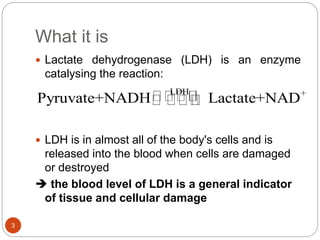
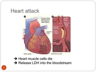
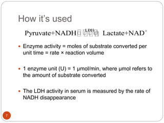
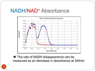
PRESENTER: DUNG TRONG TRAN
1
LACTATE DEHYDROGENASE (LDH)
BLOOD TEST OF HEART ATTACK VICTIMS
Contents
2
 What it is
 When it’s used
 How it’s used
What it is
3
 Lactate dehydrogenase (LDH) is an enzyme
catalysing the reaction:
 LDH is in almost all of the body's cells and is
released into the blood when cells are damaged
or destroyed
 the blood level of LDH is a general indicator
of tissue and cellular damage
LDH +
Pyruvate+NADH Lactate+NAD
What it is
4
 LDH exists in five forms, or isoenzymes.
 Each isoenzyme has a slightly different structure
and is found in different tissues.
 The results of each LDH isoenzyme
concentration indicates which tissue may be
damaged or injured.
Heart attack
5
 Heart muscle cells die
 Release LDH into the bloodstream
When it’s done
6
 The assay is used within 12-24 hours after the
infarction.
 The increase in enzyme activity tends to be
proportional to the extent of the myocardial lesion
 If
Total LDH increases
LDH-1 > LDH-2 (normally LDH1<LDH2)
 Heart attack
 Rarely used now
How it’s used
7
 Enzyme activity = moles of substrate converted per
unit time = rate × reaction volume
 1 enzyme unit (U) = 1 μmol/min, where μmol refers to
the amount of substrate converted
 The LDH activity in serum is measured by the rate of
NADH disappearance
LDH +
Pyruvate+NADH Lactate+NAD
How it’s used
8
 Using UV-Vis spectrometer to measure enzyme
activity
o Easy handling.
o Low susceptibility against disturbances.
o Cover UV range, where NADH shows absorption.
NADH/NAD+ Absorbance
9
 The rate of NADH disappearance can be
measured as an decrease in absorbance at 340nm
How it’s used
10
 Reagents:
 NADH solution
 Sodium Pyruvate solution
 Buffer solution: pH ~ 8.0
 Sample:
 Centrifuge the blood samples
 Serum can be separated and assayed directly
Procedures
11
1. Determining e of NADH
A = C x e
where
C is the concentration of NADH in moles/L
e is extinction coefficient
 Obtain e from calibration curves of A vs known C
 e = 6220
Procedures
12
2. Measuring blood LDH:
i. Set up the LDH reaction using the mixture
without an enzyme as a blank
ii. Put the full mixture in the spectrophotometer
and read the absorptions
Procedures
13
iii. Plot of the absorption vs time of the LDH
reaction. The slope we got for our graph
was ΔA/min
Calculations
14
1. The change in concentration of NADH was
calculated by using the equation :
∆CNADH/min = ∆A/min/e = slope / 6220 (εNADH)
2. Concentration of enzyme in unit (U):
∆CNADH/min x V(L) x 106 (μmole/mole)= μmol/min= U
Summary
15
 Lactate dehydrogenase assay is often used as a
diagnostic tool in monitoring patients after heart
attack
 UV-Vis photometric assay is applied
 Temperature and pH must be maintain in the
assay
References
16
 http://www.labtestsonline.org.uk/understanding/an
alytes/ldh/
 http://www.biopacific.net/pointe-package-
inserts/Lactate_dehydrogenase.pdf
 http://www.biotek.com/resources/docs/NADH_Ap
p_Note.pdf
 Jack E.Dixon (1975), “Determining blood enzyme
concentrations: A Unified laboratory experiment”,
Biochemical education, 3(2), pp.31-33.
 Hans Bisswanger (2014), “Enzyme assays”,
Perspectives in Science, Volume 1, pp.41-55.
Thanks for listening
17

Lactate dehydrogenase assays

  • 1.
    PRESENTER: DUNG TRONGTRAN 1 LACTATE DEHYDROGENASE (LDH) BLOOD TEST OF HEART ATTACK VICTIMS
  • 2.
    Contents 2  What itis  When it’s used  How it’s used
  • 3.
    What it is 3 Lactate dehydrogenase (LDH) is an enzyme catalysing the reaction:  LDH is in almost all of the body's cells and is released into the blood when cells are damaged or destroyed  the blood level of LDH is a general indicator of tissue and cellular damage LDH + Pyruvate+NADH Lactate+NAD
  • 4.
    What it is 4 LDH exists in five forms, or isoenzymes.  Each isoenzyme has a slightly different structure and is found in different tissues.  The results of each LDH isoenzyme concentration indicates which tissue may be damaged or injured.
  • 5.
    Heart attack 5  Heartmuscle cells die  Release LDH into the bloodstream
  • 6.
    When it’s done 6 The assay is used within 12-24 hours after the infarction.  The increase in enzyme activity tends to be proportional to the extent of the myocardial lesion  If Total LDH increases LDH-1 > LDH-2 (normally LDH1<LDH2)  Heart attack  Rarely used now
  • 7.
    How it’s used 7 Enzyme activity = moles of substrate converted per unit time = rate × reaction volume  1 enzyme unit (U) = 1 μmol/min, where μmol refers to the amount of substrate converted  The LDH activity in serum is measured by the rate of NADH disappearance LDH + Pyruvate+NADH Lactate+NAD
  • 8.
    How it’s used 8 Using UV-Vis spectrometer to measure enzyme activity o Easy handling. o Low susceptibility against disturbances. o Cover UV range, where NADH shows absorption.
  • 9.
    NADH/NAD+ Absorbance 9  Therate of NADH disappearance can be measured as an decrease in absorbance at 340nm
  • 10.
    How it’s used 10 Reagents:  NADH solution  Sodium Pyruvate solution  Buffer solution: pH ~ 8.0  Sample:  Centrifuge the blood samples  Serum can be separated and assayed directly
  • 11.
    Procedures 11 1. Determining eof NADH A = C x e where C is the concentration of NADH in moles/L e is extinction coefficient  Obtain e from calibration curves of A vs known C  e = 6220
  • 12.
    Procedures 12 2. Measuring bloodLDH: i. Set up the LDH reaction using the mixture without an enzyme as a blank ii. Put the full mixture in the spectrophotometer and read the absorptions
  • 13.
    Procedures 13 iii. Plot ofthe absorption vs time of the LDH reaction. The slope we got for our graph was ΔA/min
  • 14.
    Calculations 14 1. The changein concentration of NADH was calculated by using the equation : ∆CNADH/min = ∆A/min/e = slope / 6220 (εNADH) 2. Concentration of enzyme in unit (U): ∆CNADH/min x V(L) x 106 (μmole/mole)= μmol/min= U
  • 15.
    Summary 15  Lactate dehydrogenaseassay is often used as a diagnostic tool in monitoring patients after heart attack  UV-Vis photometric assay is applied  Temperature and pH must be maintain in the assay
  • 16.
    References 16  http://www.labtestsonline.org.uk/understanding/an alytes/ldh/  http://www.biopacific.net/pointe-package- inserts/Lactate_dehydrogenase.pdf http://www.biotek.com/resources/docs/NADH_Ap p_Note.pdf  Jack E.Dixon (1975), “Determining blood enzyme concentrations: A Unified laboratory experiment”, Biochemical education, 3(2), pp.31-33.  Hans Bisswanger (2014), “Enzyme assays”, Perspectives in Science, Volume 1, pp.41-55.
  • 17.

Editor's Notes

  • #4 First of all, Lactate dehydrogenase LDH tests or assays are laboratory method for measuring enzymatic activity. LDH is an enzyme that catalyzes the interconversion of pyruvate and lactate and also the interconversion of NADH and NAD+ LDH is found in almost all body tissues but only a small amount of it is usually detectable in the blood. When cells are damaged or destroyed, they release LDH into the bloodstream, causing blood levels to rise. For this reason, LDH is used as a general marker of injury to cells
  • #5 LDH exist in 5 isozymes, which called LDH 1 to 5. Although there is some overlap, each of the five LDH isoenzymes tends to be concentrated in specific body tissues. Because of this, measurements of the individual LDH isoenzyme levels can be used to help determine the disease or condition causing cellular damage and to help identify the organs and tissues involved.
  • #6 A heart attack happens when the flow of oxygen-rich blood to a section of heart muscle suddenly becomes blocked and the heart can't get oxygen. Heart attacks most often occur as a result of coronary heart disease, also called coronary artery disease. It is a condition in which a waxy substance called plaque (plak) builds up inside the coronary arteries. This causes a blood clot to form on the plaque's surface. If the clot becomes large enough, it can mostly or completely block blood flow through a coronary artery. If blood flow isn't restored quickly, the section of heart muscle begins to die.
  • #7 The blood LDH increases about 12 hours following the infarction and peaks at 48-72 hours, with a gradual return to normal within the next 7-12 days. So the test always conducted within 12-24h after the first symptom. The increase in enzyme activity tends to be proportional to the extent of the myocardial lesion, reaching levels approximately three times the normal value If the result of the test witness the increase in total concentration of LDH and LDH-1 level is higher than that of LDH-2 in blood, it means the patient got a heart attack. But nowadays, the use of this phenomenon to diagnose infarction has been largely replaced by the use of Troponin measurement
  • #8 LDH catalyzes the reduction of pyruvate to lactate with simultaneous oxidation of NADH to NAD+ as this reaction. We can determine the enzyme activity based on this reaction The enzyme activity is calculated by moles of substrate converted per unit time and equal rate of reaction × reaction volume. The enzyme activity generally determined in unit of International Unit, denoted by U, which refers to the amount of enzyme that catalyzes the conversion of 1 micromole of substrate per minute. Thus, 1 enzyme unit (U) = 1 μmol/min, where μmol refers to the amount of substrate converted. so we can determine the LDH activity by measuring the rate of NADH disappearance.
  • #9 There are several methods can observe the enzyme reaction but in this assays, we choose UV-Vis spectrometry to measure the change in concentration of NADH. This method has some noticeable advantages such as it covers UV range, where substances show absorptions it is relatively easy handling and has low susceptibility against disturbances
  • #10 In this figure, we can see that NADH has absorbance at 340nm while NAD has no. So the LDH could therefore be assayed by following the decrease in UV absorbance at a wavelength of 340 nm.
  • #11 To prepare for the assays, we use reagents including NADH solution, sodium pyruvate solution as reactants and buffer solution such as Tris or phosphate solution that can maintain pH of the reaction mixture at 8.0. The blood sample is withdrawn from body and centrifuged for 15 minute to separate serum and plasma. Then serum can be analyzed directly.
  • #12 The first step, we need to determine the extinction coefficient of NADH. As you know from Lambert Beer law, we have A = Cxe because most cuvettes have path length equal to 1 cm. In this case, C is the concentration of NADH and e is the molar extinction coefficient. Therefore by measuring the absorbance of several solutions having known concentrations of NADH at 340nm, it is possible to determine the molar extinction coefficient. In the most case, epsilon of NADH equal to 6220.
  • #13 The second step is determining the blood concentration of LDH Set up the LDH reaction using the mixture without an enzyme as a blank Put the full mixture in the spectrophotometer and read the absorptions
  • #14 We only takeFrom observed data, we can plot absorption vs time of the LDH reaction. And from the equation: slope equal to y2-y1/x2-x1 so we can obtain slope=delta A/min
  • #15 After that, we calculate the ……
  • #16 In conclusion, Lactate dehydrogenase assay is often used as a diagnostic tool in monitoring patients during recovery after heart attack To measure the enzyme activity, UV spectrometer is used to measure the decrease in absorbance of NADH at 340nm wavelength The conditions of the reaction should maintain constants.