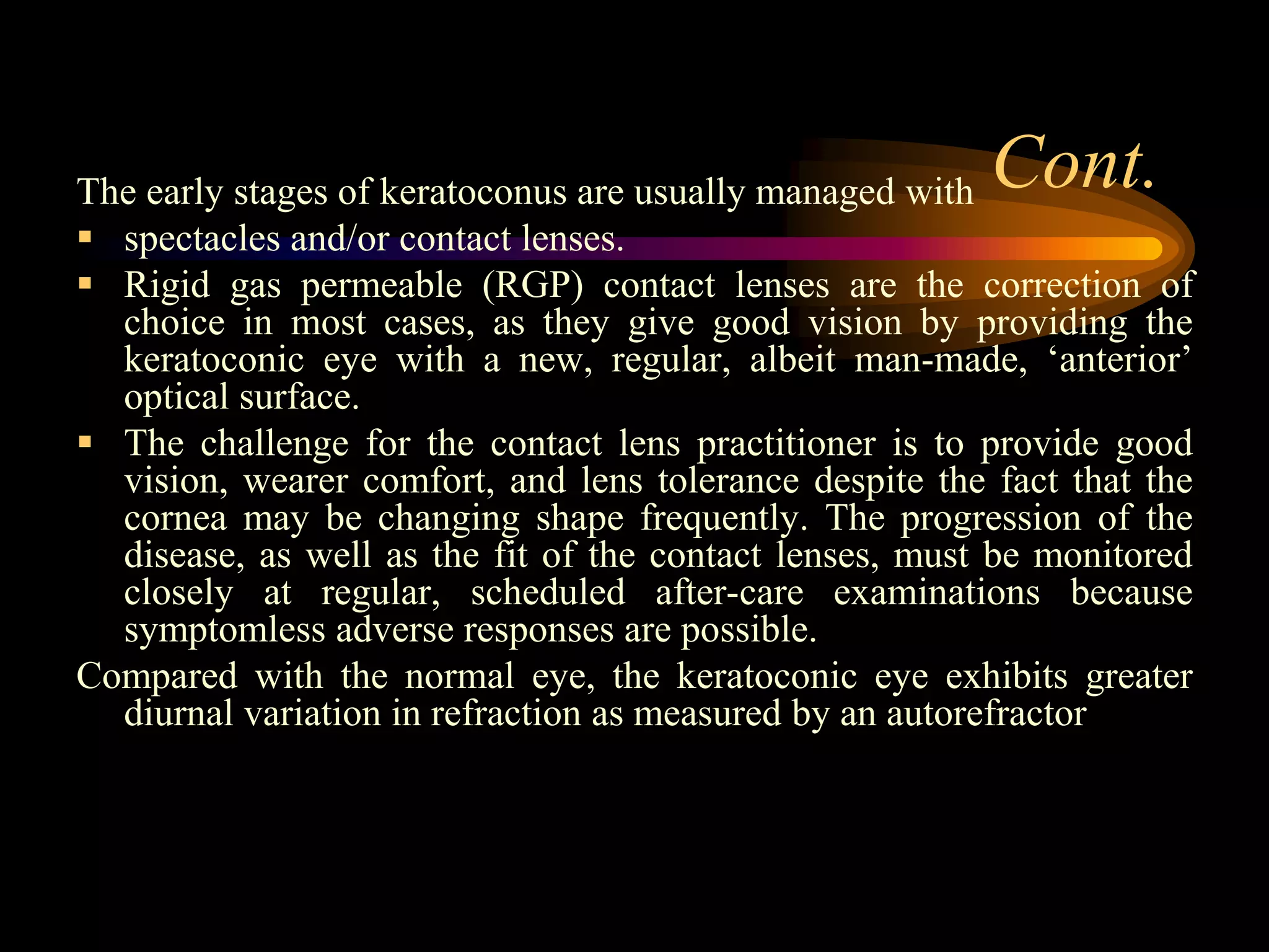
Keratoconus and Contact Lenses
 Introduction to Keratoconus
 Aetiology of Keratoconus and Associated
Conditions
 Symptoms and Signs
 Contact Lens Fitting
 Surgery for Keratoconus
 Miscellaneous ‘Treatments’ for
Keratoconus
 Conclusions
Introduction to Keratoconus
Keratoconus, conical cornea, is an interesting, complex, and challenging
condition for both the patient and contact lens practitioner alike.
The aetiology of keratoconus is still not well understood, but it would
seem to include both genetic and environmental factors. Around the
second decade of life (between 12 and 20 but cases have been reported
at birth and as late as 51 years of age [Heverly and Lowther, 2005]),
the cornea of a keratoconic patient begins to thin and protrude,
producing irregular corneal astigmatism.
Despite our ability to detect and document keratoconus, especially in the
era of videokeratoscopy, there is still no early treatment or cure, and
individual prognosis is highly variable . Optimal treatment depends on
the severity of the disease process, requires accurate diagnosis, and
may require the use of a number of therapeutic alternatives.
Cont.The early stages of keratoconus are usually managed with
 spectacles and/or contact lenses.
 Rigid gas permeable (RGP) contact lenses are the correction of
choice in most cases, as they give good vision by providing the
keratoconic eye with a new, regular, albeit man-made, ‘anterior’
optical surface.
 The challenge for the contact lens practitioner is to provide good
vision, wearer comfort, and lens tolerance despite the fact that the
cornea may be changing shape frequently. The progression of the
disease, as well as the fit of the contact lenses, must be monitored
closely at regular, scheduled after-care examinations because
symptomless adverse responses are possible.
Compared with the normal eye, the keratoconic eye exhibits greater
diurnal variation in refraction as measured by an autorefractor
If the progress of the disease is marked by the
following issues, surgical options may have to be
considered:
1. Increased RGP lens intolerance.
2. The development of stromal opacities.
3. Corneal hydrops.
4. Significant lens decentration and/or an inability to
retain lenses on the eyes.
5. Increased astigmatism/irregular astigmatism.
Keratoconus is a leading indication for corneal
transplantation (Penetrating Keratoplasty [PK]). The
reasons for performing a PK according to several
sources appear in descending order.
Description
Keratoconus is a benign, usually bilateral,
non-inflammatory thinning and ectasia of
the cornea, resulting in a high degree of
irregular myopic astigmatism
Cont.
The protrusion usually develops in the infero-
central zone of the cornea that results in a
cone-like anterior protrusion exhibiting a high
degree of irregular, myopic astigmatism, and
marked visual impairment (data in slide 7 comes from the
Collaborative Longitudinal Evaluation of Keratoconus Study [CLEK]).
The condition typically, though not always,
affects young people in the second or third
decades of their lives.
Ocular abnormalities that occur more
frequently in keratoconus include:
• Congenital cataract.
• Retinitis pigmentosa.
• Ectopia lentis.
• Pellucid marginal degeneration (PMD)
Usually, conventional RGP designs work well
in the early stages of keratoconus. Special
multi-curve, specialized keratoconic fitting
sets are required later when the cone
becomes more clearly defined and the
cornea more ‘abnormal’.
 Keratoconus and Contact lenses
 Keratoconus and Contact lenses
 Keratoconus and Contact lenses
 Keratoconus and Contact lenses
 Keratoconus and Contact lenses
 Keratoconus and Contact lenses
 Keratoconus and Contact lenses
 Keratoconus and Contact lenses
 Keratoconus and Contact lenses
 Keratoconus and Contact lenses
 Keratoconus and Contact lenses
 Keratoconus and Contact lenses
 Keratoconus and Contact lenses
 Keratoconus and Contact lenses
 Keratoconus and Contact lenses
 Keratoconus and Contact lenses
 Keratoconus and Contact lenses
 Keratoconus and Contact lenses
 Keratoconus and Contact lenses
 Keratoconus and Contact lenses
 Keratoconus and Contact lenses
 Keratoconus and Contact lenses
 Keratoconus and Contact lenses
 Keratoconus and Contact lenses
 Keratoconus and Contact lenses
 Keratoconus and Contact lenses
 Keratoconus and Contact lenses
 Keratoconus and Contact lenses
 Keratoconus and Contact lenses
 Keratoconus and Contact lenses
 Keratoconus and Contact lenses
 Keratoconus and Contact lenses
 Keratoconus and Contact lenses
 Keratoconus and Contact lenses
 Keratoconus and Contact lenses
 Keratoconus and Contact lenses
 Keratoconus and Contact lenses
 Keratoconus and Contact lenses
 Keratoconus and Contact lenses
 Keratoconus and Contact lenses
 Keratoconus and Contact lenses
 Keratoconus and Contact lenses
 Keratoconus and Contact lenses
 Keratoconus and Contact lenses

Keratoconus and Contact lenses

  • 1.
    Keratoconus and ContactLenses  Introduction to Keratoconus  Aetiology of Keratoconus and Associated Conditions  Symptoms and Signs  Contact Lens Fitting  Surgery for Keratoconus  Miscellaneous ‘Treatments’ for Keratoconus  Conclusions
  • 2.
    Introduction to Keratoconus Keratoconus,conical cornea, is an interesting, complex, and challenging condition for both the patient and contact lens practitioner alike. The aetiology of keratoconus is still not well understood, but it would seem to include both genetic and environmental factors. Around the second decade of life (between 12 and 20 but cases have been reported at birth and as late as 51 years of age [Heverly and Lowther, 2005]), the cornea of a keratoconic patient begins to thin and protrude, producing irregular corneal astigmatism. Despite our ability to detect and document keratoconus, especially in the era of videokeratoscopy, there is still no early treatment or cure, and individual prognosis is highly variable . Optimal treatment depends on the severity of the disease process, requires accurate diagnosis, and may require the use of a number of therapeutic alternatives.
  • 3.
    Cont.The early stagesof keratoconus are usually managed with  spectacles and/or contact lenses.  Rigid gas permeable (RGP) contact lenses are the correction of choice in most cases, as they give good vision by providing the keratoconic eye with a new, regular, albeit man-made, ‘anterior’ optical surface.  The challenge for the contact lens practitioner is to provide good vision, wearer comfort, and lens tolerance despite the fact that the cornea may be changing shape frequently. The progression of the disease, as well as the fit of the contact lenses, must be monitored closely at regular, scheduled after-care examinations because symptomless adverse responses are possible. Compared with the normal eye, the keratoconic eye exhibits greater diurnal variation in refraction as measured by an autorefractor
  • 4.
    If the progressof the disease is marked by the following issues, surgical options may have to be considered: 1. Increased RGP lens intolerance. 2. The development of stromal opacities. 3. Corneal hydrops. 4. Significant lens decentration and/or an inability to retain lenses on the eyes. 5. Increased astigmatism/irregular astigmatism. Keratoconus is a leading indication for corneal transplantation (Penetrating Keratoplasty [PK]). The reasons for performing a PK according to several sources appear in descending order.
  • 7.
    Description Keratoconus is abenign, usually bilateral, non-inflammatory thinning and ectasia of the cornea, resulting in a high degree of irregular myopic astigmatism
  • 8.
    Cont. The protrusion usuallydevelops in the infero- central zone of the cornea that results in a cone-like anterior protrusion exhibiting a high degree of irregular, myopic astigmatism, and marked visual impairment (data in slide 7 comes from the Collaborative Longitudinal Evaluation of Keratoconus Study [CLEK]). The condition typically, though not always, affects young people in the second or third decades of their lives.
  • 17.
    Ocular abnormalities thatoccur more frequently in keratoconus include: • Congenital cataract. • Retinitis pigmentosa. • Ectopia lentis. • Pellucid marginal degeneration (PMD)
  • 47.
    Usually, conventional RGPdesigns work well in the early stages of keratoconus. Special multi-curve, specialized keratoconic fitting sets are required later when the cone becomes more clearly defined and the cornea more ‘abnormal’.