Including Mental Health Support in Project Delivery, 14 May.pdf
Jyfyjcdhrgzdfxfdzyzuyv CT hfcf by htxth ex yfi
1. A
B
C
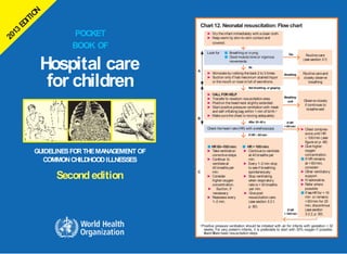
aPositive pressure ventilation should be initiated with air for infants with gestation > 32
weeks. For very preterm infants, it is preferable to start with 30% oxygen if possible.
Aand Bare basic resuscitation steps
Chart 12. Neonatal resuscitation: Flow chart
No
Yes
Breathing
Breathing
well
Not breathing, or gasping
After 30–60 s
If HR 60/min
If HR
<60/min
► Dry theinfant immediately with aclean cloth.
► Keep warm by skin-to-skin contact and
covered.
Look for ■ Breathing or crying
■ Good muscle toneor vigorous
movements
► Stimulateby rubbing theback 2 to 3 times.
► Suction only if had meconium stained liquor
or themouth or noseis full of secretions.
► CALLFORHELP
.
► Transfer to newborn resuscitationarea.
► Position thehead/neck slightly extended.
► Start positive pressure ventilation with mask
and self-inflating bag within 1 min of birth.a
► Makesurethechest is moving adequately.
Check theheart rate(HR) with astethoscope.
■ HR 60–100/min: ■ HR> 100/min:
► Takeventilation ► Continueto ventilate
correctivesteps. at 40 breaths per
► Continue to min.
ventilateat ► Every 1–2 min stop
40 breaths per to seeif breathing
min. spontaneously.
► Consider ► Stop ventilating
higher oxygen when respiratory
concentration. rateis >30 breaths
► Suction, if per min.
necessary. ► Givepost
► Reassess every resuscitation care.
1–2 min. (seesection 3.2.1,
p. 50).
Routinecare
(seesection 3.1)
Routinecareand
closely observe
breathing
Observeclosely
if continues to
breathewell
► Chest compres-
sions until HR
100/min (see
figureon p. 48)
► Givehigher
oxygen
concentration.
■ If HRremains
at < 60/min,
consider:
► Other ventilatory
support.
► IVadrenaline.
► Refer where
possible
■ If noHRfor > 10
min or remains
<60/min for 20
min, discontinue
(seesection
3.2.2, p. 50).
If HR
>100/min
GUIDELINESFORTHEMANAGEMENT OF
COMMONCHILDHOODILLNESSES
Secondedition
POCKET
BOOK OF
Hospital care
for children
2. Triage of all sick children
EMERGENCYSIGNS:
If any sign is positive, call for help, assess and resuscitate, give
treatment(s), draw blood for emergency laboratory investigations
(glucose, malaria smear, Hb)
TREAT
Do not move neck if a cervical spine
injury is possible, but open the airway.
ASSESS
Check for
severe
malnutrition
ANY SIGN
POSITIVE
SIGNS
POSITIVE
Airway and breathing
■ Obstructed or
absent breathing
or
■ Central cyanosis
or
■ Severe respiratory
distress
Circulation
Cold skin with:
■ Capillary refill
longer than 3 s
and
■ Weak and fast
pulse
If foreign body aspirated
► Manage airway in choking
child (Chart 3)
If no foreign body aspirated
► Manage airway (Chart 4)
► Give oxygen (Chart 5)
► Make sure the child is warm
► Stop any bleeding
► Give oxygen (Chart 5)
► Make sure the child is warm.
If no severe malnutrition
► Insert an IV lineand begin
giving fluids rapidly (Chart 7).
If peripheral IV cannot be
inserted, insert an intraosseous
or external jugular line
(see pp. 340–342).
If severe malnutrition:
If lethargic or unconscious:
► Give IV glucose (Chart 10).
► Insert IV lineand givefluids
(Chart 8).
If not lethargic or unconscious:
► Give glucose orally or by
nasogastric tube.
► Proceed immediately to full
assessment and treatment.
EMERGENCYSIGNS:
If any sign is positive: call for help, assess and resuscitate, give
treatment(s), draw blood for emergency laboratory investigations
(glucose, malaria smear, Hb)
PRIORITY SIGNS
These children need prompt assessment and treatment
ASSESS TREAT
Do not move neck if you suspect cervical
spine injury, but open the airway.
Coma/
convulsing
■ Coma
or
■ Convulsing
(now)
► Manage theairway (Chart 4)
► If convulsing, givediazepam rectally
(Chart 9)
► Position the unconscious child (if
head or neck traumais suspected,
stabilizetheneck first) (Chart 6).
► GiveIVglucose (Chart 10).
► Makesurethe child is warm.
If no severe malnutrition:
► Insert an IVlineand begin giving
fluids rapidly following Chart 11 and
diarrhoea treatment plan Cin hospital
(Chart 13, p. 131).
If severe malnutrition:
► Do not insert an IVline.
► Proceed immediately to full
assessment and treatment (see
section 1.4, p. 19).
IFCOMAOR
CONVULSION
DIARRHOEA
PLUS
twosigns
positive
Check for
severe
malnutrition
Severe
dehydration
(only in a child
with diarrhoea)
Diarrhoea plus
any two of these
signs:
■ Lethargy
■ Sunken eyes
■ Very slow skin pinch
■ Unableto drink or drinks
poorly
condition
■ Pallor (severe)
■ Poisoning (history of)
■ Pain (severe)
■ Respiratory distress
■ Tiny infant (< 2 months) ■ Restless, continuously irritable, or lethargic
■ Temperature very high ■ Referral (urgent)
■ Trauma or other urgent surgical ■ Malnutrition: visibleseverewasting
■ Oedema of both feet or face
■ Burns (major)
Note: If achild has traumaor other surgical problems,
get surgical help or follow surgical guidelines.
NON-URGENT
Proceed with assessment and further treatment according to the child’s priority.
5. iii
Contents
Preface
Acknowledgements
Abbreviations
Chart1: Stages in the management of asick child admitted
to hospital: key elements
xv
xviii
xxi
xxii
1. TRIAG
EANDEMERG
ENCYCO
NDITIO
NS 1
1. Triage 2
2. Summaryof steps in emergencytriageassessment and treatment 3
3. Assessment of emergencyand priority signs 4
Triageof all sickchildren 5
How to manageachokinginfant or child 7
How to managetheairway in achild with obstructedbreathing 9
How to giveoxygen 11
How to position theunconsciouschild 12
Give IV fluids for shock in a child without severe acute malnutrition 13
Give IV fluids for shock in a child with severe acute malnutrition 14
Givediazepamrectally 15
GiveIVglucose 16
Treat severedehydrationin an emergencysetting 17
4. Emergencytreatment for achildwith severemalnutrition 19
5. Diagnostic considerations for childrenwith emergencyconditions 20
21
1. Child presenting with an airway or severe breathing problem 20
2. Child presentingwith shock
3. Child presentingwith lethargy, unconsciousness
or convulsions
6. Commonpoisoning
1. Principlesfor ingested poisons
2. Principles for poisons in contact with skin or eyes
23
26
27
29
6. iv
3. Principlesfor inhaledpoisons
4. Specific poisons
Corrosivecompounds
Petroleumcompounds
Organophosphorus and carbamatecompounds
Paracetamol
Aspirinand other salicylates
Iron
Morphineand other opiates
Carbonmonoxide
5. Preventionof poisoning
29
29
29
30
30
31
31
32
32
33
33
33
34
34
34
37
38
38
38
39
1.7
1.8
1.9
Drowning
Electrocution
Commoncauses of envenoming
1. Snakebite
2. Scorpionsting
3. Other sources of envenoming
10. Traumaand injuries
1. Primary survey or initial assessment
2. Secondarysurvey
2. DIAGNOSTIC APPROACHES TOTHESICK CHILD 41
1. Relationship to the IMCIapproachand stages of hospital care
2. Taking history
3. Approachto thesick child and clinical examination
4. Laboratory investigations
5. Differential diagnoses
41
42
43
43
44
3. PROBLEMSOFTHENEONATEANDYOUNGINFANT 45
1. Essential newborn careat delivery
2. Neonatal resuscitation
1. Post resuscitationcare
2. Cessationof resuscitation
3. Routinecarefor all newborns after delivery
4. Preventionof neonatal infections
46
46
50
50
50
51
HOSPITAL CAREFORCHILDREN
7. v
3.5
3.6
3.7
3.8
3.9
Management of the infant with hypoxic ischaemic encephalopathy 51
Danger signs in newborns and young infants 52
Convulsionsor fits 53
Seriousbacterial infection 54
Meningitis 55
56
56
57
58
58
58
10. Supportivecarefor sick neonates
1. Thermal environment
2. Fluid management
3. Oxygen therapy
4. High fever
11. Pretermand low-birth-weight infants
1. Infantswith abirth weight of 2.0–2.5kg
(35–36weeks’ gestation)
2. Infants with abirth weight <2.0 kg
(< 35 weeks’ gestation)
3. Commonproblems of low-birth-weight infants
4. Dischargeand follow-up of low-birth-weight infants
12. Other commonneonatal problems
1. Jaundice
2. Conjunctivitis
3. Congential malformations
13. Infants of mothers with infectious diseases
1. Congenital syphilis
2. Infants of mothers with tuberculosis
3. Infants of mothers with HIVinfection
14. Doses of commondrugs for neonates and low-birth-weight
infants
58
59
61
63
64
64
66
67
67
67
68
68
69
4. COUGHORDIFFICULTY IN BREATHING 75
1. Child presentingwith cough
2. Pneumonia
1. Severepneumonia
2. Pneumonia
3. Complications of pneumonia
1. Pleural effusionand empyema
76
80
80
86
88
88
CONTENTS
14. A1.5 Insertion of a chest drain 348
A1.6 Supra-pubic aspiration 350
A1.7 Measuring blood glucose 350
Annex 2. Drug dosages and regimens 353
Annex 3. Equipment sizes 375
Annex 4. Intravenous fluids 377
A4.1 Choice of intravenous fluids 378
Annex 5. Assessing nutritional status 379
A5.1 Calculating achild’s weight-for-age 379
A5.2 Calculating a child’s weight-for-length or height 386
Annex 6. Job aids and charts 403
INDEX 405
CHARTS
Chart 1. Stages in the managementof asick child admitted to
hospital: key elements xxii
Chart 2. Triage of all sick children 5
Chart 3. How to manage a choking infant or child 7
Chart 4. How to managethe airways in achild with obstructed
breathing (or who has just stopped breathing) 9
Chart 5. How to give oxygen 11
Chart 6. How to position an unconscious child 12
Chart 7. Howto giveintravenous fluids rapidly to achild in shock
without severe malnutrition 13
Chart 8. How to give intravenous fluids to achild in shock with
severe malnutrition 14
Chart 9. How to give diazepam rectally 15
Chart 10. How to give glucose intravenously 16
Chart 11. How to treat severe dehydration in an emergency
after initial management of shock 17
Chart 12. Neonatal resuscitation 47
Chart 13. Diarrhoeatreatment plan C: Treat severedehydration quickly 130
Chart 14. Diarrhoeatreatment plan B: Treat some dehydration with
oral rehydration salts 135
Chart 15. Diarrhoea treatment plan A: Treat diarrhoea at home 138
Chart 16. Feeding recommendations during sickness and health 302
xii
HOSPITAL CAREFORCHILDREN
15. xiii
TABLES
Table1. Differential diagnosis in achild presenting with
an airways or severebreathingproblem
Differential diagnosis in achild presenting with shock
Differential diagnosis in achild presenting with lethargy,
unconsciousnessor convulsions
21
22
Table2.
Table3.
24
Table4. Differential diagnosis in ayoung infant (< 2 months)
presentingwith lethargy, unconsciousnessor convulsions 25
Table5.
Table6.
28
Poisoning: amount of activated charcoal per dose
Differential diagnosis in achild presenting with cough or
difficulty in breathing 77
Classificationof theseverityof pneumonia 81
Differential diagnosis in a child presenting with wheeze 93
Differential diagnosis in a child presenting with stridor 103
Differential diagnosis in achild presenting with
chroniccough
Table7.
Table8.
Table9.
Table10.
110
Table11.
Table12.
Differential diagnosis in achild presenting with diarrhoea 127
128
Table13.
130
Table14.
141
Table15.
141
151
152
153
Table16.
Table17.
Table18.
Table19.
155
Table20
194
Table21.
201
Classification of the severity of dehydration in children
with diarrhoea
Administration of intravenous fluids to aseverely
dehydratedchild
First diet for persistent diarrhoea: astarch-based,
reduced-milk (low-lactose) diet
Seconddiet for persistent diarrhoea: areduced-starch
(cereal) no-milk (lactose-free) diet
Differential diagnosis of fever without localizing signs
Differential diagnosis of feverwith localized signs
Differential diagnosis of fever with rash
Additional differential diagnosis of fever lasting longer
than 7 days
WHOcriteria for the diagnosis of rheumatic fever
(basedon the revised Jones criteria)
Timeframe for the managementof achild with
severeacutemalnutrition
Volumes of F-75 per feed for malnourished children
(approximately130 ml/kgper day) 211
WHOpaediatric clinical staging system for HIVinfection 231
Classesof antiretroviral drugs recommendedfor use
in children
Table22.
Table23.
Table24.
234
CONTENTS
16. xiv
Table 25. First-line treatment regimensfor children 234
Table 26. Common side-effects of antiretroviral drugs 236
Table 27. Recommendedsecond-line treatment regimens
for children 240
Table 28. Endotracheal tube size by age 259
Table 29. Blood volume of children by age 260
Table 30. Normal pulse rate andblood pressurein children 261
Table 31. Examples of local adaptations of feeding
recommendations on the mother’scard in Bolivia,
303
304
Table32.
Table33.
Indonesia, Nepal, South Africaand the
United Republic of Tanzania
Maintenancefluidrequirements
Primary vaccination schedule for infants recommended
in the Expanded Programme on Immunization 326
Table A2.1 Drug dosage by surface area (m2) of the child 354
Table A5.1.1 Weight-for-age from birth to 5 years: Boys 379
Table A5.1.2 Weight-for-age from birth to 5 years: Girls 381
Table A5.2.1 Weight-for-length from birth to 2 years: Boys 386
Table
A5.2.2
Weight-for-length from birth to 2 years: Girls 391
Table
A5.2.3
Weight-for-height from 2 to 5 years: Boys 395
Table
A5.2.4
Weight-for-height from 2 to 5 years: Girls 399
HOSPITAL CAREFORCHILDREN
17. This is the second edition of the World Health Organization (WHO) Pocket book
of hospital care for children, which was first published in 2005. It is acompila-
tion of the updated WHOguidelines for the management of common childhood
illnesses at the first-referral level in low-resource countries. It presents relevant,
up-to-date, evidence-based clinical guidelines that can be used by clinicians
in their daily work in hospitals with basic laboratory facilities and inexpensive
medicines. The guidelines focus on inpatient management of children who are
severely ill with conditions that are major causes of childhood mortality, such
as neonatal illness, pneumonia, diarrhoea, fever (mainly malaria, meningitis
and septicaemia), severe acute malnutrition and HIV/AIDS. It also includes
guidance on common surgical problems, appropriate supportive care and
monitoringof patientson theward.
The Pocket book is part of a series of tools for improving the quality of care
for severely ill children and is consistent with the Integrated Management of
Childhood Illness (IMCI) guidelines for outpatient management of sick chil-
dren. It is for use by doctors, senior nurses and other senior health workers
who are responsible for the care of young children at the first referral level in
developingcountries.
Thefirst edition of the Pocket book was reviewed by aWHOguidelines steering
committee,which identifiedthosechaptersthatrequiredupdating,comprising:
• revisions to align the Pocket book with recently published, WHO-approved
guidelines; and
• priorities for which new information had become available, which was col-
lated, analysed and synthesized beforeupdating.
In the first category, recommendations approved by the WHO Guidelines Re-
view Committee were incorporated. The second category required synthesis
of evidence and updates consistent with new recommendations. The changes
made are therefore based on published WHOguidelines and recommendations
as of 2012, which are listed in the bibliography on p. 329; in addition, certain
subsections were added or removed, others reorganized and some editorial
changes madeon the basis of feedback from Pocket book users. In response to
users’feedbackandthepopularityof thefirstedition,thepresentationis similar.
xv
Preface
18. xvi
All the changes were reviewed by external clinical experts and were approved
by the WHO Guidelines Review Committee. A web version of the Pocket book
will be updated regularly as new evidence with clinical implications emerges.
Printed editions will be published every 5 years if there are substantial new
changes. Users are therefore advised to check the WHOweb site regularly for
Pocketbookupdates(http://www.who.int/maternal_child_adolescent/en/).
The main changesin the secondedition are listed below.
Chapters unchangedfrom the first edition of the Pocket book(2005):
Chapterswith only editorialchangesor reorganizationbutwith nomajor update
of previous information:
• Chapter 1. Triageand emergencyconditions
• Chapter 2. Diagnostic approaches to the sick child
• Chapter 5. Diarrhoea
• Chapter 9. Commonsurgical problems
• Chapter 11. Monitoringthechild’s progress
• Chapter 12. Counsellinganddischarge from hospital
• Annexes1, 3 and 6
Chapters substantially changed from the first edition of the
Pocket book (2005):
Chapters with substantial changes to clinical guidance or which have been
restructuredare:
• Chapter 3. Problems of theneonateand young infant
• Chapter 4. Cough or difficultyin breathing
• Chapter 6. Fever
• Chapter 7. Severeacutemalnutrition
• Chapter 8. Childrenwith HIV/AIDS
• Chapter 10. Supportivecare
• Annexes2, 4 and 5
19. xvii
Additional sections or subsections in this secondedition
Several sections of some chapters were added or substantially expanded in
responseto demandfrom users:
• Chapter 1, section 1.10. Trauma andinjuries
• Chapter 3, section 3.7. Convulsionsor fits
• Chapter 3, section 3.11.3. Respiratory distress syndrome
• Chapter 4, section 4.6.3. Epiglottitis
• Chapter 4, section4.6.4. Anaphylaxis
• Chapter 4, section 4.9. Rheumatic heart disease
• Chapter 6, section 6.11. Rheumatic fever
• Chapter 8, section 8.5. Prevention of mother to child HIV transmission,
and infant feeding
ThePocketbook is presentedin aformat that could becarried bydoctors, nurses
and other health workers during their daily work and be available to help guide
the management of sick children. Although some newtopics have been added,
standard textbooks of paediatrics should be consulted for rarer conditions not
covered in the Pocket book. These guidelines are applicable in most areas of
theworld andmaybeadaptedbycountriestosuittheir specificcircumstances.
WHOrecommends that countries should locally adapt the Pocket book to include
important conditions not covered and believes its widespread adoption would
improve the care of children in hospital and lead to lower casefatality rates.
20. WHO expresses its gratitude to the following members of the group that up-
dated the guidelines, people who made original contributions, and reviewers,
institutions and consultants for their contributions to updating the Pocket book
of hospital carefor children.
Guideline developmentgroup
WHO thanks the members of the guideline development group who reviewed
most of the evidence and made recommendations for updating the Pocket book
and also those who reviewed the chapters: Dr Fizan Abdullah, Johns Hopkins
University School of Medicine, USA; Shinjini Bhatnagar, All India Institute of
Medical Sciences, India; Bridget Wills, Clinical Research Unit, University of
Oxford Centre for Tropical Diseases, Viet Nam; Harry Campbell, University
of Edinburgh Medical School, United Kingdom; Leonila Dans, University of
Philippines, Philippines; Trevor Duke, Centre for International Child Health,
University of Melbourne, Australia; Michael English, University of Nairobi and
Kenya Medical Research Institute, Kenya; Andy Gray, University of KwaZulu-
Natal, South Africa; Sandra Grisi, São Paulo University, Brazil; Stuart Macleod,
University of British Columbia, Canada; Hilda Mujuru, University of Zimbabwe,
Zimbabwe; Susan Niermeyer, University of Colorado, USA; Jesca Nsungwa,
Ministry of Health, Uganda; Vinod Paul, All India Institute of Medical Sci-
ences, India; Haroon Saloojee, Witwatersrand University, South Africa; Mathu
Santosham, Johns Hopkins School of Public Health, USA; Giorgio Tamburlini,
Institute of Child Health, Italy; and Anita Zaidi, Aga Khan University, Pakistan.
Special gratitude is owed to Rhona MacDonald, Maternal Child Health Advo-
cacyInternational, who incorporatedthe changesandpreparedthe first draft.
Originalcontributors andexternal reviewers
WHO coordinated the international contributions for the 2005 edition of the
Pocket book and thanks the original contributors to chapters: Dr Ann Ashworth
(United Kingdom), Dr Stephen Bickler (USA), Dr Jacqueline Deen (Philippines),
Dr Trevor Duke (Papaua New Guinea and Australia), Dr Greg Hussey (South
Africa), Dr Michael English (Kenya), Dr Stephen Graham (Malawi), Dr Eliza-
beth Molyneux (Malawi), Dr Nathaniel Pierce (USA), Dr Barbara Stoll (USA),
xviii
Acknowledgements
21. xix
Dr Giorgio Tamburlini (Italy), Dr Bridget Wills (Viet Nam) and Fabienne Jäger
(Switzerland).
WHO wishes to acknowledge the following for comments and contributions
made at various stages of the Pocket book updating: Sabrina Bakeere-Kitaka,
Makerere Medical School, Uganda; Zulfiqar Bhutta, Aga Khan University,
Pakistan; Stephen W. Bickler, University of California-San Diego, USA; Uday
Bodhankar, Commonwealth Association for Health and Disability, United
Kingdom; Adegoke Falade, College of Medicine, University of Ibadan, Nigeria;
Jeremy Farrar, Centre for Tropical Medicine, Ho Chi Minh City, Viet Nam;
Julian Kelly, Royal Children’s Hospital, Centre for International Child Health,
Melbourne, Australia; Carolyn Maclennan, Flinders University, Australia; Rhona
MacDonald, David Southall and Barbara Phillips, Maternal Child Health Advo-
cacy International; Amha Mekasha, Addis Ababa University, Ethiopia; Elizabeth
Molyneux, College of Medicine, Malawi; Maria Asuncion Silvestre, University
of the Philippines, Manila, Philippines, Joan Skinner, Victoria University of
Wellington, New Zealand and Andrew Steer, Royal Children’s Hospital, Centre
for International Child Health, Melbourne, Australia.
Valuable input was provided by several WHO clusters and the departments of
Family,Women’s andChildren’s Health, Health Systems andServices, HIV/AIDS,
Tuberculosis, Neglected Tropical Diseases, Noncommunicable Diseases, and
Mental Health. We particularly acknowledge the WHO staff who participated
as members of the Guidelines Steering Committee or who contributed to and
reviewed various draft chapters: Desta Teshome, WHO Regional Office for
Africa; Meena Cherian, Essential Health Technologies; Tarun Dua, Mental Health
and Substance Abuse; Lisa Nelson, Martina Penazzato, and Sandra Gove, HIV/
AIDS; Malgorzata Grzemska, Stop TB; Emmalita Manalac, WHO Regional Of-
fice for the Western Pacific; Peter Olumese, Global Malaria Programme; Ma
del Carmen Casanovas, Zita Weise Prinzo and Chantal Gegout, Nutrition for
Health and Development; Susan Hill and Clive Ondari, Essential Medicines and
Pharmaceutical Policies; Raman Velayudhan, Neglected Tropical Diseases; and
MartinWeber, WHOCountryOffice, Indonesia.
Special thanks to Rami Subhi at the Centre for International Child Health
in Australia, who helped in collating the evidence for recommendations for
updating thePocket book.
The updating of the Pocket book was coordinated by Wilson Were, supported
by Rajiv Bahl, Lulu Muhe, Olivier Fontaine, Severin Ritter Von Xylander, Nigel
Rollins and Shamim Qazi of the Department of Maternal, Newborn, Child and
Adolescent Health.
22. xx
Institutions
We are grateful to the following institutions for providing input and support
during the review of the Pocket book: Centre for International Child Health,
University of Melbourne, Australia; University of Edinburgh, Scotland; Kenya
Medical Research Institute, Kenya; Asociación Colaboración Cochrane Iber-
oamericana, Spain; Aga Khan University, Pakistan; Institute of Child Health
Burlo Garofolo, Italy; University of Malawi, Malawi; Capital Institute of Pae-
diatrics, China; University of Western Australia, Australia; and Instituto de
MedicinaIntegral Professor FernandoFigueira, Brazil.
WHOacknowledges the financial support for this second edition of the Pocket
book provided by the Bill and Melinda Gates Foundation through the medicines
for children project, and the Russian Federation through the quality of care
improvement initiative.
23. xxi
Abbreviations
AIDS
ART
AVPU
acquiredimmunodeficiency syndrome
antiretroviraltherapy
alert, responding to voice, responding to pain, unconscious
(simpleconsciousnessscale)
BCG bacilleCalmette-Guérin
CSF cerebrospinal fluid
DPT diphtheria, pertussis, tetanus
EVF erythrocytevolumefraction (haematocrit)
Hb haemoglobin
HIV humanimmunodeficiencyvirus
IM intramuscular(injection), intramuscularly
IMCI IntegratedManagement of ChildhoodIllness
IV intravenous(injection), intravenously
MDR multidrug-resistant
NNRTI non-nucleoside reverse transcriptase inhibitor
NRTI nucleosidereversetranscriptase inhibitor
NSAID non-steroidal anti-inflammatory drug
ORS oral rehydrationsalt(s)
PCP Pneumocystiscarinii pneumonia
ReSoMal rehydration solution for malnutrition
standarddeviation
tuberculosis
WorldHealthOrganization
SD
TB
WHO
Symbols
■diagnosticsignor symptom
►treatment recommendation
24. xxii
Chart 1. Stagesin the management of a sick child
admittedtohospital: keyelements
TRIAGE
(present)
• Check for emergency signs Give emergency
treatment until stable
(absent)
• Check for priority signs or conditions
HISTORY ANDEXAMINATION
(including assessment of vaccination status, nutritional status and feeding)
• Check children with emergency and priority conditions first.
Laboratory andother investigations, if required
List and consider differential diagnoses
Select maindiagnoses (and secondary diagnoses)
Plan and begin inpatient treatment
(including supportive care)
Monitor for signs of
— improvement
— complications
— failure of treatment.
Plan and begin outpatient
treatment.
Arrange follow-up, if
required.
(not improving or new problem) (improving)
Reassess
for causes of failure of
treatment.
Revise treatment.
Continue treatment.
Plandischarge.
Discharge home.
Arrange continuing care or
follow-up at hospital or in
the community.
25. 1. Triage 2
2. Summary of steps in emergency triage assessment and treatment 3
3. Assessment of emergency and priority signs 4
Triage of all sick children 5
How to manage a choking infant or child 7
How to manage the airway in a child with obstructed breathing 9
How to give oxygen
1
1
How to position the unconscious child
1
2 Give IV fluids for shock in a child without severe acute malnutrition
13 Give IV fluids for shock in achild with severe acute malnutrition 14
Give diazepam rectally
1
5
Give IV glucose 16
Treat severe dehydration in an emergency setting 17
4. Emergency treatment for a child with severe malnutrition 19
5. Diagnostic considerations for children with emergency conditions 20
1. Child presenting with an airway or severe breathing
problem 20
2. Child presenting with shock 21
3. Child presenting with lethargy, unconsciousness
or convulsions 23
6. Common poisoning 26
1.6.1 Principles for ingested poisons 27
1.6.2 Principles for poisons in contact with skin or eyes 29
1.6.3 Principles for inhaled poisons 29
1.6.4 Specific poisons 29
Corrosive compounds 29
Petroleum compounds 30
Organophosphorus and carbamate compounds 30
Paracetamol 31
Aspirin and other salicylates 31
Iron 32
Morphine and other opiates 32
Carbon monoxide 33
1.
TRIAGE
CHAPTER1
Triage and emergency
conditions
26. 1.1 Triage
Triage is the process of rapidly screeningsick children soonafter their arrival
in hospital, in order to identify:
– thosewith emergencysigns,who requireimmediateemergencytreatment;
– thosewith priority signs, who should begivenpriority in the queueso that
they can beassessedandtreated without delay; and
– non-urgent cases, who haveneither emergency nor priority signs.
Emergencysignsinclude:
■obstructedor absent breathing
■severerespiratory distress
■central cyanosis
■signs of shock (cold hands, capillary refill time longer than 3 s, high heart
rate with weak pulse, and low or unmeasurableblood pressure)
■coma (or seriously reduced level of consciousness)
■convulsions
■signsof severedehydration inachildwith diarrhoea(lethargy,sunkeneyes,
very slow return after pinching the skin or any two of these).
Children with these signs require immediate emergency treatment to avert
death.
The priority signs (see p. 6) identify children who are at higher risk of dying.
These children should be assessed without unnecessary delay. If a child has
one or more emergency signs, don’t spend time looking for priority signs.
2
1.
TRIAGE TRIAGE
7. Drowning
8. Electrocution
9. Common causes of envenoming
1. Snakebite
2. Scorpion sting
3. Other sources of envenoming
10. Trauma and injuries
1. Primary survey or initial assessment
2. Secondary survey
33
34
34
34
37
38
38
38
39
27. 1.2 Summaryof stepsin emergencytriage assessment
andtreatment
Steps in emergency triage assessment and treatment are summarized in the
charts on pp. 5–17.
First checkfor emergency signs in three steps:
• Step 1. Check whether there is any airway or breathing problem; start im-
mediatetreatmentto restorebreathing. Managetheairway andgiveoxygen.
• Step 2. Quickly check whether the child is in shock or has diarrhoea with
severe dehydration. Give oxygen and start IV fluid resuscitation. In trauma,
if thereisexternal bleeding,compressthewoundto stopfurtherbloodloss.
• Step 3. Quickly determine whether the child is unconscious or convulsing.
GiveIVglucosefor hypoglycaemiaand/orananti-convulsantfor convulsing.
If emergencysignsare found:
• Call for help from an experienced health professional if available, but do
not delay starting treatment. Stay calm and work with other health workers
who may be required to give the treatment, because a very sick child may
need several treatments at once. The most experienced health professional
should continue assessing the child (see Chapter 2, p. 41), to identify all
underlying problems andprepareatreatment plan.
• Carry out emergency investigations (blood glucose, blood smear, haemoglo-
bin [Hb]). Send blood for typing and cross-matching if the child is in shock,
appears to beseverely anaemicor is bleedingsignificantly.
• After giving emergency treatment, proceed immediately to assessing,
diagnosing andtreating the underlying problem.
Tables of common differential diagnoses for emergency signs are provided
from p. 21 onwards.
If no emergency signs are found, check for priority signs:
■Tiny infant: any sick child aged < 2 months
■Temperature: child is veryhot
■Traumaor other urgent surgical condition
■Pallor (severe)
■Poisoning(historyof)
■Pain(severe)
■Respiratorydistress
■Restless, continuouslyirritableor lethargic
3
1.
TRIAGE
SUMMARY OFSTEPSINEMERGENCY TRIAGEASSESSMENT AND TREATMENT
28. ASSESSMENT OFEMERGENCY AND PRIORITYSIGNS
■Referral (urgent)
■Malnutrition: visibleseverewasting
■Oedemaof both feet
■Burns (major)
The abovecan berememberedfrom the mnemonic3TPR MOB.
Thesechildren need prompt assessment (no waiting in the queue) to determine
what further treatment is needed. Move a child with any priority sign to the
front of the queue to be assessed next. If a child has trauma or other surgical
problems, get surgical help whereavailable.
1.3 Assessment of emergencyandpriority signs
■Assessthe airwayandbreathing(A, B)
Does the child’s breathing appear to be obstructed? Look at the chest wall
movement, and listen to breath sounds to determine whether there is poor air
movement during breathing. Stridor indicates obstruction.
Is there central cyanosis? Determine whether there is bluish or purplish dis-
coloration of the tongue andthe inside of the mouth.
Is the child breathing? Look and listen to determine whether the child is
breathing.
Is there severe respiratory distress? The breathing is very laboured, fast or
gasping, with chest indrawing, nasal flaring, grunting or the use of auxiliary
muscles for breathing (head nodding). Child is unable to feed because of
respiratory distress andtires easily.
■Assesscirculation(for shock) (C)
Children in shock who require bolus fluid resuscitation are lethargic and have
cold skin, prolonged capillary refill, fast weak pulse andhypotension.
Check whether the child’s hand is cold. If so, determine whether the child is
in shock.
Check whether the capillary refill time is longer than 3 s. Apply pressure to
whiten the nail of the thumb or the big toe for 5 s. Determine the time from the
moment of releaseuntil total recovery of the pink colour.
If capillary refill is longer than 3 s, check the pulse. Is it weak and fast? If the
radial pulse is strong and not obviously fast, the child is not in shock. If you
cannot feel the radial pulse of an infant (< 1 year old), feel the brachial pulse
or, if the infant is lying down, the femoral pulse. If you cannot feel the radial
pulseof achild, feel thecarotid.
4
1.
TRIAGE
29. 5
1.
TRIAGE
CHART2. TRIAGEOFALL SICKCHILDREN
Chart 2. Triage of all sick children
Emergency signs:
If anysignis positive, call for help, assess andresuscitate, give
treatment(s), draw blood for emergency laboratory investigations
(glucose, malaria smear, Hb)
TREAT
Do not move neck if acervical spine
injury is possible, but open the airway.
ASSESS
Check for
severe
malnutrition
ANY SIGN
POSITIVE
SIGNS
POSITIVE
Airway and breathing
■ Obstructed or
absent breathing
or
■ Central cyanosis
or
■ Severe respiratory
distress
Circulation
Cold skin with:
■ Capillary refill
longer than 3 s
and
■ Weak and fast
pulse
If foreign body aspirated
► Manage airway in choking
child (Chart 3)
If noforeignbody aspirated
► Manage airway (Chart 4)
► Give oxygen (Chart 5)
► Make sure the child is warm
► Stop any bleeding
► Give oxygen (Chart 5)
► Make sure the child is warm.
If nosevere malnutrition
► Insert an IV line and begin
giving fluids rapidly (Chart 7).
If peripheral IV cannot be
inserted, insert an intraosseous
or external jugular line
(see pp. 340, 342).
If severe malnutrition:
If lethargic or unconscious:
► Give IV glucose (Chart 10).
► Insert IV line and givefluids
(Chart 8).
If not lethargic or unconscious:
► Give glucose orally or by
nasogastric tube.
► Proceed immediately to full
assessment and treatment.
30. 1.
TRIAGE CHART2. TRIAGEOFALL SICKCHILDREN
Chart 2. Triage of all sick children
Emergency signs:
If anysignis positive: call for help, assess andresuscitate, give
treatment(s), draw blood for emergency laboratory investigations
(glucose, malaria smear, Hb)
ASSESS TREAT
Do not move neck if you suspect cervical
spineinjury, but open the airway.
Coma/
convulsing
■ Coma
or
■ Convulsing
(now)
► Manage theairway (Chart 4)
► If convulsing, give diazepam rectally
(Chart 9)
► Position the unconscious child (if
head or neck trauma is suspected,
stabilize theneck first) (Chart 6).
► GiveIVglucose(Chart 10).
► Makesurethechild is warm.
If nosevere malnutrition:
► Insert an IVlineand begin giving
fluids rapidlyfollowing Chart 11 and
diarrhoeatreatment plan Cin hospital
(Chart 13, p. 131).
If severe malnutrition:
► Do not insert an IVline.
► Proceed immediatelyto full
assessment and treatment (see
section 1.4, p. 19).
IFCOMAOR
CONVULSION
DIARRHOEA
PLUS
twosigns
positive
Check for
severe
malnutrition
Severe
dehydration
(only in achild
with diarrhoea)
Diarrhoeaplus
any two of these
signs:
■ Lethargy
■ Sunken eyes
■ Very slowskin pinch
■ Unableto drink or drinks
poorly
condition
■ Pallor (severe)
■ Poisoning (historyof)
■ Pain (severe)
■ Respiratory distress
PRIORITY SIGNS
These children need prompt assessment and treatment
■ Tiny infant (< 2 months) ■ Restless, continuously irritable, or lethargic
■ Temperature very high ■ Referral (urgent)
■ Trauma or other urgent surgical ■ Malnutrition: visible severe wasting
■ Oedema of both feet or face
■ Burns (major)
Note:If achild has trauma or other surgical problems,
get surgical help or follow surgical guidelines.
NON-URGENT
Proceedwith assessmentand further treatment accordingto the child’s priority.
6
31. 7
1.
TRIAGE
CHART3. HOWTOMANAGEACHOKINGINFANT
Chart 3. Howtomanage a chokinginfant
Chest thrusts
► Lay the infant on your arm
or thigh in a head-down
position.
► Give fiveblows to the
middleof the infant’s back
with the heel of the hand.
► If obstruction persists, turn
the infant over and give
five chest thrusts with two
fingers on the lower half of
the sternum.
► If obstruction persists,
check infant’s mouth for
any obstruction that can be
removed.
► If necessary, repeat
sequence with back slaps.
Backslaps
32. 8
1.
TRIAGE CHART3. HOWTOMANAGEACHOKINGCHILD
Chart 3. Howto managea chokingchild (> 1 year of age)
Heimlich manoeuvre for
a choking older child
Administer back blows to clear airway
obstruction in a choking child.
► Give fiveblows to the middleof the
child’s back with the heel of the hand,
with the child sitting, kneeling or
lying.
► If the obstruction persists, go behind
the child and pass your arms around
the child’s body; form a fist with one
hand immediately below the child’s
sternum; place the other hand over
the fist and pull upwards into the
abdomen (see diagram); repeat this
Heimlich manoeuvre fivetimes.
► If the obstruction persists, check the
child’s mouth for any obstruction that
can be removed.
► If necessary, repeat this sequence
with back blows.
Backblows to clear airway
obstruction ina choking child
33. 9
1.
TRIAGE
CHART4. HOWTOMANAGETHEAIRWAYINACHILD
Chart 4. Howtomanage the airwayin a childwith
obstructedbreathing (or whohas just stopped
breathing)
A: When noneck trauma is suspected
Look, listen andfeel for breathing
Child conscious
1. Inspect mouth and
removeforeign body,
if present.
2. Clear secretions from
the throat.
3. Let child assume
position of maximal
comfort.
Child unconscious
1. Tilt the head as shown,
keep it tilted and lift
chin to open airway.
2. Inspect mouth and
removeforeign body
if present and easily
visible.
3. Clear secretions from
the throat.
4. Check the airway
by looking for chest
movements, listening
for breath sounds and
feeling for breath (see
diagram).
■INFANT
Neutral position toopen the airway in an
infant
■OLDERCHILD
Tilting position to
open the airway in
anolder child
34. 10
1.
TRIAGE
Usejaw thrust if airway are still notopen. Place the fourth andfifth fingers
behind the angle ofthe jaw andmoveit upwardssothat the bottom ofthe
jaw is thrust forwards, at 90° tothe body
CHART4. HOWTOMANAGETHEAIRWAYINACHILD
Chart 4. Howtomanage the airwayin a childwith
obstructed breathing (or whohas just stopped
breathing)
B:When neck trauma or cervical spine injury is suspected: jaw thrust
1. Stabilize the neck as shown in Chart 6, and open the airway.
2. Inspect mouth and remove foreign body, if present.
3. Clear secretions from throat under direct vision.
4. Check the airway by looking for chest movements, listening for breath
sounds and feeling for breath.
If the child is still not breathing
after the above, ventilate with bag
andmask, ideally with a reservoir
bag andoxygen
35. 11
1.
TRIAGE
Chart 5. Howtogive oxygen
Give oxygen through nasal
prongs or a nasal catheter.
■NASALPRONGS
► Place the prongs just inside
the nostrils and secure with
tape.
CHART5. HOWTOGIVEOXYGEN
■NASALCATHETER
► Use an 8 French gauge size
tube
► Measure the distance from
the sideof the nostril to the
inner eyebrow margin with
the catheter.
► Insert the catheter as shown
in the diagram.
► Secure with tape.
Start oxygen flow at
1–2 litres/min to aim for an
oxygen saturation > 90%
(see section 10.7, p. 312).
36. 12
1.
TRIAGE
Chart 6. Howto position an unconscious child
■If neck trauma is suspected:
► Stabilize the child’s neck and keep the child lying on the back.
► Tapethe child’s forehead and chin
to the sides of a firm board to
secure this position.
► Prevent the neck from moving
by supporting the child’s head
(e.g. using litre bags of IV
fluid on each side).
► If the child is vomiting,
turn on the side, keeping
the head in line with
the body.
CHART 6. HOWTOPOSITIONAN UNCONSCIOUSCHILD
■If neck trauma is not suspected:
► Turn the child on the sideto reduce risk of aspiration.
► Keep the neck slightly extended, and stabilize by placing cheek on one
hand.
► Bend one leg to stabilize the body position.
37. 13
1.
TRIAGE
Age (weight)
Volume of Ringer’s lactate or normal
saline solution (20 ml/kg)
2 months (< 4 kg) 50 ml
2–< 4 months (4–< 6 kg) 100 ml
4–< 12 months (6–< 10 kg) 150 ml
1–< 3 years (10–< 14 kg) 250 ml
3–< 5 years (14–19 kg) 350 ml
Reassess the child after the appropriate volume has run in.
Reasse
ss after
first
infusion:
• If no improvement, repeat 10–20 ml/kg as
rapidly as possible.
• If bleeding, give blood at 20 ml/kg over 30 min, and
observe closely.
Reasse
ss after
second
infusion:
• If no improvement with signs of dehydration (as in profuse
diarrhoea or cholera), repeat 20 ml/kg of Ringer’s lactate
or normal saline.
• If no improvement, with suspected septic shock, repeat
20 ml/kg and consider adrenaline or dopamine if available
(see Annex 2, p. 353).
• If no improvement, see disease-specific
treatment guidelines. You should have established a
provisional diagnosis by now.
After improvement at anystage (pulse volume increases, heart rate slows,
blood pressure increases by 10% or normalizes, faster capillary refill < 2 s),
go to Chart 11, p. 17.
Note: In children with suspected malaria or anaemia with shock, rapid fluid
infusion must be administered cautiously, or blood transfusion should be
given in severe anaemia instead.
CHART 7.HOWTOGIVEINTRAVENOUS FLUIDSRAPIDLY TOACHILD IN SHOCK
Chart 7. Howto give intravenous fluids to a child in
shock without severe malnutrition
► Check that the child is not severely malnourished, as the fluid volume
and rate are different. (Shock with severe malnutrition, see Chart 8.)
► Insert an IV line (and draw blood for emergency laboratory
investigations).
► Attach Ringer’s lactate or normal saline; make sure the infusion is
running well.
► Infuse 20 ml/kg as rapidly as possible.
38. 1.
TRIAGE
Weight
Volume of IV fluid
Give over 1 h (15
ml/kg)
Weight
Volume of IV fluid
Give over 1 h (15
ml/kg)
4 kg 60 ml 12 kg 180 ml
6 kg 90 ml 14 kg 210 ml
8 kg 120 ml 16 kg 240 ml
10 kg 150 ml 18 kg 270 ml
► Measure the pulse rate and volume and breathing rate at the start and every 5–10
min.
If there are signs of improvement (pulse rate falls, pulse volume increases or
respiratory rate falls) and no evidence of pulmonary oedema
– repeat IVinfusion at 15 ml/kg over 1 h; then
– switch to oral or nasogastric rehydrationwith ReSoMal at 10 ml/kg per h up to
10 h (see p. 204);
– initiate re-feeding with starter F-75(seep. 209).
If the child fails to improve after two IV boluses of 15 ml/kg,
– give maintenance IV fluid (4 ml/kg per h) while waiting for blood;
– when blood is available, transfusefresh whole blood at 10 ml/kg slowly over 3 h
(usepacked cells if thechild is in cardiac failure); then
– initiate re-feeding with starter F-75(seep. 209);
– start IVantibiotic treatment (seep. 207).
If the child deteriorates during IV rehydration (breathing rate increases by 5/min and
pulse rate increases by 15/min, liver enlarges, fine crackles throughout lung fields,
jugular venous pressure increases, galloping heart rhythm develops), stop the
infusion, because IV fluid can worsen the child’s condition by inducing pulmonary
oedema.
14
CHART 8. HOWTOGIVEINTRAVENOUS FLUIDSTOACHILD IN SHOCK
Chart 8. Howto give intravenous fluids to a child in
shock withsevere malnutrition
Givethis treatment only if the child has signs of shock (usually there will also be a
reduced level of consciousness, i.e. lethargy or loss of consciousness):
► Insert an IV line (and draw blood for emergency laboratory investigations).
► Weighthe child (or estimate the weight) to calculate the volume of fluid to be given.
► GiveIV fluid at 15 ml/kg over 1 h. Use one of the following solutions according to
availability:
– Ringer’s lactatewith 5% glucose (dextrose);
– Half-strength Darrow’s solution with 5% glucose (dextrose);
– 0.45% NaCl plus 5% glucose (dextrose).
39. 1.
TRIAGE
Age (weight)
Diazepam given
rectally 10 mg/2 ml
solution
Dose 0.1 ml/kg
2 weeks to 2 months (< 4 kg)a 0.3 ml
2–< 4 months (4–< 6 kg) 0.5 ml
4–< 12 months (6–< 10 kg) 1.0 ml
1–< 3 years (10–< 14 kg) 1.25 ml
3–< 5 years (14–19 kg) 1.5 ml
a Use phenobarbital (200 mg/ml solution) at a dose of 20 mg/kg to control convulsions
in infants < 2 weeks of age:
Weight 2 kg – initial dose, 0.2 ml; repeat 0.1 ml after 30 min If convulsions
Weight3 kg – initial dose, 0.3 ml; repeat 0.15ml after 30 min continue
If convulsions continue after 10 min, give a second dose of diazepam
(or give diazepam IV at 0.05 ml/kg = 0.25 mg/kg if IV infusion is running).
Donot givemorethan two doses of diazepam.
If convulsions continue after another 10 min, suspect status epilepticus:
► Give phenobarbital IM or IV at 15 mg/kg over 15 min;
or
► Phenytoin at 15–18 mg/kg IV (through a different line from diazepam)
over 60 min. Ensure a very good IV line, as the drug is caustic and will
cause local damage if it extravasates.
■If high fever:
► Undress the child to reduce the fever.
► Do not give any oral medication until the convulsion has been controlled
(danger of aspiration).
► After convulsions stop and child is able to take orally, give paracetamol
or ibuprofen.
Warning:Always have aworking bag and mask of appropriate size available in
case the patient stops breathing, especially when diazepam is given.
15
CHART 9. HOWTOGIVEDIAZEPAMRECTALLY
Chart 9. Howtogive diazepamrectally
■Give diazepam rectally:
► Draw up the dose from an ampoule of diazepam into a tuberculin (1-ml)
syringe. Base the dose on the weight of the child, when possible. Then
removethe needle.
► Insert the syringe 4–5 cm into the rectum, and inject the diazepam
solution.
► Hold the buttocks together for a few minutes.
40. 16
1.
TRIAGE
Age (weight)
Volume of 10% glucose
solution as bolus (5
ml/kg)
< 2 months (< 4 kg) 15 ml
2–< 4 months (4–< 6 kg) 25 ml
4–< 12 months (6–< 10 kg) 40 ml
1–< 3 years (10–< 14 kg) 60 ml
3–< 5 years (14–< 19 kg) 80 ml
► Recheck the blood glucose in 30 min. If it is still low, repeat 5 ml/kg of
10% glucose solution.
► Feed the child as soon as he or sheis conscious.
If the child is unableto feed without danger of aspiration, give:
– milk or sugar solution via a nasogastric tube (to make sugar solution,
dissolve four level teaspoons of sugar (20 g) in a 200-ml cup of clean
water), or
– IV fluids containing 5–10% glucose (dextrose) (see Annex 4, p. 377)
Note: 50% glucose solution is the same as 50% dextrose solution.
If only 50% glucose solution is available: dilute one part 50% glucose solution in four
parts sterile water, or dilute one part 50% glucose solution in nine parts 5% glucose
solution. For example, 10 ml 50% solution with 90 ml 5% solution gives 100 ml of
approximately a10% solution.
Note: To use blood glucose stick tests, refer to instructions on box. Generally, the strip
must be stored in its box at 2–3 °C, avoiding sunlight or high humidity. A drop of blood
should be placed on the strip (it should cover all the reagent area). After 60 s, the blood
should be washed off gently with drops of cold water and the colour compared with
the key on the bottle or on the blood glucose reader. (The exact procedure varies for
different strips.)
Note: Sublingual sugar may be used as animmediate ‘first aid’ measurein managing
hypoglycaemia if IV access is impossible or delayed. Place one level teaspoonful of sugar
moistened with water under the tongue every 10–20 min.
CHART10. HOWTOGIVEGLUCOSEINTRAVENOUSLY
Chart 10. Howtogive glucose intravenously
► Insert an IV line, and draw blood for emergency laboratory
investigations.
► Check blood glucose with a glucose monitoring stick. If the level is
< 2.5 mmol/litre (45 mg/dl) in a well-nourished or < 3 mmol/litre
(54 mg/dl) in aseverely malnourished child or if blood glucose cannot be
measured as no stick test is available, treat as for hypoglycaemia:
► Give 5 ml/kg of 10% glucose solution rapidly by IV injection
41. 1.
TRIAGE
Total volume IV fluid (volume per hour)
Weight
Age < 12
months
Give over 5 h
Age 12 months to 5
years Give over
2.5 h
< 4 kg 200 ml (40 ml/h) –
4–6 kg 350 ml (70 ml/h) –
6–10 kg 550 ml (110 ml/h) 550 ml (220 ml/h)
10–14 kg 850 ml (170 ml/h) 850 ml (340 ml/h)
14–19 kg – 1200 ml (480 ml/h)
Reassess the child every 1–2 h. If the hydration status is not improving,
givethe IV drip morerapidly.
Also give oral rehydration salt (ORS) solution (about 5 ml/kg per h) as soon
as the child can drink, usually after 3–4 h (in infants) or 1–2 h (in children).
Weight Volume of ORS solution per hour
< 4 kg 15 ml
4–6 kg 25 ml
6–10 kg 40 ml
10–14 kg 60 ml
14–19 kg 85 ml
Reassess after 6 h for infants and after 3 h for children. Classify
dehydration. Then choose the appropriate plan A, Bor C(pp. 138, 135, 131)
to continue treatment.
If possible, observe the child for at least 6 h after rehydration to be sure
that the mother can maintain hydration by giving the child ORSsolution by
mouth.
17
CHART11. HOWTOTREATSEVEREDEHYDRATION INAN EMERGENCY
Chart11.Howto treat severe dehydration in an
emergency after initial managementof shock
For children with severe dehydration but without shock, refer to diarrhoea
treatment plan C, p. 131.
If the child is in shock, first follow the instructions in Charts 7 and 8 (pp. 13
and 14). Switch to the chart below when the child’s pulse becomes slower
or capillary refill is faster.
► Give 70 ml/kg of Ringer’s lactate (Hartmann’s) solution (or, if not
available, normal saline) over 5 h to infants (aged < 12 months) and over
2.5 h to children (aged 12 months to 5 years).
42. 18
1.
TRIAGE ASSESSMENT OFEMERGENCY AND PRIORITYSIGNS
If the room is very cold, rely on the pulse to determine whether the child is
in shock.
Check whether the systolic blood pressure is low for the child’s age (see Table
below). Shock may be present with normal blood pressure, but very low blood
pressuremeansthe child is in shock.
Normal blood pressure ranges in infants and children
Age Systolic blood pressure
Premature 55–75
0–3 months 65–85
3–6 months 70–90
6–12 months 80–100
1–3 years 90–105
3–6 years 95–110
■Assessfor comaor convulsions or other abnormalmentalstatus (C)
Is the child in coma?Checkthe level of consciousness on the ‘AVPU’ scale:
A alert,
V respondsto voice,
P respondsto pain,
U unconscious.
If the child is not awake and alert, try to rouse the child by talking or shaking
the arm. If the child is not alert but responds to voice, he or she is lethargic. If
there is no response, ask the mother whether the child has been abnormally
sleepy or difficult to wake. Determine whether the child responds to pain or
is unresponsive to a painful stimulus. If this is the case, the child is in coma
(unconscious) andneedsemergencytreatment.
Is the child convulsing? Are there spasmodic repeated movements in an
unresponsivechild?
■Assessthe child for severedehydration if he or she has diarrhoea
Does the child have sunken eyes? Ask the mother if the child’s eyes are more
sunkenthan usual.
Does a skin pinch go back very slowly (longer than 2 s)? Pinch the skin of the
abdomen halfway between the umbilicus and the side for 1 s, then release
andobserve.
43. EMERGENCY TREATMENT FORACHILD WITHSEVERE MALNUTRITION
■Assessfor prioritysigns
While assessing the child for emergency signs, you will have noted several
possible priority signs:
Is there any respiratory distress (not severe)?
Is the child lethargic or continuously irritable or restless?
This was noted when you assessedfor coma.
Note the other priority signs (seep. 6).
1.4 Emergencytreatment for a childwith
severe malnutrition
During triage, all children with severemalnutritionwill be identified as having
prioritysigns,whichmeansthattheyrequirepromptassessmentandtreatment.
Afew childrenwith severemalnutritionwill befound during triageassessment
to haveemergencysigns.
Thosewith emergencysignsfor ‘airway andbreathing’or‘comaorconvulsions’
should receiveemergency treatment accordingly (seecharts on pp. 5–17).
• Those with signs of severe dehydration but not in shock should not be rehy-
drated with IV fluids, because severe dehydration is difficult to diagnose in
severe malnutrition and is often misdiagnosed. Giving IV fluids puts these
children at risk of over-hydration and death from heart failure. Therefore,
these children should be rehydrated orally with the special rehydration solu-
tion for severemalnutrition (ReSoMal). SeeChapter 7 (p. 204).
• In severe malnutrition, individual emergency signs of shock may be pre-
sent even when there is no shock. Malnourished children with many signs
of shock: lethargy, reduced level of consciousness, cold skin, prolonged
capillary refill and fast weak pulse, should receive additional fluids for
shock as above.
• Treatment of a malnourished child for shock differs from that for a well-
nourished child, because shock from dehydration and sepsis are likely to
coexist, and these are difficult to differentiate on clinical grounds alone, and
because children with severe malnutrition may not cope with large amounts
of water and salt. The amount of fluid given should be guided by the child’s
response. Avoid over-hydration. Monitor the pulse and breathing at the start
and every 5–10 min to check whether they are improving. Note that the type
of IV fluid differs for severemalnutrition, andthe infusion rate is slower.
All severelymalnourishedchildren requireprompt assessment and treatment
to deal with serious problems such as hypoglycaemia, hypothermia, severe
19
1.
TRIAGE
44. DIAGNOSTIC CONSIDERATIONS FORCHILDREN WITHEMERGENCY CONDITIONS
infection, severe anaemia and potentially blinding eye problems. It is equally
important to take prompt action to prevent some of these problems, if they were
not present at thetimeof admissionto hospital.
5. Diagnostic considerations for childrenwith
emergencyconditions
The following text provides guidance for approaches to the diagnosis and dif-
ferential diagnosis of presenting conditions for which emergency treatment
has been given. After you have stabilized the child and provided emergency
treatment, determine the underlying cause of the problem, in order to provide
specific curative treatment. The following lists and tables are complemented
by the tables in the disease-specific chapters.
1. Childpresentingwithan airwayor severe breathingproblem
History
• Onset of symptoms: slow or sudden
• Previoussimilar episodes
• Upper respiratory tract infection
• Cough and durationin days
• Historyof choking
• Present since birth or acquired
• Vaccination history: diphtheria, pertussis, tetanus (DPT), measles
• Known HIVinfection
• Family historyof asthma
Examination
• Cough and quality of cough
• Cyanosis
• Respiratorydistress
• Grunting
• Stridor, abnormal breathsounds
• Nasal flaring
• Swelling of theneck
• Crepitations
• Wheezing
– generalized
– focal
• Reducedair entry
– generalized
– focal
20
1.
TRIAGE
45. 21
1.
TRIAGE
CHILDPRESENTINGWITHSHOCK
Table 1. Differential diagnosis in a child presenting with an airway or severe
breathing problem
Diagnosisor underlying cause Infavour
Pneumonia – Cough with fast breathing and fever
– Grunting or difficulty in breathing
– Development over days, getting worse
– Crepitations on auscultation
– Signs of consolidation or effusion
Asthma – History of recurrent wheezing
– Prolonged expiration
– Wheezing or reduced air entry
– Response to bronchodilators
Foreign body aspiration – History of sudden choking
– Sudden onset of stridor or respiratory
distress
– Focal reduced air entry or wheeze
Retropharyngeal abscess – Slow development over days, getting worse
– Inability to swallow
– High fever
Croup – Barking cough
– Hoarse voice
– Associated with upper respiratory tract
infection
– Stridor on inspiration
– Signs of respiratory distress
Diphtheria – ‘Bull neck’ appearance due to enlarged lymph
nodes
– Signs of airway obstruction with stridor and
recession
– Grey pharyngeal membrane
– No DPTvaccination
1.5.2 Childpresentingwithshock
History
• Acuteor suddenonset
• Trauma
• Bleeding
• Historyof congenital or rheumaticheart disease
• Historyof diarrhoea
• Any febrileillness
46. 22
1.
TRIAGE CHILDPRESENTINGWITHSHOCK
• Known dengueoutbreak
• Known meningitis outbreak
• Fever
• Ableto feed
Examination
• Consciousness level
• Any bleedingsites
• Cold or warm extremities
• Neck veins (elevatedjugular venouspressure)
• Pulsevolumeand rate
• Blood pressure
• Liver sizeincreased
• Petaechiae
• Purpura
Table 2. Differential diagnosis in a child presenting with shock
Children with shock are lethargic, have fast breathing, cold skin, prolonged
capillary refill, fast weak pulse and may have low blood pressure as a late sign.
To help make a specific diagnosis of the cause of shock, look for the signs below.
Diagnosisor underlying cause Infavour
Bleeding shock – History of trauma
– Bleeding site
Dengue shock syndrome – Known dengue outbreak or season
– History of high fever
– Purpura
Cardiac shock – History of heart diseaseor heart murmur
– Enlarged neck veins and liver
– Crepitations in both lung fields
Septic shock – History of febrile illness
– Very ill child
– Skin may bewarm but blood pressure low,
or skin may be cold
– Purpura may be present or history of
meningococcal outbreak
Shock associated with severe
dehydration
– History of profuse diarrhoea
– Known cholera outbreak
47. CHILDPRESENTINGWITHLETHARGY, UNCONSCIOUSNESS ORCONVULSIONS
1.5.3 Childpresentingwithlethargy, unconsciousness
or convulsions
History
• Fever
• Headinjury
• Drug overdoseor toxin ingestion
• Convulsions: How long do they last? Havethere been previous febrile
convulsions? Epilepsy?
In the caseof an infant < 1 week old, consider history of:
• birth asphyxia
• birth injuryto thebrain
Examination
General
• Jaundice
• Severepalmar pallor
• Peripheral or facial oedema(suggestingrenal failure)
• Level of consciousness
• Petaechial rash
• Blood pressure
• DetermineAVPUscore(seep. 18).
Headand neck
• Stiff neck
• Signs of head trauma or other injuries
• Pupil sizeand reactionsto light
• Tenseor bulgingfontanelle
• Abnormal posture, especially opisthotonus (arched back).
The coma scale score should be monitored regularly. In young infants < 1
week old, note the time between birth and the onset of unconsciousness.
Other causes of lethargy, unconsciousness or convulsions in some regions of
the world include malaria, Japanese encephalitis, dengue haemorrhagic fever,
measlesencephalitis, typhoid and relapsingfever.
Laboratoryinvestigations
• If meningitis is suspected and the child has no signs of raised intracranial
pressure (unequal pupils, rigid posture, paralysis of limbs or trunk, irregular
breathing), perform alumbar puncture.
23
1.
TRIAGE
48. 24
1.
TRIAGE CHILDPRESENTINGWITHLETHARGY, UNCONSCIOUSNESS ORCONVULSIONS
• In a malarious area, perform a rapid malaria diagnostic test and prepare
ablood smear.
• If the child is unconscious, check the blood glucose. If not possible, then
treat as hypoglycaemia; if the level of consciousness improves, presume
hypoglycaemia.
• Carry out urinemicroscopyif possible.
Table 3. Differential diagnosis in a child presenting with lethargy,
unconsciousness or convulsions
Diagnosisor underlying cause Infavour
Meningitisa,b – Very irritable
– Stiff neck or bulging fontanelle
– Petaechial rash (meningococcal
meningitis only)
– Opisthotonous
Cerebral malaria(only in children
exposed to P. falciparum; often
seasonal)
– Blood smear or rapid diagnostic test
positive for malariaparasites
– Jaundice
– Anaemia
– Convulsions
– Hypoglycaemia
Febrile convulsions (not likely to be
the cause of unconsciousness)
– Prior episodes of short convulsions
when febrile
– Associated with fever
– Age6 months to 5 years
– Blood smear normal
Hypoglycaemia (always seek
the cause, e.g. severe malaria,
and treat the cause to prevent a
recurrence)
– Blood glucose low (< 2.5 mmol/litre
(< 45 mg/dl) or < 3.0 mmol/litre
(< 54 mg/dl) in a severely
malnourished child); responds to
glucose treatment
Head injury – Signs or history of head trauma
Poisoning – History of poison ingestion or drug
overdose
Shock (can cause lethargy or
unconsciousness, but is unlikely to
cause convulsions)
– Poor perfusion
– Rapid, weak pulse
49. 25
1.
TRIAGE
CHILDPRESENTINGWITHLETHARGY, UNCONSCIOUSNESS ORCONVULSIONS
Table 3. Continued
Diagnosisor underlying cause Infavour
Acute glomerulonephritis with
encephalopathy
– Raised blood pressure
– Peripheral or facial oedema
– Blood and/or protein in urine
– Decreased or no urine
Diabetic ketoacidosis – High blood sugar
– History of polydipsia and polyuria
– Acidotic (deep, laboured) breathing
a The differential diagnosis of meningitis may include encephalitis, cerebral abscess or tuber-
culous meningitis. Consult astandard textbook of paediatrics for further guidance.
b A lumbar puncture should not be done if there are signs of raised intracranial pressure (see
section 6.3, p. 167 and A1.4,p. 346). Apositive lumbar puncture may show cloudy cerebrospinal
fluid (CSF)on direct visual inspection, or CSFexamination shows anabnormal number of white
cells (usually > 100 polymorphonuclear cells per ml in bacterial meningitis). Confirmation is
given by a low CSF glucose (< 1.5 mmol/litre), high CSF protein (> 0.4 g/litre), organisms
identifiedby Gramstaining or apositive culture.
Table 4. Differential diagnosis in a young infant (< 2 months) presenting with
lethargy, unconsciousness or convulsions
Diagnosis or underlying cause In favour
Birth asphyxia
Hypoxic ischaemic
encephalopathy Birth trauma
– Onset in first 3 days of life
– History of difficult delivery
Intracranial haemorrhage – Onset in first 3 days of life in a
low- birth-weight or preterm
infant
Haemolytic disease of
the newborn,
kernicterus
– Onset in first 3 days of life
– Jaundice
– Pallor
– Serious bacterial infection
– No vitamin K given
Neonatal tetanus – Onset at age 3–14 days
– Irritability
– Difficulty in breastfeeding
– Trismus
– Muscle spasms
– Convulsions
50. For poisoning andenvenomationseebelow and p. 34.
1.6 Commonpoisoning
Suspect poisoning in any unexplained illness in a previously healthy child.
Consult standard textbook of paediatrics for management of exposure to
specific poisons and/or any local sources of expertise in the management of
poisoning, for example apoison centre. Only the principles for managing inges-
tion of few common poisons are given here. Note that traditional medicines
can beasourceof poisoning.
Diagnosis
Adiagnosis is based on ahistory from the child or carer, aclinical examination
andthe results of investigations, where appropriate.
■Obtain full details of the poisoning agent, the amount ingested and the time
of ingestion. Attempt to identify the exact agent involved and ask to see
the container, when relevant. Check that no other children were involved.
The symptoms and signs depend on the agent ingested and therefore vary
widely – seebelow.
■Check for signs of burns in or around the mouth or of stridor (upper airway
or laryngeal damage), which suggest ingestion of corrosives.
►Admit all children who have deliberately ingested iron, pesticides, par-
acetamol or aspirin, narcotics or antidepressant drugs; and those who may
havebeen giventhe drug or poison intentionally by another child or adult.
►Children who have ingested corrosives or petroleum products should not
be sent home without observation for at least 6 h. Corrosives can cause
26
1.
TRIAGE
Diagnosis or underlying cause In favour
Meningitis – Lethargy
– Apnoeic episodes
– Convulsions
– High-pitched cry
– Tense or bulging fontanelle
Sepsis – Fever or hypothermia
– Shock (lethargy, fast breathing,
cold skin, prolonged capillary refill,
fast weak pulse, and sometimes
low blood pressure)
– Seriously ill with no apparent cause
COMMONPOISONING
Table 4. Continued
51. 27
1.
TRIAGE
PRINCIPLES FORINGESTED POISONS
oesophageal burns, which may not be immediately apparent, and petroleum
products, if aspirated, can cause pulmonary oedema, which may take some
hours to develop.
1.6.1 Principlesfor ingested poisons
All children who present as poisoning cases should quickly be assessed for
emergency signs (airway, breathing, circulation and level of consciousness),
as some poisons depress breathing, cause shock or induce coma. Ingested
poisons must beremovedfrom thestomach.
Gastric decontamination is most effective within 1 h of ingestion. After this
time, there is usually little benefit, except for agents that delay gastric empty-
ing or in patients who are deeply unconscious. Adecision to undertake gastric
decontamination must weigh the likely benefits against the risks associated
with each method. Gastric decontamination does not guarantee that all the
substancehas been removed, so the child may still be in danger.
Contraindications to gastric decontamination are:
– an unprotected airway in an unconscious child, except when the airway
has been protected by intubation with an inflated tube by the anaesthetist
– ingestionof corrosives or petroleum products
►Checkthechild for emergencysigns (seep. 2) andfor hypoglycaemia; if blood
glucose is not available and the child has a reduced level of consciousness,
treat as if hypoglycaemia(p. 16).
►Identify the specific agent and remove or adsorb it as soon as possible.
Treatment is most effective if given as quickly as possible after the poison-
ing event, ideallywithin 1 h.
• If the child swallowed kerosene, petrol or petrol-based products (note that
most pesticides are in petrol-based solvents) or if the child’s mouth and
throat have been burnt (for example with bleach, toilet cleaner or battery
acid), do not make the child vomit but give water or, if available, milk, orally.
Call ananaesthetist to assess the airway.
• If the child has swallowed other poisons, never use salt as an emetic, as
this can befatal.
►Give activated charcoal, if available, and do not induce vomiting; give by
mouth or nasogastric tube at the doses shown in Table 5. If a nasogastric
tube is used, be particularly careful that the tube is in the stomach and not
in theairway or lungs.
52. • Mix the charcoal in 8–10 volumes of water, e.g. 5 g in 40 ml of water.
• If possible, give the whole amount at once; if the child has difficulty in
tolerating it, the charcoal dose can be divided.
►If charcoal is not available, then induce vomiting, but only if the child is
conscious, and give an emetic such as paediatric ipecacuanha (10 ml for
children aged 6 months to 2 years and 15 ml for those > 2 years). Note:
Ipecacuanha can cause repeated vomiting, drowsiness and lethargy, which
can confuse a diagnosis of poisoning. Never induce vomiting if a corrosive
or petroleum-basedpoisonhas been ingested.
Gastriclavage
Undertake gastric lavage only if staff have experience in the procedure, if inges-
tion was less than 1 h previously and is life-threatening and if the child did not
ingest corrosives or petroleum derivatives. Make sure a suction apparatus is
available in case the child vomits. Place the child in the left lateral head-down
position. Measure the length of tube to beinserted. Pass a24–28 French gauge
tube through the mouth into the stomach, as asmaller nasogastric tube is not
sufficient to let particles such as tablets pass. Ensurethe tube is in the stomach.
Perform lavage with 10 ml/kg of normal saline (0.9%). The volume of lavage
fluid returned should approximate the amount of fluid given. Lavage should
becontinued until the recoveredlavagesolution is clear of particulate matter.
Note that tracheal intubation by an anaesthetist may be required to reduce
therisk of aspiration.
►Giveaspecific antidoteif this is indicated.
►Givegeneral care.
►Keep the child under observation for 4–24 h, depending on the poison
swallowed.
►Keepunconscious children in the recovery position.
►Consider transferring the child to next level referral hospital only when
appropriate and when this can be done safely, if the child is unconscious
or has a deteriorating level of consciousness, has burns to the mouth and
throat, is in severerespiratory distress, is cyanosedor is in heart failure.
28
1.
TRIAGE PRINCIPLES FORINGESTED POISONS
Table 5. Poisoning: Amount of activated charcoal per dose
Children 1 year of age 1 g/kg
Children 1–12 years of age 25–50 g
Adolescents and adults 25–100 g
53. PRINCIPLES FORPOISONS IN CONTACTWITHSKIN OREYES
1.6.2 Principlesfor poisonsincontact withskin or eyes
Skin contamination
►Remove all clothing and personal effects, and thoroughly clean all exposed
areas with copious amounts of tepid water. Use soap and water for oily
substances. Attending staff should take care to protect themselves from
secondary contamination by wearing gloves and aprons. Removed clothing
and personal effects should be stored safely in a see-through plastic bag
that can besealed,for later cleansing or disposal.
Eye contamination
►Rinse the eye for 10–15 min with clean running water or normal saline, tak-
ing care that the run-off does not enter the other eye if the child is lying on
the side, when it can run into the inner canthus and out the outer canthus.
The use of anaesthetic eye drops will assist irrigation. Evert the eyelids
and ensure that all surfaces are rinsed. When possible, the eye should be
thoroughly examined under fluorescein staining for signs of corneal damage.
If there is significant conjunctival or corneal damage, the child should be
seen urgentlyby an ophthalmologist.
1.6.3 Principlesfor inhaledpoisons
►Removethe child from the source of exposure.
►Urgently call for help.
►Administer supplementary oxygen if the child has respiratory distress, is
cyanosedor has oxygen saturation 90%.
►Inhalation of irritant gases may cause swelling and upper airway obstruction,
bronchospasm and delayed pneumonitis. Intubation, bronchodilators and
ventilatorysupport may berequired.
1.6.4 Specific poisons
Corrosive compounds
Examples: sodium hydroxide, potassium hydroxide, acids, bleaches or disin-
fectants
►Donot induce vomiting or use activated charcoal when corrosives have been
ingested, as this may cause further damage to the mouth, throat, airway,
lungs, oesophagusand stomach.
►Givemilk or water as soon as possible to dilute the corrosive agent.
►Then give the child nothing by mouth and arrange for surgical review to
checkfor oesophagealdamageor rupture, if severe.
29
1.
TRIAGE
54. SPECIFICPOISONS
Petroleum compounds
Examples: kerosene, turpentinesubstitutes, petrol
►Do not induce vomiting or give activated charcoal, as inhalation can cause
respiratory distress with hypoxaemia due to pulmonary oedema and lipoid
pneumonia. Ingestion can causeencephalopathy.
►Specific treatment includes oxygen therapy if there is respiratory distress
(seep. 312).
Organophosphorusand carbamate compounds
Examples: organophosphorus compounds (malathion, parathion, tetra ethyl
pyrophosphate, mevinphos(Phosdrin)); carbamates(methiocarb, carbaryl)
These compounds can be absorbed throughthe skin, ingested or inhaled.
The child may complain of vomiting, diarrhoea, blurred vision or weakness.
The signs are those of excess parasympathetic activation: excessive bronchial
secretion, salivation, sweating, lachrymation, slow pulse, small pupils, convul-
sions, muscle weakness or twitching, then paralysis and loss of bladder control,
pulmonaryoedema andrespiratory depression.
Treatment
►Removethe poison by irrigating eyeif in eyeor washing skin if on skin.
►Giveactivated charcoal within 4 h of ingestion if ingested.
►Donotinducevomitingbecausemostpesticidesareinpetrol-basedsolvents.
►In a serious case of ingestion, when activated charcoal cannot be given,
consider careful aspiration of stomach contents by nasogastric tube (the
airway shouldbeprotected).
►If the child has signs of excess parasympathetic activation (see above), one
of the main risks is excessive bronchial secretion. Give atropine at 20 µg/
kg (maximum dose, 2000 µg or 2 mg) IM or IV every 5–10 min, depending
on the severity of the poisoning, until there is no sign of secretions in the
chest, the skin becomes flushed and dry, the pupils dilate and tachycardia
develops. Doses may be repeated every 1–4 h for at least 24 h to maintain
atropine effects. Themain aim is to reduce bronchial secretions while avoid-
ing atropine toxicity. Auscultate the chest for signs of respiratory secretions,
andmonitor respiratory rate, heart rate andcoma score (if appropriate).
►Check for hypoxaemia by pulse oximetry if atropine is given, as it can cause
heart irregularities (ventricular arrhythmia) in hypoxic children. Give oxygen
if theoxygensaturationis 90%
30
1.
TRIAGE
55. SPECIFICPOISONS
►If there is muscle weakness, give pralidoxime (cholinesterase reactivator)
at 25–50 mg/kg diluted in 15 ml water by IV infusion over 30 min, repeated
onceortwice or followedbyIVinfusionof 10–20mg/kgperh,asnecessary.
Paracetamol
In paracetamol poisoning:
►If within 4 h of ingestion, give activated charcoal, if available, or induce
vomiting unlessan oral or IV antidote is required (seebelow).
►Decide whether an antidote is required to prevent liver damage: ingestion
of 150 mg/kg or more or toxic 4-h paracetamol level when this is available.
An antidote is more often required for older children who deliberately ingest
paracetamolor when parents overdosechildren by mistake.
►If within 8 h of ingestion, give oral methionine or IV acetylcysteine. Methio-
nine can be used if the child is conscious and not vomiting (< 6 years: 1 g
every4 h for four doses; 6 years: 2.5 g every4 h for four doses).
►If more than 8 h after ingestion, or the child cannot take oral treatment, give
IV acetylcysteine. Note that the fluid volumes used in the standard regimen
aretoo largefor young children.
• For children < 20 kg give the loading dose of 150 mg/kg in 3 ml/kg of 5%
glucose over 15 min, followed by 50 mg/kg in 7 ml/kg of 5% glucose over
4 h, then 100 mg/kg IV in 14 ml/kg of 5% glucose over 16 h. The volume
of glucose can be increased for larger children. Continue infusion of ace-
tylcysteine beyond 20 h if presentation is late or there is evidence of liver
toxicity. If liver enzymes can be measured and are elevated, continue IV
infusion until enzymelevelsfall.
Aspirin and othersalicylates
Ingestion of these compounds can be very serious in young children because
they rapidly become acidotic and are consequently more likely to suffer the
severe central nervous system effects of toxicity. Salicylate overdose can be
complexto manage.
■These compounds causeacidotic-like breathing, vomiting and tinnitus.
►Give activated charcoal if available. Note that salicylate tablets tend to
form a concretion in the stomach, resulting in delayed absorption, so it is
worthwhile giving several doses of charcoal. If charcoal is not available and
a severely toxic dose has been ingested, perform gastric lavage or induce
vomiting, as above.
31
1.
TRIAGE
56. SPECIFICPOISONS
►GiveIV sodium bicarbonate at 1 mmol/kg over 4 h to correct acidosis and to
raise the pH of the urine above 7.5 so that salicylate excretion is increased.
Give oral supplementary potassium too (2–5 mmol/kg per day in three or
four divideddoses). Monitor urinepHhourly.
►Give IV fluids at maintenance requirements unless the child shows signs
of dehydration, in which casegive adequaterehydration (seeChapter 5).
►Monitor blood glucose every 6 h, and correct as necessary (seep. 350).
►GivevitaminKat 10 mg IM or IV.
Iron
Check for clinical features of iron poisoning: nausea, vomiting, abdominal pain
and diarrhoea. The vomit and stools are often grey or black. In severe poison-
ing, there may be gastrointestinal haemorrhage, hypotension, drowsiness,
convulsions and metabolic acidosis. Gastrointestinal features usually appear
within the first 6 h, and a child who has remained asymptomatic for this time
probably does not require anantidote.
►Activated charcoal does not bind to iron salts; therefore, consider a gastric
lavage if potentially toxic amounts of iron were taken. This also allows
deferoxamine, the antidote, to remain in the stomach to counteract any
remainingiron.
►Decide whether to give the antidote. As this can have side-effects, it should
begivenonly if there is clinical evidenceof poisoning (seeabove).
►Give deferoxamine, preferably by slow IV infusion: initially 15 mg/kg per
h, reduced after 4–6 h so that the total dose does not exceed 80 mg/kg in
24 h. Maximumdose, 6 g/day.
►If deferoxamine is givenIM: 50 mg/kg every 6 h. Maximum dose, 6 g/day.
►More than 24 h therapy for acute iron overdose is uncommon. Therapeutic
end-points for ceasing infusion may be aclinically stable patient and serum
iron < 60 µmol/litre.
Morphineand otheropiates
Check for reduced consciousness, vomiting or nausea, respiratory depression
(slowing or absence of breathing), slow response time and pin-point pupils.
Clear the airway; if necessary assist breathing with a bag-valve-mask and
provideoxygen.
►Givethe specific antidote naloxone IV 10 µg/kg; if no response, give another
dose of 10µg/kg. Further doses may berequired if respiratory function dete-
riorates. If theIV routeis not feasible, giveIM, but theactionwill beslower.
32
1.
TRIAGE
57. DROWNING
Carbonmonoxide
►Give 100% oxygen to accelerate removal of carbon monoxide (Note: patient
can look pink but still behypoxaemic) until signs of hypoxia disappear.
►Monitor with a pulse oximeter, but be aware that it can give falsely high
readings. If in doubt, be guided by the presence or absence of clinical signs
of hypoxaemia.
1.6.5 Preventionof poisoning
►Teach parents to keep drugs and poisons in proper containers and out of
reachof children.
►Adviseparentson first aid if poisoningoccursagain.
• Do not induce vomiting if the child has swallowed kerosene, petrol or petrol-
basedproducts, if the child’s mouth and throat havebeenburnt or if the child
is drowsy. If the child swallowed bleach or another corrosive, give milk or
water to drink as soon as possible.
• Take the child to a health facility as soon as possible, together with infor-
mation about the substance concerned, e.g. the container, label, sample
of tablets, berries.
1.7 Drowning
Initial assessment should include ensuring adequate airway patency, breath-
ing, circulation and consciousness (the ‘ABCs’). Check if there are any injuries,
especially after diving or an accidental fall. Facial, head and cervical spine
injuriesarecommon.
Management
►Giveoxygenandensureadequateoxygenation.
►Removeall wet clothes.
►Use a nasogastric tube to remove swallowed water and debris from the
stomach, and when necessary bronchoscopy to remove foreign material,
such as aspirateddebris or vomitusplugs, from theairway.
►Warm the child externally if the core temperature is > 32 °C by using radi-
ant heaters or warmed dry blankets; if the core temperature is < 32 °C, use
warmedIVfluid(39°C)or conductgastriclavagewith warmed0.9%saline.
►Check for hypoglycaemia and electrolyte abnormalities, especially hy-
ponatraemia, which increasetherisk of cerebral oedema.
►Giveantibiotics for possible infection if there are pulmonary signs.
33
1.
TRIAGE
58. ELECTROCUTION
1.8 Electrocution
►Provide emergency care by ensuring airway patency, breathing and circula-
tory support. Provide oxygen, especially for children with severe hypoxia,
facial or oral burns, loss of consciousness or inability to protect the airway,
or respiratory distress.
►Assess for traumatic injuries such as pneumothorax, peritonitis or pelvic
fractures.
►Begin normal saline or Ringer’s lactate fluid resuscitation, and titrate to
urine output of at least 2 ml/kg per h in any patient with significant burns
or myoglobinuria.
►Considerfurosemide or mannitol for further diuresis of myoglobin.
►Give tetanus vaccine as indicated, and provide wound care. Treatment may
includeearly fasciotomywhen necessary.
9. Commoncausesof envenoming
Accidents caused by venomous and poisonous animals may be relatively com-
mon in some countries. Management of these cases may be complex because
of the variety of such animals, differences in the nature of the accidents and the
course of envenoming or poisoning. It is important to have some knowledge of
the common poisonous animals, early recognition of clinically relevant enven-
omingorpoisoning,andsymptomaticandspecificformsoftreatmentavailable.
1. Snake bite
Snake bite should be considered in any case of severe pain or swelling of
a limb or in any unexplained illness presenting with bleeding or abnormal
neurological signs. Some cobras spit venom into the eyes of victims, causing
pain and inflammation.
Diagnosis
■Generalsignsincludeshock,vomitingandheadache.Examinebitefor signs
such as local necrosis, bleeding or tender local lymph node enlargement.
■Specific signs dependon the venomand its effects. These include:
– shock
– local swelling that may gradually extend up the bitten limb
– bleeding: external from gums, wounds or sores; internal, especially
intracranial
34
1.
TRIAGE
59. COMMON CAUSES OFENVENOMING
– signs of neurotoxicity: respiratory difficulty or paralysis, ptosis, bulbar
palsy (difficulty in swallowing andtalking), limb weakness
– signs of musclebreakdown: musclepains andblack urine
■CheckHb (when possible, blood clotting should be assessed).
Treatment
First aid
►Splint the limb to reduce movement and absorption of venom. If the bite is
likely to have been by a snake with neurotoxic venom, apply afirm bandage
to the affected limb, from fingers or toes to near the site of the bite.
►Cleanthewound.
►If any of the above signs are present, transport the child to a hospital that
has antivenom as soon as possible. If the snake has been killed, take it with
thechild to hospital.
►Avoidcuttingthewound or applying atourniquet.
Hospital care
Treatmentof shock or respiratory arrest
►Treat shock, if present (seepp. 4, 13, 17).
►Paralysis of respiratory muscles can last for days andrequires intubation and
mechanical ventilation or manual ventilation (with a mask or endotracheal
tube and bag-valve system) by relays of staff and/or relatives until respira-
tory function returns. Attention to carefully securing the endotracheal tube
is important. An alternative is to perform anelective tracheostomy.
Antivenom
■If there are systemic or severe local signs (swelling of more than half the
limb or severenecrosis), giveantivenom, if available.
►Prepare IM adrenaline 0.15ml of 1:1000 solution IM and IV chlorphenamine,
andbe ready to treat an allergic reaction (seebelow).
►Givemonovalent antivenom if the species of snakeis known. Givepolyvalent
antivenom if thespeciesisnot known. Followthedirections givenon prepara-
tion of the antivenom. The dose for childrenis the same as that for adults.
• Dilute the antivenom in two to three volumes of 0.9% saline and give
intravenously over 1 h. Give more slowly initially, and monitor closely for
anaphylaxisor other serious adversereactions.
35
1.
TRIAGE
60. 36
1.
TRIAGE SNAKEBITE
►If itching or an urticarial rash, restlessness, fever, cough or difficult breathing
develop, then stop antivenom and give adrenaline at 0.15 ml of 1:1000 IM
(see anaphylaxis treatment, p. 109. Possible additional treatment includes
bronchodilators, antihistamines (chlorphenamine at 0.25 mg/kg) and ster-
oids. Whenthe child is stable, re-start antivenom infusion slowly.
►More antivenom should be given after 6 h if there is recurrence of blood
clotting disorder or after 1–2 h if the patient is continuing to bleed briskly
or has deteriorating neurotoxic or cardiovascularsigns.
►Blood transfusion should not be required if antivenom is given. Clotting
function returns to normal only after clotting factors are produced by the
liver. The response of abnormal neurological signs to antivenom is more
variableand depends on thetypeof venom.
►If there is no responseto antivenom infusion, it should berepeated.
►Anticholinesterases can reverse neurological signs in children bitten by some
speciesof snake(seestandard textbooks of paediatricsfor further details).
Other treatment
►Surgicalopinion:Seekasurgicalopinionif thereis severeswellingin alimb,
it ispulselessor painful or thereislocal necrosis.Surgicalcarewill include:
– excisionof deadtissuefrom wound
– incision of fascial membranes (fasciotomy) to relieve pressure in limb
compartments, if necessary
– skin grafting, if thereis extensivenecrosis
– tracheostomy (or endotracheal intubation) if the muscles involved in
swallowing areparalysed
Supportivecare
►Give fluids orally or by nasogastric tube according to daily requirements
(seep. 304). Keepaclose record of fluid intake andoutput.
►Provideadequatepain relief.
►Elevatethelimb if swollen.
►Giveantitetanusprophylaxis.
►Antibiotic treatment is not required unless there is tissue necrosis at the
wound site.
►AvoidIM injections.
61. 37
1.
TRIAGE
SCORPIONSTING
►Monitor the patient very closely immediately after admission, then hourly
for at least 24 h, as envenomingcan developrapidly.
1.9.2 Scorpionsting
Scorpion stings canbevery painful for days.Systemic effects of venomare
much commonerin children than adults.
Diagnosis
Signs of envenoming can develop within minutes and are due to autonomic
nervoussystem activation. They include:
■shock
■high or low blood pressure
■fast and/or irregular pulse
■nausea, vomiting, abdominal pain
■breathingdifficulty (due to heart failure) or respiratory failure
■muscletwitches and spasms.
►Checkfor low bloodpressureor raised bloodpressureandtreat if there are
signs of heart failure(seep. 120).
Treatment
First aid
►Transport to hospital as soon as possible.
Hospital care
►If therearesignsof severeenvenoming,givescorpionantivenom,if available
(as abovefor snakeantivenominfusion).
Other treatment
►Treat heart failure, if present (seep. 120).
►Consider useof prazosin if there is pulmonary oedema(seestandard
textbooksof paediatrics).
Supportivecare
►Giveoral paracetamolor oral or IM morphine according to severity. If very
severe,infiltrate site with 1% lignocaine, without adrenaline.
62. TRAUMA AND INJURIES
1.9.3 Other sources of envenoming
►Follow the same principles of treatment as above. Give antivenom, when
available, if there are severelocal or any systemic effects.
In general, venomous spider bites can be painful but rarely result in systemic
envenoming. Antivenom is available for some species such as widow and
banana spiders. Venomous fish can give very severe local pain, but, again,
systemic envenoming is rare. Box jellyfish stings are occasionally rapidly
life-threatening. Apply vinegar on cotton-wool to denature the protein in the
skin. Adherent tentacles should be carefully removed. Rubbing the sting may
cause further discharge of venom. Antivenom may be available. The dose of
antivenom to jellyfish andspider venoms should bedetermined by the amount of
venom injected. Higher doses are required for multiple bites, severe symptoms
or delayedpresentation.
10. Trauma andinjuries
Severe multiple injuries or major trauma are life-threatening problems that
children may present with to hospital. Multiple organs and limbs may be
affected, and the cumulative effects of these injuries may cause rapid dete-
rioration of the child’s condition. Management requires urgent recognition of
thelife-threateninginjuries.
Basic techniques of emergency triage and assessment are most critical in the
first hour of the patient’s arrival at hospital. When there is more than one life-
threatening state, simultaneous treatment of injuries is essential and requires
effective teamwork.
1. Primarysurvey or initial assessment
Theinitialrapidassessment,alsocommonlyreferredtoas‘theprimarysurvey’,
should identify life-threatening injuries such as:
• airway obstruction
• chest injuries with breathing difficulty
• severeexternal or internal haemorrhage
• head and cervical spineinjuries
• abdominal injuries.
The primary survey should be systematic, as described in section 1.2. If there
is arisk of neck injury, try to avoid moving the neck, and stabilize as appropri-
ate(seep. 12).
During the primary survey, any deterioration in the patient’s clinical condition
shouldbemanagedbyreassessmentfrom thestart of theprotocol; asaprevi-
38
1.
TRIAGE
63. TRAUMA AND INJURIES
ously undiagnosed injury may become apparent. Expose the child’s whole body
to look for injuries. Start with assessment andstabilization of the airway, assess
breathing, circulation and level of consciousness, and stop any haemorrhage.
The systematic approachshould comprise assessment of:
■airway patency
■breathingadequacy
■circulation and control of haemorrhage
■centralnervous system (assesscomascale),cervicalspineimmobilization
■exposureof the whole body andlooking for injuries.
►Note all the key organ systems and body areas injured during the primary
assessment, and provideemergencytreatment.
►Resuscitate the patient as appropriate; give oxygen by bag or mask if neces-
sary; stop any haemorrhage; gain circulatory access in order to support the
circulation by infusion of crystalloids or blood if necessary. Draw blood for
Hb andgroup andcross-matchingas you set up IV access.
►Document all proceduresundertaken.
1.10.2 Secondarysurvey
Conductasecondarysurveyonlywhenthepatient’sairway patency,breathing,
circulation and consciousness are stable.
►Undertakeahead-to-toe examination, noting particularly the following:
• Head: scalp and ocular abnormalities, external ears and periorbital soft
tissueinjuries
• Neck: penetrating wounds, subcutaneous emphysema, tracheal deviation
and neck veinappearance
• Neurological: brain function (level of consciousness, AVPU), spinal cord
motor activityand sensationand reflex
• Chest: clavicles and all ribs, breathsounds and heart sounds
• Abdominal:penetrating abdominalwound requiring surgical exploration,
blunt traumaand rectal examinationwhen necessary
• Pelvis and limbs: fractures, peripheral pulses, cuts, bruises and other
minor injuries
Investigations
After the child is stabilized and when indicated, investigations can be performed
(see details in section 9.3, p. 269). In general, the following investigations may
beuseful, dependingon the type of injury:
39
1.
TRIAGE
64. 40
1.
TRIAGE TRAUMA AND INJURIES
• X-rays: depending on the suspected injury (may include chest, lateral neck,
pelvis, cervical spine, with all sevenvertebrae, long bones and skull).
• Ultrasound scan: ascan of the abdomen may be useful in diagnosing internal
haemorrhageor organ injury.
Treatment
Once the child is stable, proceed with management, with emphasis on achiev-
ing and maintaining homeostasis, and, if necessary arrange transfer to an
appropriate ward or referral hospital.
►In the absence of head injury, give morphine 0.05–0.1 mg/kg IV for pain
relief, followed by 0.01–0.02 mg/kg increments at 10-min intervals until an
adequate response is achieved. Pain relief and patient reassurance should
beprovidedduringall stages of care.
►If there are signs of shock, give 20 ml/kg of normal saline, and re-assess
(seep. 13).
►If blood is required after haemorrhage, give initially 20 ml/kg of whole blood
or 10 ml/kgof packed red cells.
►Managehypoglycaemia(seep. 16).
►For management of specific injuries, seesection 9.3, p. 269.
Notes