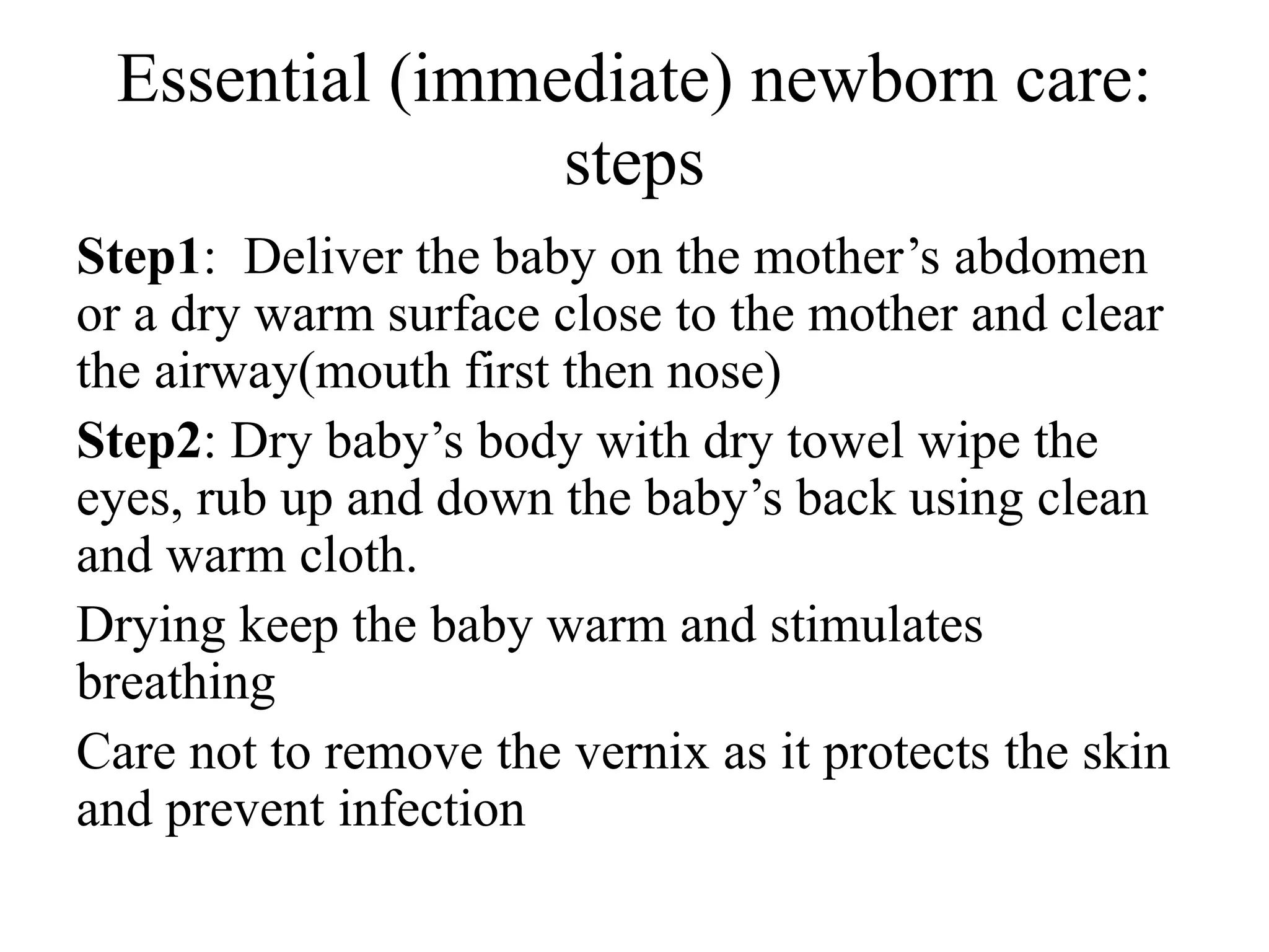
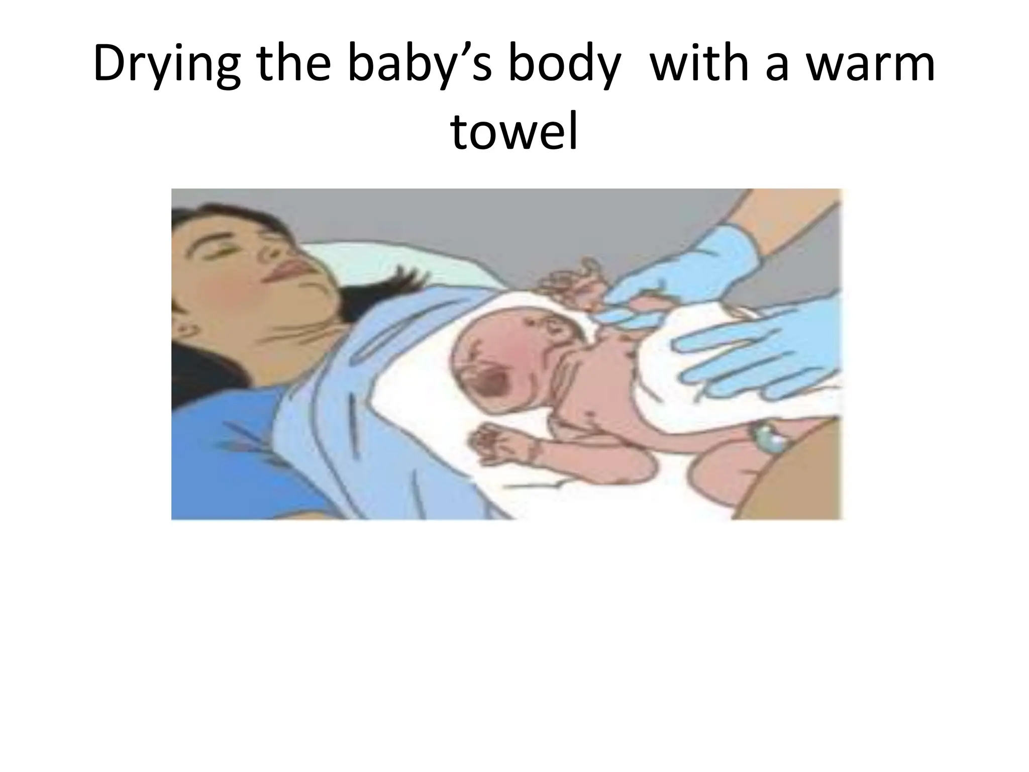
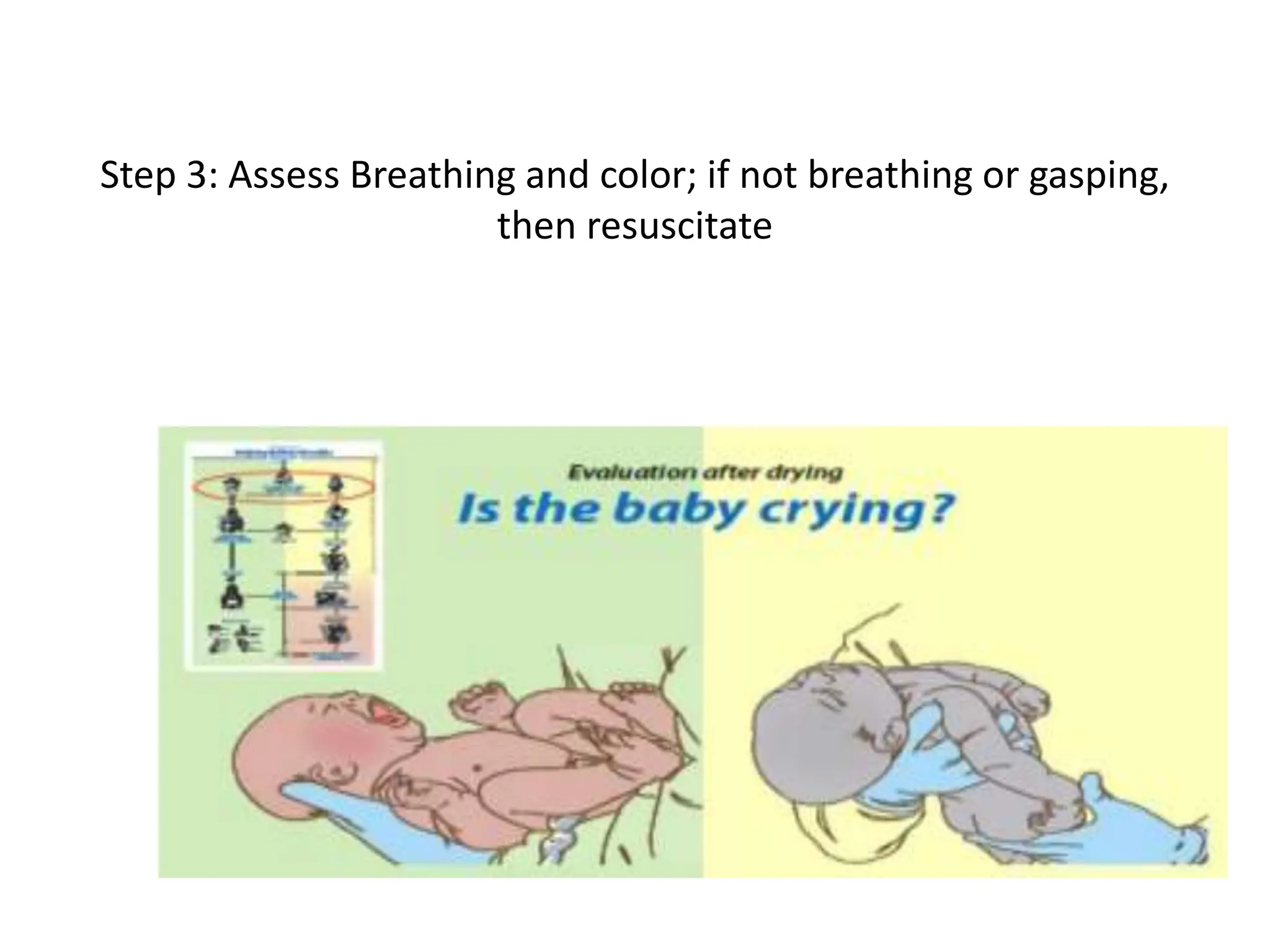
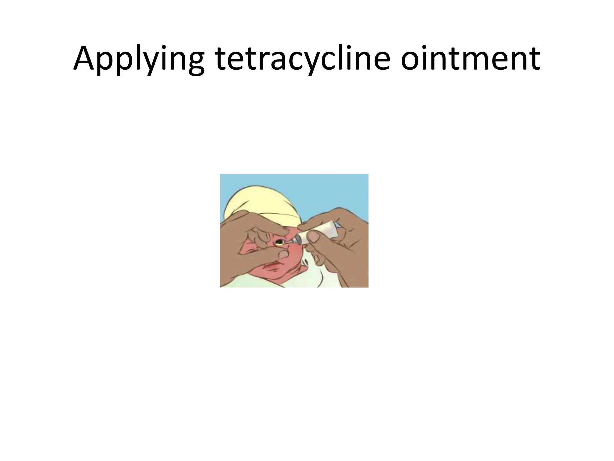
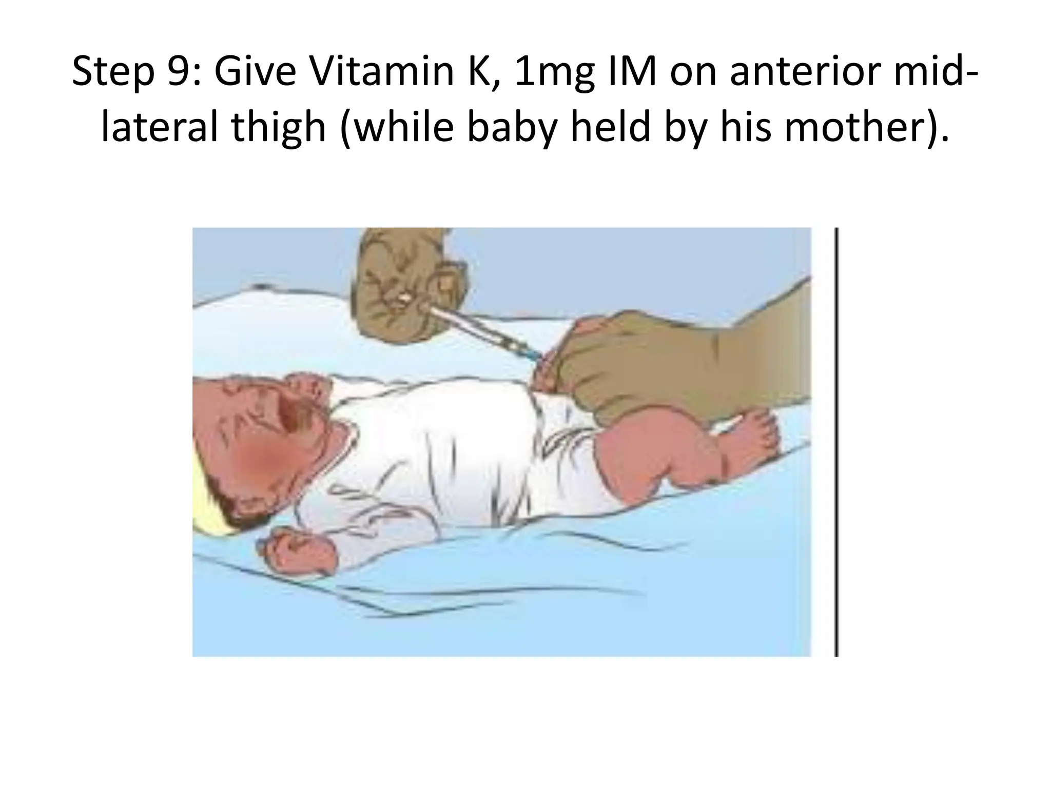
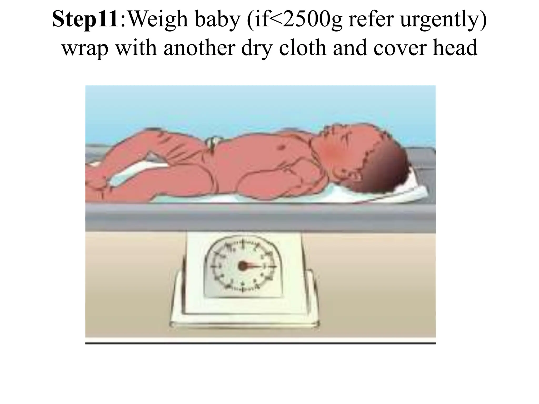
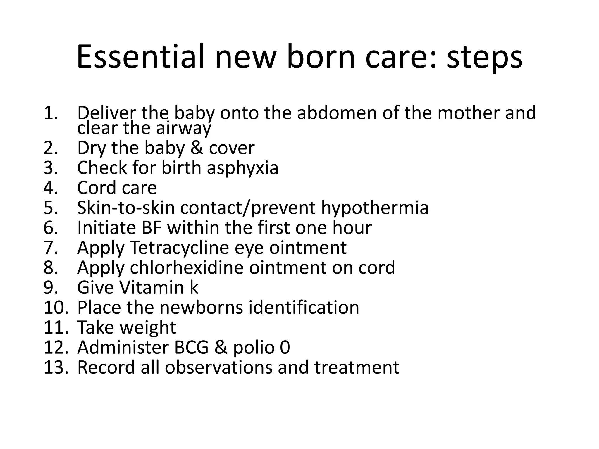
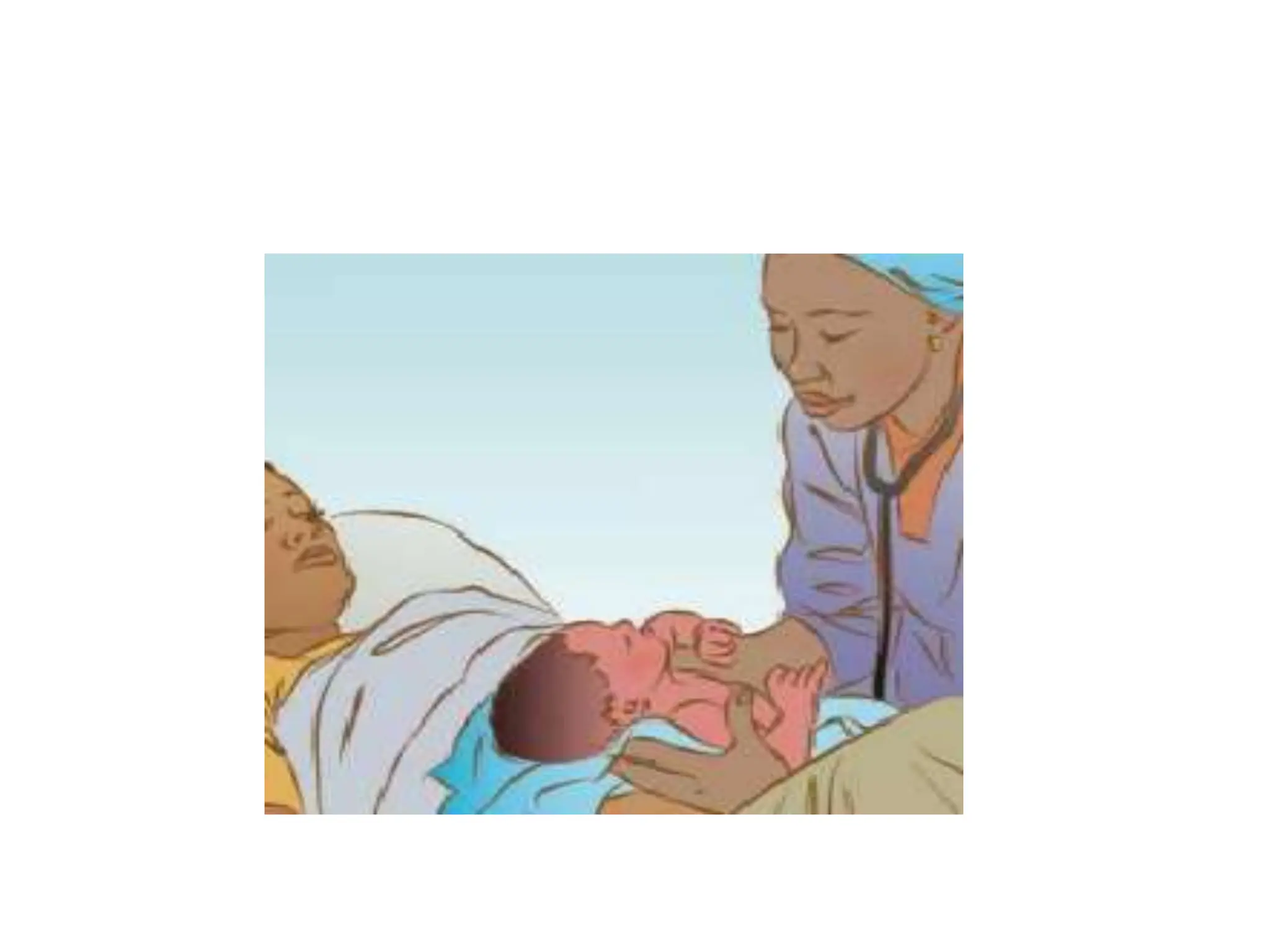
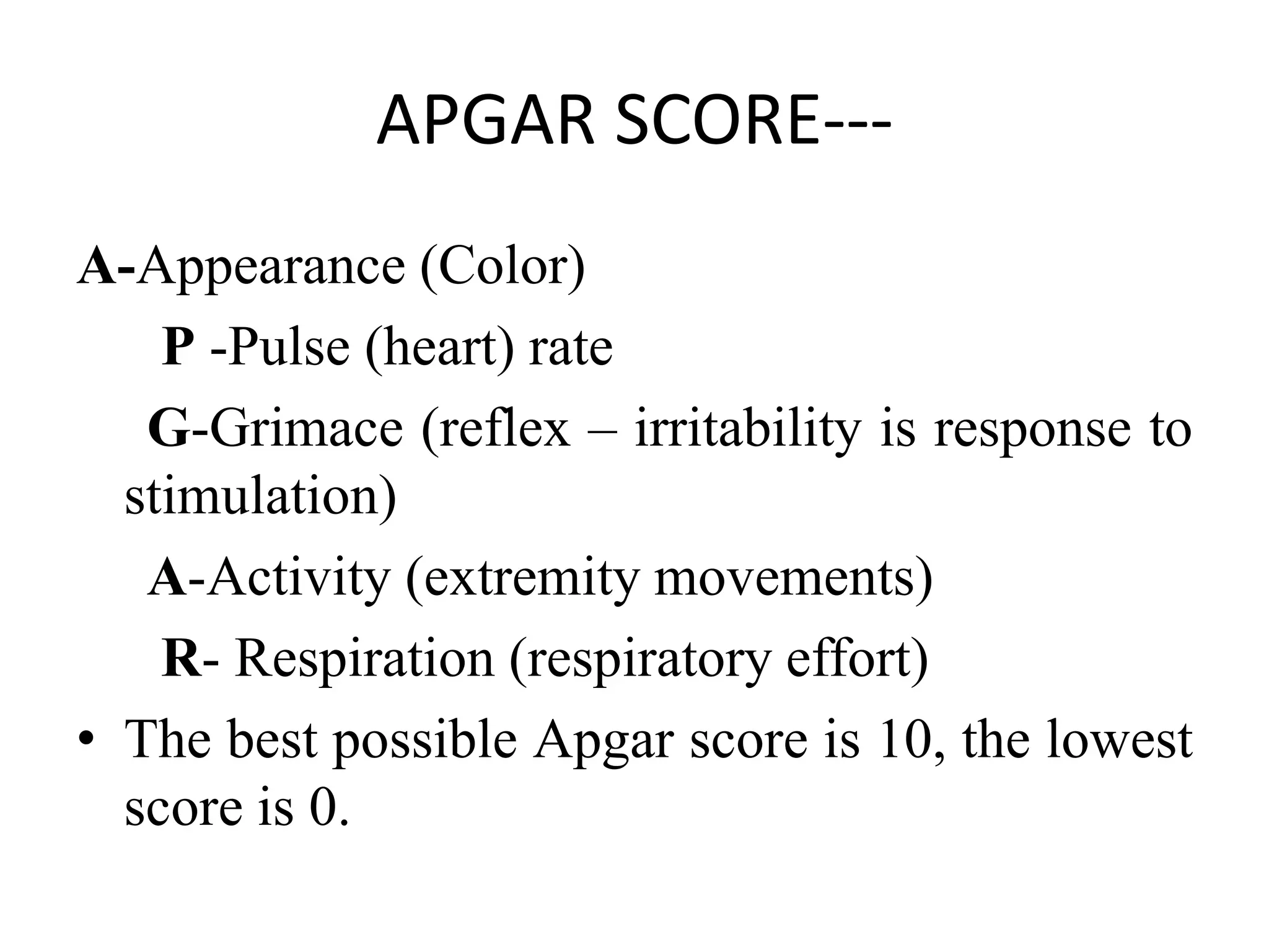
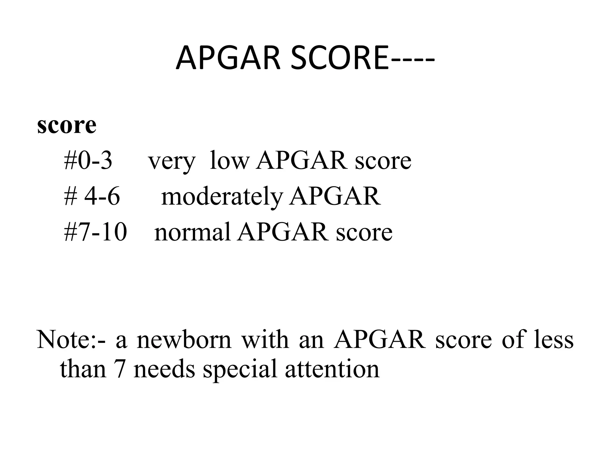
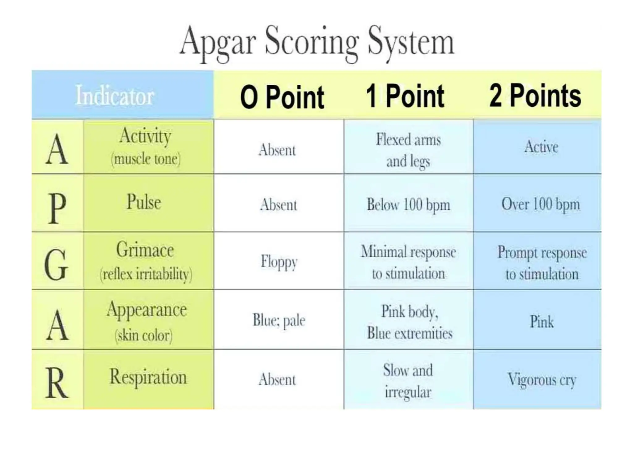
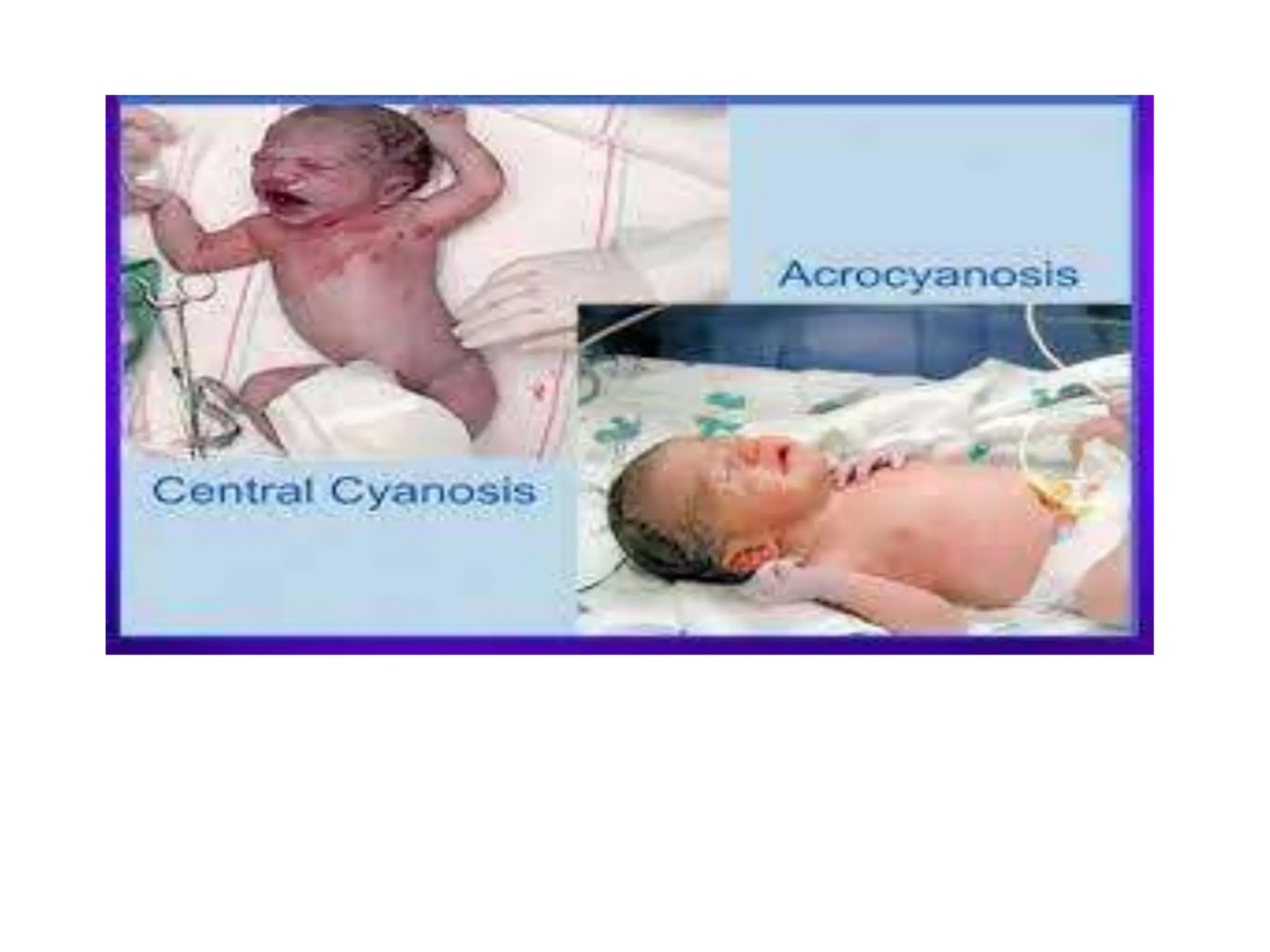
2. Definition of terms
• Neonatology :- is a branch of pediatrics which studies on
neonates
• The neonatal period is birth to 28 days of life and may
be further subdivided into the
Very early (birth to 24 hr)
Early (birth to 7 days)
late neonatal periods (7 days to 28 days)
3. • Newborn: the age range from birth to
seven days
• Neonate: the age range from birth to
twenty eight days
4. Essential new born care
• Definition: Essential Newborn Care is a
package of basic care provided to newborns to
support their survival and wellbeing
5. Essential (immediate) newborn care:
steps
Step1: Deliver the baby on the mother’s abdomen
or a dry warm surface close to the mother and clear
the airway(mouth first then nose)
Step2: Dry baby’s body with dry towel wipe the
eyes, rub up and down the baby’s back using clean
and warm cloth.
Drying keep the baby warm and stimulates
breathing
Care not to remove the vernix as it protects the skin
and prevent infection
7. Step 3: Assess Breathing and color; if not breathing or gasping,
then resuscitate
8. • Call for help & Start resuscitation if any of the
following signs are seen
• 1) breathing less than 30/min
• 2) having trouble breathing
• 3) not breathing at all
This step is used for assessing and classifying the
newborn for birth asphyxia. If the baby needs.
9. Step 4: If the baby does cry or breaths well - clamp/tie
and cut the cord
Tie the cord two fingers from abdomen and another tie
two fingers from the first tie.
-Tie the cord securely in two places:
-Tie the first two fingers away the baby’s abdomen
-Tie the second four fingers away the baby’s abdomen
-Make sure that tie is well secured
-Make sure that the tread used to tie the cord is clean
and safe
10. Use a new razor blade or sterile scissors
-Use a small piece of cloth or gauze to cover the
cord you are cutting to prevent blood splashes on
you or others
-Be careful not to cut or injure the baby
If bleeding or oozing occurs, place a second
clamp or tie between the first one and the
baby’s skin and retie if necessary
13. Step6:Place the baby in skin-to-skin contact and on
the breast to initiate breast feeding
-The warmth of the mother passes easily to the
baby and helps to stabilize the baby’s temperature.
-Put the baby on the mother’s chest for skin-to-skin
warmth
-Cover both the mother and baby together with
warm cloth or blanket
-Cover the baby’s head
14. • Initiate breastfeeding within the first hour. Select the
appropriate method of feeding for the HIVinfected mother,
based on informed choice.
• To encourage early breastfeeding, keep mother and baby
together unless a problem separates them. Babies are often
alert immediately after birth and will move toward the
mother’s breast but may not suck.
• Signs of readiness to feed include: 1) Licking movements 2)
Eyes open 3) The baby’s head slightly back 4) Tongue down
and forward 5) Mouth open
• Teach mothers how to recognize these signs and initiate
breastfeeding early.
• Skin to skin contact & early initiation of breast feeding are the
2 most important measures to prevent hypothermia after
delivery.
18. Step7: Give eye care; after breast feeding within 1hour of
age, give the newborn eye care with antimicrobial
medication to prevent from serious eye infection.
-Steps for giving eye care
-Wash your hands
-Tetracycline 1% eye ointment
-Hold one eye open and apply a rice grain size of
ointment along the inside of the lower eyelid.
-Repeat this step to put medication into the other eye
-Do not rinse out the medication
20. Step 8. Apply Chlorhexidine gel (4%) on the
cord Within 30 minutes after birth
• Application of Chlorhexidine on the cord
prevents babies from getting infection
• Gel on the cord once per day for seven
consecutive days (for six additional days if the
first dose is given at the health facility in the
first 30 minutes after birth).
23. Step 9: Give Vitamin K, 1mg IM on anterior mid-
lateral thigh (while baby held by his mother).
24. • Vitamin K protects babies from serious
bleeding that may result in death or brain
damage. Every newborn should be given
vitamin K. Because this treatment is painful, it
should not be given during the first hour after
birth, a time when the mother and baby
should not be disturbed.
25. • Step 10: Place the baby identification bands
on the wrist and ankle Within 90 minutes
after birth, place the baby identification bands
on the wrist and ankle
• Note that, at a minimum, the names of the
mother and, if available, the father, and the
date and time of birth should be written on
the identification bands. Putting the
identification bands on the hands and ankle
will save you from misshaping babies in busy
labour room.
27. Weighing the newborn when he/she is stable
• Place a clean linen or paper on the pan of the
weighing scale.
• Adjust the pointer to zero on the scale with the
linen/paper on the pan.
• Place the naked baby on the paper/linen. If the
linen is large, cover the baby with the cloth.
• Note the weight of the baby when the scale stops
moving.
• Never leave the baby unattended on the scale.
• Record the baby’s weight in partograph/maternal/
newborn charts .
29. Essential new born care: steps
1. Deliver the baby onto the abdomen of the mother and
clear the airway
2. Dry the baby & cover
3. Check for birth asphyxia
4. Cord care
5. Skin-to-skin contact/prevent hypothermia
6. Initiate BF within the first one hour
7. Apply Tetracycline eye ointment
8. Apply chlorhexidine ointment on cord
9. Give Vitamin k
10. Place the newborns identification
11. Take weight
12. Administer BCG & polio 0
13. Record all observations and treatment
30. Routine care of new born
• A complete exam should be performed within
90 minutes of birth or whenever a baby
appears unwell. During the exam, evaluate a
baby by looking, listening and feeling. Focus
on the following features:
• Breathing: A baby should breathe easily
between 40-60 times per minute. Count a
baby’s breathing rate for one minute.
31.
32. • Movement and tone: When active, well babies
have spontaneous movements of arms and
legs that are equal on both sides. Limbs are
flexed at rest. The tone should be neither
floppy nor rigid
33. • Skin color: The normal skin color of a newborn
is pink, but hands and feet may still look pale
or blue soon after delivery. Pink color may be
difficult to detect in dark-skinned babies. The
inside of the mouth should be pink in all
babies. Babies with jaundice may have yellow
skin. Recognizing jaundice is important
because severe jaundice may require
advanced care
34. • Cord appearance: On the initial exam, there
should be no drainage or bleeding from the
cord. Other features of a general exam:
Inspect the baby’s entire body for
abnormalities. Document the results of this
exam even if all findings are normal
35. • Measure Temperature Within 90 minutes after
birth measure temperature to identify babies
who require special care
• The normal temperature range is 36.5-37.5 °C. A
temperature 35.5 °C-36.4 °C requires improved
thermal care. A temperature below 35.5 °C is a
Danger Sign. A temperature above 37.5 °C not
due to over-warming (for example being placed in
direct sunlight) is a Danger Sign
41. New born Assessment
The main objective of routine examination
To ensure and assess that the lungs have expanded
and that air passages are not obstructed
To make an early diagnosis of life threatening ,
congenital mal formations and birth injuries
To assess the gestational age to classify as term or
preterm and as appropriate or not appropriate for
gestational age based on the birth weight.
To assess whether the baby has any sign of
infection or metabolic diseases
42. Neonatal assessment
Neonatal history
Demographic and social data:
socioeconomic status, age, sex
C/C- failure to suck, fever, breathing difficulty...
Past medical illnesses in the mother and family, previous
siblings:
cardiopulmonary disorders
infectious diseases
genetic disorders
diabetes mellitus
Previous maternal reproductive problems:
Stillbirth
Prematurity
blood group sensitization
43. Neonatal assessment ----
Events occurring in the present pregnancy:
preterm labor
fetal assessments
vaginal bleeding
medications,
acute illness
duration of rupture of membranes
Description of the labor
Duration
fetal presentation
fetal distress
fever
44. Neonatal assessment ----
Description of delivery
cesarean section
anesthesia or sedation
use of forceps
Apgar scores (cyanosis, pallor)
need for resuscitation
45. APGAR SCORE
• 1953 Virginia Apgar describes her scoring
system
Dr. Virginia Apgar (1909–1974)
46. APGAR SCORE----
APGAR: - a tool that can be used objectively to
assess the status of an infant
• APGAR score assessed at 1st & 5th minutes
the 1- minute APGAR score measures how well
the newborn tolerated the berthing process.
The 5- minute APGAR score assesses how well the
newborn is adapting the new external environment.
47. APGAR SCORE----
• Scoring system provides points between 0 and 2 for
each of five categories i.e.
• The score may be recorded every 5 minutes until a
score of ≥7 is reached.
• Note: the APGAR score is not used to determine the
need for resuscitation!
– but to assess the response for resuscitation
48. APGAR SCORE---
A-Appearance (Color)
P -Pulse (heart) rate
G-Grimace (reflex – irritability is response to
stimulation)
A-Activity (extremity movements)
R- Respiration (respiratory effort)
• The best possible Apgar score is 10, the lowest
score is 0.
49. APGAR SCORE----
score
#0-3 very low APGAR score
# 4-6 moderately APGAR
#7-10 normal APGAR score
Note:- a newborn with an APGAR score of less
than 7 needs special attention
52. PHYSICAL EXAMINATION
Initial examination -immediately
Temperature N(36.5-37.5 oc) axillary
temperature
Respiratory rate N(30-60/ min)
Type of respiration
pulse rate N(120-160 /min)
Check for capillary refill
Color
Activity
Tone
Level of consciousness
53. PHYSICAL EXAMINATION
• BP: Depends on birth weight, GA, postnatal age
• Normal systolic BP shouldn’t be less than
60mmHg
• Note: taking BP is not an easy task in the newborn
babies.
• Moreover, the apparatus may not be available in
most of our set ups.
• Therefore, in our set ups, we will be left with only
assessing the capillary refill time. The normal
capillary refill time should be less than 3 seconds
54. PHYSICAL EXAMINATION
Anthropometric measurement
Weight, length, head circumference, chest
circumference
Normal values of term neonates at birth
# weight 2500- 3999gm
# length 48-53cm
# HC 33-38cm
# CC= 3cm less than HC
# upper/lower segment ration= 1.7:1
55. PHYSICAL EXAMINATION
2nd or detailed physical examination
Head to toe
After stabilization
In the first 24 hrs.
56. PHYSICAL EXAMINATION
General appearance
Observation is the most important part of neonatal
examination
Therefore, special attention must be given to the
general appearance, particularly to the colour,
position and activity of the neonate.
A normal newborn has pink color and only a few
of them may have acrocyanosis
The neonate normally, flexes all the extremities,
and is always in spontaneous motion
Listen to the cry whether it is normal or abnormal,
high pitched ( shrill cry)
57.
58. PHYSICAL EXAMINATION
- Observe whether the baby in cardiorepiratory
distress or not
- Watch if the baby is irritable or consolable
- Level of consciousness, gross anomalies,
activity( kicking, limp, crying,), cyanosis,
jaundice and pallor
Then proceed to systemic examination
59. PHYSICAL EXAMINATION
G/A...
• Inspect for
congenital anomalies
• Down syndrome
• Cleft lip/ palate
• Hypoplasia
• Talipes equinovarus ( club foot)...
number of vessels on the umbilical cord:
two arteries and one vein
60. PHYSICAL EXAMINATION
• HEENMN:
Head – look for
caput succedaneum
sub galial hemorrhage
cephalo hematoma
Size- macro/microcephaly
Shape – Dolichocephalic , molded, rounded
Fontanel: anterior/posterior – size, whether bulged
or flat, palpate the size and fullness
Palpate suture; look for any ridging
63. PHYSICAL EXAMINATION
Ear – look for shape and size ( large/small or
malformed ear)- low set ear, ear tag
The pinna is considered ‘’low set ear’’ when
the top lies completely below the imaginary
line (outer canthus)
72. PHYSICAL EXAMINATION
Cardiovascular system:
it is always advisable to start neonatal
examination with the examination of the heart
inspect for cyanosis ( central cyanosis)
Count the pulse rate ( 120-160)
- Feel the pulses- whether it is weak or bounding
- Auscultate for murmurs and additional heart
sounds
74. PHYSICAL EXAMINATION
Gastro intestinal system:
Abdomen :
normally it is flat and not distended.
Scaphoid abdomen is suggestive of
diaphramatic hernia in most of the cases.
Distended abdomen is sugestive of paralytic
illeus, necrotizing entercolotis, congenital
upper GI obstruction due to atressia and
stenosis.
75. PHYSICAL EXAMINATION
Liver: it may normally be palpable up to 2 cm
below right costal margin
- Enlarged liver is suggestive of TORCH infection,
CHF, Neonatal syphillis, congenital tuberculosis,
Spleen: Enlarged spleen is suggestive of TORCH
infection
Kidneys: Enlarged kidney is suggestive of :
polysystic kidney, renal vein thrombosis, and
congenital hydronephrosis
76.
77. PHYSICAL EXAMINATION
Skin:
observe the color. Normally pink
In few cases there may be acrocyanosis
pigmnetation: mongolian spot ( brown pigmented
naevi, scatered around any area of the body, most
commonly on the back), may normally present
Vernix caseosa and lanugo are also the usual normal
finding on the skin
- Skin lesions which can normally be detected in some
neonates are:- Milia ( plugged sweat glands) on the
nose, Erythema toxicum and at times you may observe
petichia
81. PHYSICAL EXAMINATION
Nervous system: routine examination in a healthy
term baby is unnecessary
- At first assess for skull and spine abnormalities
- sensorium
level of consciousness/ level of alertness tone
activity
Look for facial deviation and abnormal movement
of the eye
Tend to neonatal reflexes ( primitive reflexes)
86. Assessing neonatal reflexes
• Neonatal reflexes are inborn reflexes which are
present at birth and occur in a predictable
fashion.
• Normally developing newborn should respond
to certain stimuli with these reflexes, which
eventually become inhibited as the child
matures
87. Assessing neonatal reflexes
Moro reflex
Infants will respond to sudden movements by throwing
their arms and legs out, and throwing their heads back
Hold supine infant by arms a few inches above bed.
Gently drop infant back to elicit
Baby throws Arms out in extension and baby grimace.
Disappears by 3 – 4 months
88. Assessing neonatal reflexes
Palmar grasp
Babies will grasp anything that is placed in their
palm.
The strength of this grip is strong
Most babies can support their entire weight in
their grip.
Disappears at 3 to 4 months.
89. Assessing neonatal reflexes
Plantar reflex ( plantar grasp)
Infant reflexively grasp with bottom of foot
when pressure is applied to plantar surface
Disappears at 9 months
90. Assessing neonatal reflexes
Rooting Reflex
The rooting reflex is most evident when an
infant's cheek is stroked.
The baby responds by turning his or her head in
the direction of the touch and opening their mouth
for feeding.
Disappears at 3-4 months.
91. Assessing neonatal reflexes
Sucking Reflex
Reflexive sucking when the nipple or fingure
placed in the infants mouth
Disappears at 2-5 months
92. Assessing neonatal reflexes
Tonic neck reflex
While lying supine, extremities are extended
on the side of the body to which the head is
turned and opposite extremities are flexed (
also called fencing position)
Disappeared by 4 month
93. Assessing neonatal reflexes
Babinski reflex
When the inner sole of a baby’s foot is stroked,
the infant will respond by curl his or her toes.
When the outer sole of a baby’s foot is stroked,
the infant will respond by spreading out their
toes.
Disappears at 12 months
94. Assessing neonatal reflexes
Stepping Reflex
When an infant is held upright with his or her
feet placed on a surface, he or she will lift their
legs as if they are marching or stepping.
Extension of the legs whenever the ball of the
feet touch a solid surface
Disappears by 4 months
95. Assessing neonatal reflexes
Doll’s eye: With neonate supine, slowly turn
the neonates head to either side
• Neonate’s eyes remain stationary ( normal)
Galant : Using a fingernail, gently stroke one
side of the neonate’s spinal column from the
head to the buttocks
• Neonate’s trunk curves toward the stimulated
side ( normal)
96. Assessing neonatal reflexes
Pupillary (light): Darken the room and shine a
pen light directly into the neonate’s eye for
several seconds
Startle reflex
• Make a loud noise near the neonate
• Neonate cries and abducts and flexes all
extremities
98. definition
• Postpartum hemorrhage (PPH) is defined as
vaginal bleeding in excess of 500 ml after
childbirth (Or in excess of 1000 ml after Cesarean
section.
• The importance of a given volume of blood loss
varies with the woman’s hemoglobin level.
• A woman with a normal hemoglobin level will
tolerate blood loss that would be fatal for an
anaemic woman
99. types
• Increased vaginal bleeding within the first 24
hours after childbirth is called immediate or
primary PPH.
• Increased vaginal bleeding following the first
24 hours after childbirth is called delayed PPH
100. causes
• Atonic uterus
• Genital trauma
• Retained placenta
• Coagulation failure
• Acute inversion of the uterus
101. General management
• SHOUT FOR HELP. Urgently mobilize all available personnel.
• Make a rapid evaluation of the general condition of the
woman including vital signs (pulse, blood pressure,
respiration, temperature).
• If shock is suspected, immediately begin treatment. Even if
signs of shock are not present, keep shock in mind as you
evaluate the woman further because her status may
worsen rapidly. If shock develops, it is important to begin
treatment immediately.
• Massage the uterus to expel blood and blood clots. Blood
clots trapped in the uterus will inhibit effective uterine
contractions.
• Give oxytocin 10 units IM
102. • Start an IV infusion and infuse IV fluids. Establish
two IV lines if necessary.
• Take blood (5 mL) for hemoglobin (Hg)/
hematocrit (Hct) and cross matching.
• Catheterize the bladder.
• Check to see if the placenta has been expelled
and examine the placenta to be certain it is
complete.
• Examine the cervix, vagina and perineum for
tears.
103. • Provide specific treatment for the specific cause
identified .
• After bleeding is controlled (24 hours after
bleeding stops), determine haemoglobin or
hematocrit to check for anemia:
• If haemoglobin is below 7 g/dL or hematocrit is
below 20% (severe anemia):
• → Treat with iron if clinically stable.
• → Refer immediately for transfusion if signs and
symptoms of de-compensation develop
104. • If haemoglobin is between 7–11 g/dL, treat with
iron.
• Where hookworm is endemic (prevalence of 20%
or more), give one of the following anthelmintics:
→ Albendazole 400 mg PO stat
• → Mebendazole 500 mg PO stat or 100mg PO bid
for three days.
• If hookworm is highly endemic (prevalence of
50% or more), repeat the anthelmintic treatment
12 weeks after the first dose.
105. MANAGEMENT OF SPECIFIC CAUSES
ATONIC UTERUS An atonic uterus fails to
contract after delivery.
Atonic uterus is the most common cause of
primary PPH.
• Continue to massage the uterus.
• Use oxytocic drugs which can be given
together or sequentially
108. • If bleeding continues:
• Check placenta again for completeness; o If
there are signs of retained placental fragments
(absence of a portion of maternal surface or
torn membranes with vessels), remove
remaining placental tissue;
• Assess clotting status using a bedside clotting
test.
• Failure of a clot to form after 7 minutes or a
soft clot that breaks down easily suggests
coagulopathy.
109. BIMANUAL COMPRESSION OF UTERUS
• If bleeding continues in spite of management
above:
• Perform bimanual compression of the uterus
• Wearing high-level disinfected gloves, insert a
hand into the vagina and form a fist;
• Place the fist into the anterior fornix and apply
pressure against the anterior wall of the uterus;
With the other hand, press deeply into the
abdomen behind the uterus, applying pressure
against the posterior wall of the uterus
112. • Apply downward pressure with a closed fist
over the abdominal aorta directly through the
abdominal wall:
• → The point of compression is just above the
umbilicus and slightly to the left;
• → Aortic pulsations can be felt easily through
the anterior abdominal wall in the immediate
postpartum period.
113. • With the other hand, palpate the femoral
pulse to check the adequacy of compression:
→ If the pulse is palpable during compression,
the pressure exerted by the fist is inadequate;
→ If the femoral pulse is not palpable, the
pressure exerted is adequate; - Maintain
compression until bleeding is controlled
115. • If bleeding continues in spite of compression:
refer immediately accompanying her and
continuing providing the above measures or
perform intrauterine balloon tamponade
118. TEARS OF CERVIX, VAGINA OR
PERINEUM
• Tears of the birth canal are the second most frequent
cause of PPH. Tears may coexist with atonic uterus.
Postpartum bleeding with a contracted uterus is usually
due to a cervical or vaginal tear.
• Examine the woman carefully and repair tears to the cervix
or vagina and perineum.
• If bleeding continues, assess clotting status using a bedside
clotting test. Failure of a clot to form after 7 minutes or a
soft clot that breaks down easily suggests coagulopathy.
• If coagulopathy is diagnosed refer urgently accompanying
her and providing the above basic support.
119.
120. RETAINED PLACENTA
• There may be no bleeding with retained placenta.
• If you can see the placenta, ask the woman to push it
out. If you can feel the placenta in the vagina, remove
it.
• Ensure that the bladder is empty. Catheterize the
bladder, if necessary.
• If the placenta is not expelled and no active bleeding,
give oxytocin 10 units IM if not already done for active
management of the third stage, attempt controlled
cord traction.
Note: Avoid forceful cord traction and fundal pressure as
they may cause uterine inversion.
121. • If there is active bleeding or the placenta is
undelivered after 30 minutes of oxytocin
stimulation and controlled cord traction,
attempt manual removal of placenta.
124. procedure
• Review for indications.
• Review general care principles and start an IV
infusion.
• Provide emotional support and encouragement.
Give pethidine and diazepam IV slowly (do not mix
in the same syringe) or use Ketamine.
• Give a single dose of prophylactic antibiotics: -
Ampicillin 2 g - OR cefazolin 1 g IV.
• Hold the umbilical cord with a clamp. Pull the
cord gently until it is parallel to the floor.
• Wearing high-level disinfected long sleeve gloves
or similar modification, insert a hand into the
vagina and up into the uterus
125.
126. • Let go of the cord and move the hand up over the
abdomen in order to support the fundus of the
uterus and to provide counter-traction during
removal to prevent inversion of the uterus.
• Note: If uterine inversion occurs, reposition the
uterus.
• Move the fingers of the hand laterally until the
edge of the placenta is located.
• If the cord has been detached previously, insert a
hand into the uterine cavity. Explore the entire
cavity until a line of cleavage is identified between
the placenta and the uterine wall.
127. • Detach the placenta from the implantation site by
keeping the fingers tightly together and using the edge
of the hand to gradually make a space between the
placenta and the uterine wall.
• Proceed slowly all around the placental bed until the
whole placenta is detached from the uterine wall.
NOTE: If the placenta does not separate from the uterine
surface by gentle lateral movement of the fingertips at
the line of cleavage, suspect adherent placenta, stop the
procedure and urgently refer providing supportive care.
• Hold the placenta and slowly withdraw the hand from
the uterus, bringing the placenta with it.
• With the other hand, continue to provide counter-
traction to the fundus by pushing it in the opposite
direction of the hand that is being withdrawn.
128. RETAINED PLACENTAL FRAGMENTS
• There may be no bleeding with retained placental
fragments.
• When a portion of the placenta—one or more
lobes—is retained, it prevents the uterus from
contracting effectively.
• Feel inside the uterus for placental fragments.
Manual exploration of the uterus is similar to the
technique described for removal of the retained
placenta.
• Remove placental fragments by hand, ovum
forceps or large curette.
129. • Note: Very adherent tissue may be placenta
accreta. Efforts to extract fragments that do
not separate easily may result in heavy
bleeding or uterine perforation, therefore stop
the procedure and refer urgently providing
supportive care.
130. Post-procedure care
• Observe the woman closely in labor ward or where she
can be monitored closely for at least 6 hours or until
stable.
• Check and rub the uterus every 15 minute for the next
two hours.
• Monitor the vital signs (pulse, blood pressure,
respiration) every 30 minutes for the next 6 hours
• Continue with IV fluid and oxytocin drip for next 4-6
hours.
• Continue infusion of IV fluids.
• If patient is stabilized, assist her to initiate breast
feeding if appropriate
131. INVERTED UTERUS
• Repositioning the uterus should be
performed immediately. With the passage of
time the constricting ring around the inverted
uterus becomes more rigid and the uterus
more engorged with blood.
132. • If the woman is in severe pain, give pethidine
1 mg/kg body weight (but not more than 100
mg) IM or IV slowly or give morphine 0.1
mg/kg body weight IM.
• Note: Do not give oxytocic drugs until the
inversion is corrected.
133. • Immediately after diagnosis, it is often possible to
replace the uterus by applying gentle transvaginal
pressure.
• The Johnson technique calls for lifting the uterus and
the cervix into the abdominal cavity with the fingers in
the fornix and the inverted uterine fundus on the palm.
The fundus is then gently pushed back through the
cervix. The operator’s hand should be kept in the
uterus until the fundus begins to climb up. If the
placenta is still attached, it should not be removed until
after the uterus is replaced through the cervix
134. • Give a single dose of prophylactic antibiotics
after correcting the inverted uterus: -
Ampicillin 2 g IV; - OR cefazolin 1 g IV.
• If there are signs of infection (fever, foul-
smelling vaginal discharge), give antibiotics as
for metritis.
• If necrosis is suspected, referral to a tertiary
care center.
135. Causes of secondary PPH
Bleeding in the first 3 weeks after the first day of
delivery is mainly due to:
• Sub-involution of the uterus
• Infection
• Retained pieces of placental tissue or clot
• Breakdown of the uterine wound after Cesarean
delivery or ruptured uterus
• After obstructed labor, bleeding may occur due to
sloughing of dead vaginal tissue (cervix, vagina,
bladder, and rectum)
136. - Bleeding from the third week to sixth week is
mainly due to:
- • Choriocarcinoma (rare)
- • Local causes (vaginal or cervical) such as
severe infection, malignancies, trauma
- • Early onset menstruation
137. Complications of PPH
• Immediate and late complications of primary
postpartum hemorrhage include hypovolemic
shock, cerebral anoxia, renal failure, anemia,
puerperal sepsis, and Sheehan's syndrome.
The antepartum hemoglobin status and the
rate of blood loss influence hemorrhage
outcome.
142. During the postpartum period, anaemia is defined as Hb
level < 10 g/dL
• The primary aetiologies for postpartum anaemia are;
• haemodilution,
• iron deficiency,
• anaemia during pregnancy, and
• ante-partum and postpartum haemorrhage..
143. • Postpartum anemia is associated with an
impaired quality of life, reduced cognitive
abilities, emotional instability, and depression
and constitutes a significant health problem in
women of reproductive age.
144. • Anaemia symptoms include tiredness,
shortness of breath and dizziness. Women
may bleed severely at childbirth and many
pregnant women already have anaemia,
which can worsen as a result of bleeding
145. • Postpartum anemia is treated with oral iron
supplementation and/or blood transfusion.
Recent studies have evaluated the use of
parenteral iron as a better tolerated treatment
modality.
• Ferric carboxy maltose as infusion
• Iron sucrose as oral dose
146. Dietary management
• A regular diet should be offered as soon as the woman
requests food and is conscious. Intake should be increased
by 10% (not physically active) to 20% (moderately or very
active) to cover energy cost of lactation.
• Women should be advised to eat a diet that is rich in
proteins and fluids.
• Eating more of staple food (cereal or tuber)
• Greater consumption of non-saturated fats
• Encourage foods rich in iron (e.g., liver, dark green leafy
vegetables, etc.)
• Avoid all dietary restriction
149. Puerperal sepsis
• Definition
It is defined as the infection of the genital tract
during or after labour
Cause;
Bacteria
Aerobic (gram positive/gram negative)
Anaerobic (gram positive/gram negative)
Mode of transmission;
1.endogenous origin;it may be present in genital
tract as anaerobic streptococci that becomes
pathogenic in presence of devitalized tissue.
150. 2.Exogenous origin; from infected attendants, dust,
instruments etc
Predisposing factors;
Bad general condition of the mother such as
anemia, diabetes, debilitating diseases
Improper asceptic practices
Intrapartum factors such as;
PROM
Instrumental delivery
Laceration
Marked blood losss
Retained placenta
152. • Diagnosis;
Vaginal swab culture
Urine culture and microscopy
Blood culture
Ultrasound scanning
General management;
Ice packs may be helpful for perineal wounds
Rest and adequate fluid intake
Refer the case if fever greater than 38 degree
Tachycardia, diarrhoea,vomitting,uterine
tenderness etc
154. Cystitis
Cystitis is infection of the bladder (lower urinary
tract).
s/s are;
• Dysuria
• Increased frequency and urgency of urination
Rare symptoms are;
• Retropubic/suprapubicpain
• Abdominalpain
155. • Treat with an antibiotic - amoxicillin 500 mg by
mouth every eight hours for three
days;nitrofurantoin 100 mg by mouth every
eight hours for three days.
• Note: Avoid nitrofurantoin at term as it can
cause neonatal haemolysis.
• If treatment fails or if infection recurs two or
more times during pregnancy, check urine
culture and sensitivity, if available, and treat
with an antibiotic appropriate for the
organism.
156. ACUTE PYELONEPHRITIS
• Acute pyelonephritis is an infection of the
upper urinary tract, mainly of the renal pelvis,
which may also involve the renal parenchyma.
Acute pyelonephritis can cause significant
illness in pregnant women and should be
promptly investigated and treated in every
pregnant woman with fever, urinary
symptoms and flank pain.
157. Presenting Symptom and Other Symptom and Signs Typically Present
Dysuria
Spiking fever/chills
Increased frequency and urgency of urination,
Abdominal pain
Symptoms and Signs Sometimes Present ;
Retro pubic/suprapubic pain
Loin pain/tenderness
Tenderness in ribcage
Anorexia
Nausea/vomiting
158. If shock is present or suspected, initiate
immediate treatment.
• Start an IV infusion and infuse IV fluids at 150
mL per hour
• Check urine dipstick and urine culture if
possible and begin empiric antibiotic treatment
promptly (pending results of urine culture, if
available).
• Treat with an IV antibiotic until the woman is
fever-free for 48 hours: - ampicillin 2 g IV every
six hours; - PLUS gentamicin 5 mg/kg body
weight IV every 24 hours.
159. • Ensure adequate hydration by mouth or IV.
• Give paracetamol 500–1000 mg by mouth
three to four times daily as needed for pain
and to lower temperature (4000 mg maximum
in 24 hours).
160. Uncomplicated malaria
Symptom and Signs Typically Present
• Fever
• Chills/rigors
• Headache
• Muscle/joint pain
Symptoms and Signs Sometimes Present
• Enlarged spleen
161. • Give quinine salt (dihydrochloride or sulfate)
10 mg/kg body weight by mouth three times
daily PLUS clindamycin 300 mg every six hours
for seven days.
162. POSTPARTUM ENDOMETRITIS
Postpartum endometritis is a major cause of
maternal death.
Delayed or inadequate treatment of postpartum
endometritis may result in pelvic abscess,
peritonitis, septic shock, deep vein thrombosis,
pulmonary embolism, chronic pelvic infection
with recurrent pelvic pain and dyspareunia,
tubal blockage, or infertility.
163. Symptom and Signs Typically Present
Fever/chills
Lower abdominal pain
Purulent, foul-smelling lochia
Tender uterus
s/s sometimes present
Light vaginal bleeding
Shock
164. • Transfuse as necessary. Use packed cells, if
available.
• Give the woman a combination of antibiotics for
24–48 hours after complete resolution of clinical
signs and symptoms (fever, uterine tenderness,
purulent lochia, leukocytosis: -
Clindamycin phosphate 600 mg IV every eight
hours;
PLUS gentamicin 5 mg/kg body weight IV every 24
hours.
If clindamycin is not available administer: -
Ampicillin 2 g IV every 6 hours; - PLUS gentamicin 5
mg/kg body weight IV every 24 hours +
metronidazole 500mg iv TID
165. Pelvic abscess
s/s typically present
• Persistent spiking fever/chills
• Lower abdominal pain and distension
• Tender uterus
s/s sometime present
• Poor response to antibiotics
• Swelling in adnexa or pouch of Douglas
• Pus obtained upon
166. Treatment
• Give a combination of antibiotics before
draining the abscess; continue antibiotics until
the woman is fever-free for 48 hours ():
- Ampicillin 2 g IV every six hours;
- PLUS gentamicin 5 mg/kg body weight IV
every 24 hours;
- PLUS metronidazole 500 mg IV every eight
hours.
168. Management;
• Provide nasogastric suction.
• Start an IV infusion and infuse IV fluids.
Referral to a higher center and further management
include:
• Give the woman a combination of antibiotics until she is
fever-free for 48 hours :
- Ampicillin 2 g IV every six hours;
- - PLUS gentamicin 5 mg/kg body weight IV every 24
hours PLUS metronidazole 500 mg IV every eight hours.
- • Identify and treat the underlying cause of the
peritonitis.
- • Perform additional diagnostics such as X-ray or
ultrasound to assist in identifying the underlying cause.
169. Breast engorgement
Breast engorgement is an exaggeration of the
lymphatic and venous engorgement that occurs
before lactation. It is not the result of over
distension of the breast with milk.
s/s typically present
• Breast pain and tenderness three to six days
after giving birth
s/s sometimes present
Hard, enlarged breasts Both breasts affected
170. Relief measures are;
• Applying warm compresses to the breasts just before
breastfeeding, or encouraging the woman to take a warm shower;
- Massaging the woman’s neck and back; and
- Having the woman express some milk manually before
breastfeeding, and wetting the nipple area to soften the areola to
help the baby latch on properly and easily.
- • Relief measures after feeding or expression may include: -
Supporting breasts with a binder or bra;
- Applying cold compresses to the breasts between feedings to
reduce swelling and pain.
• Give ibuprofen 200–400 mg every six to eight hours orally (maximum
dose of 1200 mg in 24 hours);
- OR paracetamol 500–1000 mg every six to eight hours orally as an
appropriate alternative (maximum 4000 mg in 24 hours).
- • Follow up in three days to ensure response.
171. NOT BREASTFEEDING
• If the woman is not breastfeeding:
- Encourage her to support breasts with a binder or
bra.
- Apply cold compresses to the breasts to reduce
swelling and pain.
- Avoid massaging or applying heat to the breasts. -
Avoid stimulating the nipples.
- Give ibuprofen 200–400 mg every six to eight
hours (maximum dose 1200 mg in 24 hours);
– OR paracetamol 500–1000 mg every six to eight
hours orally as an appropriate alternative
(maximum dose 4000 mg in 24 hours).
- Follow up in three days to ensure response.
172. Breast infection
Mastitis
s/s typically present
• Breast pain and tenderness
• Reddened, wedge-shaped area on breast
• Inflammation preceded by engorgement
• Usually only one breast affected
173. • Treat with antibiotics: - Cloxacillin 500 mg by
mouth every six hours for 10 days; OR
erythromycin 250 mg every eight hours for 10
days.
• Encourage the woman to:
- continue breastfeeding;
- support the breasts with a binder or bra; and
- apply cold compresses to the breasts between
feedings to reduce swelling and pain.
• Give the woman ibuprofen 200–400 mg every six
to eight hours
(maximum dose 1200 mg in 24 hours)
174. • paracetamol 500–1000 mg every six to eight
hours as an appropriate alternative (maximum
dose 4000 mg in 24 hours).
• Follow up in three days to ensure response.
175. BREAST ABSCESS
s/s typically present
• Firm, very tender breast
• Overlying erythema
s/s sometimes present
• Fluctuant swelling in breast
• Draining pus
176. Antibiotic Treatment
• Treat with antibiotics: - cloxacillin 500 mg by
mouth every six hours for 10 days; - OR
erythromycin 250 mg every eight hours for 10
days.
Surgical Treatment (at a higher center or by
experienced professional at health center)
Pus must be drained either by incision and
drainage or ultrasound-guided needle aspiration
(which may need to be repeated)under local
anaesthesia.
177. INFECTION OF PERINEAL AND
ABDOMINAL WOUNDS
• If there is pus or fluid, open and drain the wound.
• Remove infected skin or subcutaneous sutures
and debride the wound. Do not remove fascial
sutures.
• If there is an abscess without cellulitis, antibiotics
are not required.
• Place a damp dressing on the wound and have the
woman return to change the dressing every 24
hours.
• Advise the woman to practice good hygiene and
to wear clean pads or cloths that she changes often.
178. WOUND CELLULITIS AND
NECROTIZING FASCIITIS
• If there is fluid or pus, open and drain the wound.
• Remove infected skin or subcutaneous sutures
and debride the wound. Do not remove fascial
sutures.
• If infection is superficial and does not involve
deep tissues, monitor for development of an
abscess and give a combination of antibiotics
(: - ampicillin 500 mg by mouth every six hours for
five days.
179. • If the infection is deep;
give a combination of antibiotics until necrotic tissue has
been removed and the woman is fever-free for 48 hours
- penicillin G 2 million units IV every six hours (or
Ampiciline 2gm Iv qid);
- PLUS gentamicin 5 mg/kg body weight IV every 24
hours;
- PLUS metronidazole 500 mg IV every eight hours;
- Note: Necrotizing fasciitis requires wide surgical
debridement
182. Deep vein thrombosis
• s/s typically present
• Spiking fever despite
• Swelling in the affected leg
• Calf muscle tenderness Warmth and redness
of the affected leg
Treat with antibiotics
183. Pulmonary embolism
• s/s typically present
• Abrupt onset of pleuritic chest
• pain •
• Shortness of breath
• Tachypnea
• Hypoxia •
• Tachycardia Dry cough Cough with bloody sputum
Swollen leg or arm
Dizziness or syncope
185. • Postpartum psychosis (PPP) is a reversible — but
severe — mental health condition that affects
• s/s typically present are;
1 Hallucination
• A hallucination is when your brain acts as if it’s
getting input from your senses (usually your eyes
or ears, but occasionally touch hallucinations can
happen, too), but without any actual input
people after they give birth
186. • Delusions. Delusions are false beliefs that you
hold onto very strongly.
If you have a delusion, you hold these beliefs so
strongly that you won’t change them even if you
have convincing evidence that what you believe
isn’t true.
Examples include ;
persecutory delusions (believing someone is out
to get you),
control delusions (feeling that someone else is
controlling your body) or
somatic delusions (insisting you didn’t have a
child or weren’t pregnant).
187. • Other symptoms that are common with postpartum
psychosis include:
• Mood changes, such as mania (an increase in activity and
mood) and hypomania, or depression (a decrease in mood).
• Depersonalization (some people describe this as an out-of-
body experience).
• Disorganized thinking or behavior.
• Insomnia.
• Irritability or agitation.
• Thoughts of self-harm or harming others (especially their
newborn).
188. Risk factors are;
• personal h/o mental health condition such as bipolar
disorders
• Family h/o mental health condition
• PPP is more common in people who just gave birth to
their first child
• Hormonal changes(especially estrogen and prolactin,
might play a role)
• Sleep deprivation
• medical causes include autoimmune and inflammatory
diseases, electrolyte imbalances, vitamin deficiencies
(B1 and B12), thyroid disorders, stroke, etc. Eclampsia
and preeclampsia also may be contributing conditions.
189. • Some of the most common tests include:
• Tests on blood, urine or other body fluids. These
look for signs of a medical problem, especially
with your body’s internal chemistry processes.
These can identify infections, electrolyte
imbalances, vitamin and mineral deficiencies or
excesses, kidney or liver function problems and
more.
• Imaging scans. These tests look for changes in
your brain structure that might explain your
symptoms. The most common imaging scans for
this are computerized tomography (CT)
scans and magnetic resonance imaging (MRI)
scans.
190. • Treatment methods
• The possible treatment methods include:
• Medications.
• Electroconvulsive therapy (ECT).
• Medications
• Many different medication types can help PPP. The
types include:
• Antipsychotic medications.
• Mood stabilizers.
• Antiseizure drugs.
• Lithium.
191. • If you notice a loved one showing signs of PPP, there are things you
can do to try to help them.
• Don’t judge or argue. Avoid judging them or arguing with them
about what’s real and what isn’t, even if you have evidence.
• Stay calm. Paranoia and anxiety are common symptoms of PPP.
Staying calm, speaking slowly and keeping your tone lower can help
keep a situation from escalating.
If someone with PPP is agitated or angry, don’t react in kind. Keep calm
and try to make them feel safe and unthreatened. Never make
someone with PPP feel as if they’re trapped or in danger.
• Don’t leave them unsupervised either by themselves or with their
child. People with PPP have a higher risk of dying by suicide or
harming their children.
• Get emergency help. The only way to keep them and their newborn
safe is to get them emergency medical care immediately.
• Seek out support
193. • Postpartum preeclampsia is a rare condition
that occurs when you have high blood
pressure and excess protein in your urine soon
after childbirth.
• Preeclampsia is a similar condition that
develops during pregnancy and typically
resolves with the birth of the baby
194. • Most cases of postpartum preeclampsia
develop within 48 hours of childbirth.
• But, postpartum preeclampsia sometimes
develops up to six weeks or later after
childbirth. This is known as late postpartum
preeclampsia
195. • Exact cause unknown
Some times the tissue fluid enters into the
artery from the swollen tissue ,iv fluids,certain
pain medications,uterotonic drugs may add on
to causing high blood pressure.
Risk factors are;
-hypertesion during recent pregnancy
-multiple pregnancy
- Diabetes
- chronic hypertension
- obesity
196. s/s are;
• High blood pressure (hypertension) — 140/90
millimeters of mercury (mm Hg) or greater
• Excess protein in your urine (proteinuria)
• Severe headaches
• Changes in vision, including temporary loss of
vision, blurred vision or light sensitivity
• Pain in your upper belly, usually under the ribs on
the right side
• Nausea and vomiting
• Shortness of breath
• Decreased urination
197. • Blood tests. Liver and kidney function tests,
platelet count etc
• Urinalysis. 24 hrs urine specimen can be
tested for the total amount of protein
198. • Postpartum preeclampsia may be treated with
medication, including:
• Medication to lower high blood
pressure. (antihypertensive medication).
• Medication to prevent seizures. Magnesium
sulfate can help prevent seizures in women
with postpartum preeclampsia who have
severe signs and symptoms. Magnesium
sulfate is typically taken for 24 hours.
199. • Supportive care includes;
-low salt diet
-meditation
-take food rich in magnesium and pottassium such
as beans,spinach,potato,banana,citrus juice,fish etc
Should seek emergency care ;
If she experiences sudden hike of blood
pressure,head ache,blurred vision,nausea,
vomitting,epigastric pain( may be a signal of
convulsion)
200. • Newborn resuscitation
The great majority of infants with asphyxia can be
successfully managed by appropriate ventilation
without drugs, volume expanders or other
interventions. Applying the basic principles of
resuscitation to all infants at all levels of care will
substantially improve newborn health and decrease
deaths. Timely and correct resuscitation will not
only revive them but will enable them to develop
normally. Most will need no further special care
after resuscitation.