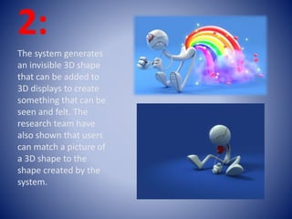
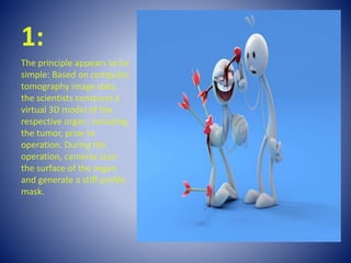
Ethan and José
Explication :
Technology has changed
rapidly over the last few
years with touch feedback,
known as haptics, being
used in entertainment,
rehabilitation and even
surgical training. New
research, using ultrasound,
has developed an invisible
3D haptic shape that can
be seen and felt.
1:
The research, led by Dr
Ben Long and colleagues
Professor Sriram
Subramanian, Sue Ann
Seah and Tom Carter from
the University of Bristol's
Department of Computer
Science, could change the
way 3D shapes are used.
The new technology could
enable surgeons to explore
a CT scan by enabling them
to feel a disease, such as a
tumour, using haptic
feedback.
2:
The system generates
an invisible 3D shape
that can be added to
3D displays to create
something that can be
seen and felt. The
research team have
also shown that users
can match a picture of
a 3D shape to the
shape created by the
system.
3:
"In the future, people
could feel holograms of
objects that would not
otherwise be touchable,
such as feeling the
differences between
materials in a CT scan or
understanding the shapes
of artefacts in a museum.
ORGAN ROBOT
During minimally invasive
operations, a surgeon has to trust
the information displayed on the
screen: A virtual 3D model of the
respective organ shows where a
tumor is located and where
sensitive vessels can be found.
Soft tissue, such as the tissue of
the liver, however, deforms during
breathing or when the scalpel is
applied. Endoscopic cameras
record in real time how the
surface deforms, but do not show
the deformation of deeper
structures such as tumors. Young
scientists have now developed a
real-time capable computation
method to adapt the virtual organ
to the deformed surface profile.
1:
The principle appears to be
simple: Based on computer
tomography image data,
the scientists construct a
virtual 3D model of the
respective organ, including
the tumor, prior to
operation. During the
operation, cameras scan
the surface of the organ
and generate a stiff profile
mask.
2:
Simulations and
experiments using a close-
to-reality phantom liver
have demonstrated that
the electrostatic-elastic
method even works when
only parts of the deformed
surface profile are
available. This is the usual
situation at the hospital
3:
• https://www.youtube.com/
watch?v=1k7XjnGVcG8
The human liver is
surrounded by other
organs and, hence, only
partly visible by
endoscopic cameras. "Only
those structures that are
clearly identified as parts
of the liver by our system
are assigned an electric
charge," says Dr.

Virtual Reality

  • 1.
  • 2.
    Explication : Technology haschanged rapidly over the last few years with touch feedback, known as haptics, being used in entertainment, rehabilitation and even surgical training. New research, using ultrasound, has developed an invisible 3D haptic shape that can be seen and felt.
  • 3.
    1: The research, ledby Dr Ben Long and colleagues Professor Sriram Subramanian, Sue Ann Seah and Tom Carter from the University of Bristol's Department of Computer Science, could change the way 3D shapes are used. The new technology could enable surgeons to explore a CT scan by enabling them to feel a disease, such as a tumour, using haptic feedback.
  • 4.
    2: The system generates aninvisible 3D shape that can be added to 3D displays to create something that can be seen and felt. The research team have also shown that users can match a picture of a 3D shape to the shape created by the system.
  • 5.
    3: "In the future,people could feel holograms of objects that would not otherwise be touchable, such as feeling the differences between materials in a CT scan or understanding the shapes of artefacts in a museum.
  • 6.
    ORGAN ROBOT During minimallyinvasive operations, a surgeon has to trust the information displayed on the screen: A virtual 3D model of the respective organ shows where a tumor is located and where sensitive vessels can be found. Soft tissue, such as the tissue of the liver, however, deforms during breathing or when the scalpel is applied. Endoscopic cameras record in real time how the surface deforms, but do not show the deformation of deeper structures such as tumors. Young scientists have now developed a real-time capable computation method to adapt the virtual organ to the deformed surface profile.
  • 7.
    1: The principle appearsto be simple: Based on computer tomography image data, the scientists construct a virtual 3D model of the respective organ, including the tumor, prior to operation. During the operation, cameras scan the surface of the organ and generate a stiff profile mask.
  • 8.
    2: Simulations and experiments usinga close- to-reality phantom liver have demonstrated that the electrostatic-elastic method even works when only parts of the deformed surface profile are available. This is the usual situation at the hospital
  • 9.
    3: • https://www.youtube.com/ watch?v=1k7XjnGVcG8 The humanliver is surrounded by other organs and, hence, only partly visible by endoscopic cameras. "Only those structures that are clearly identified as parts of the liver by our system are assigned an electric charge," says Dr.