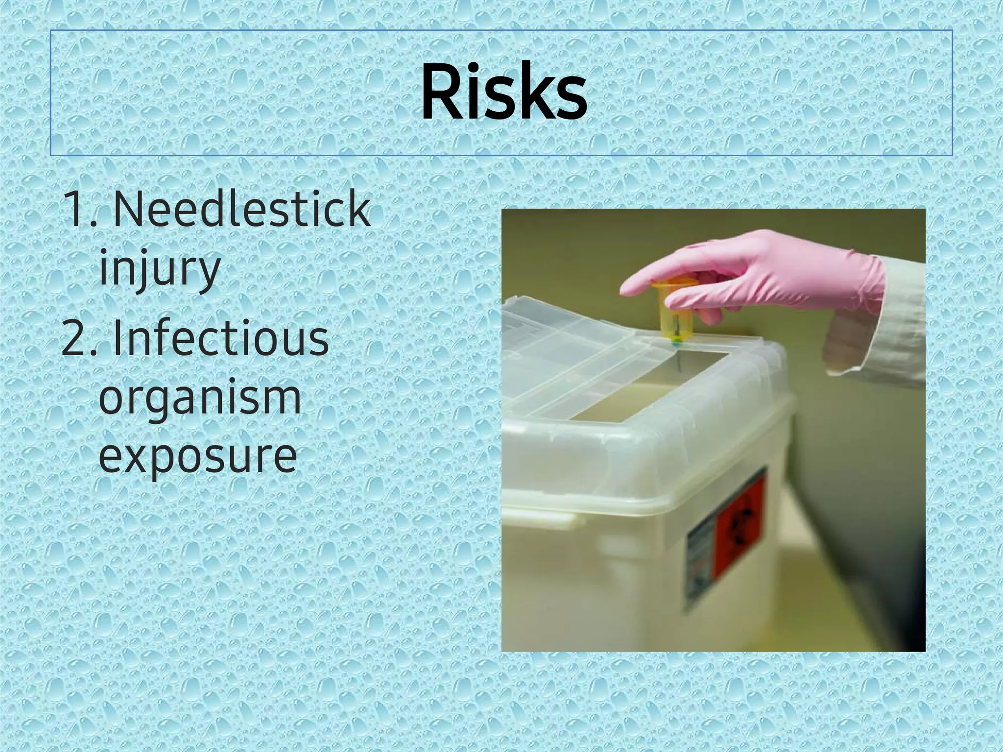
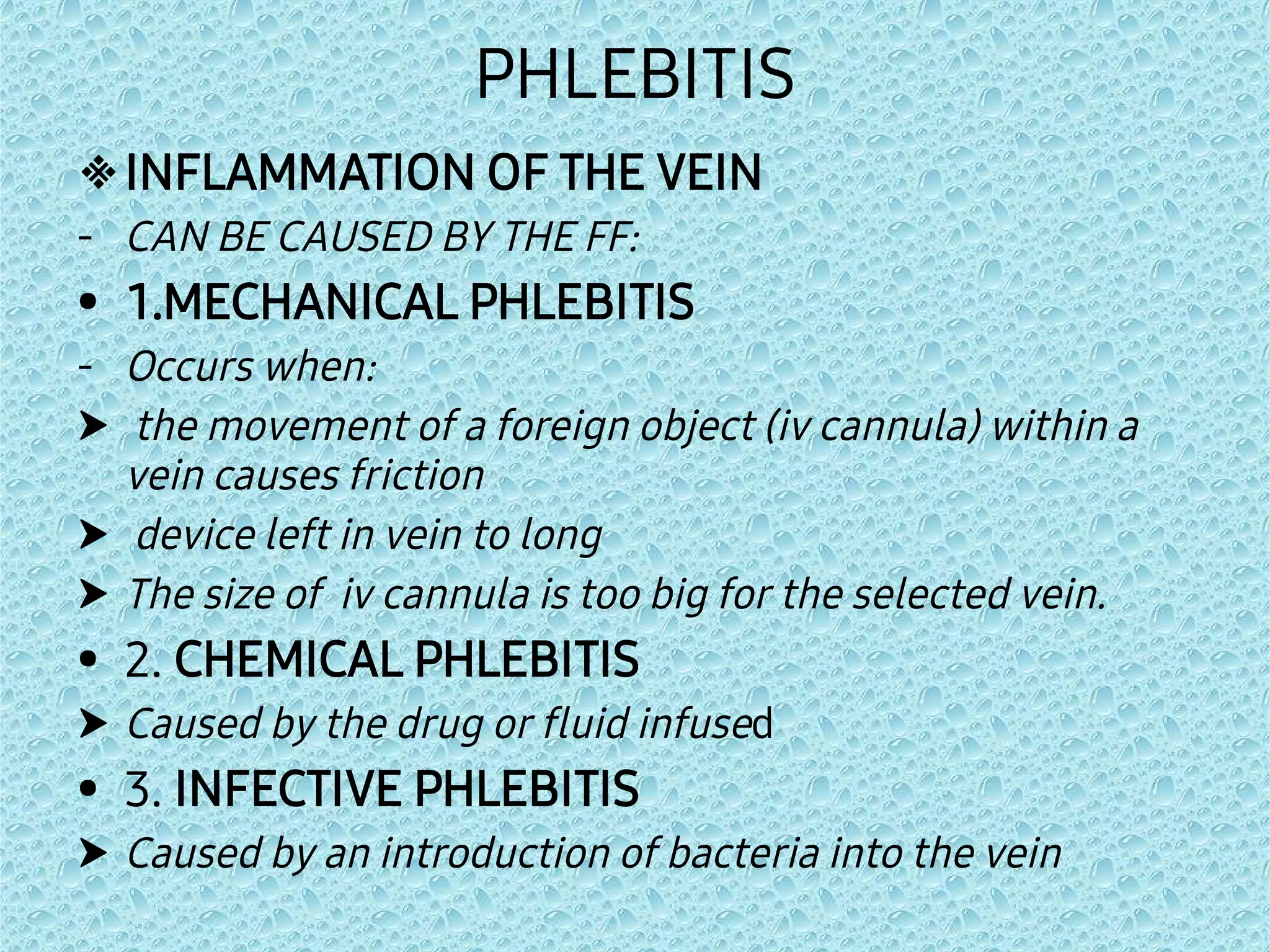
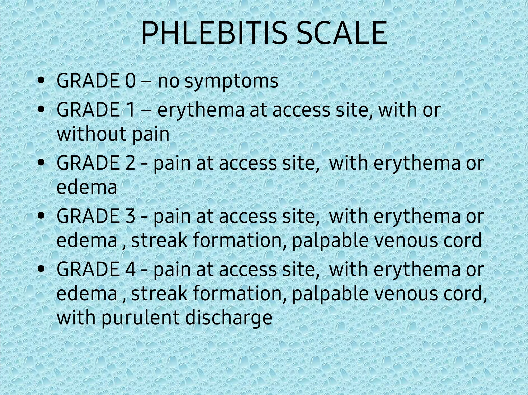
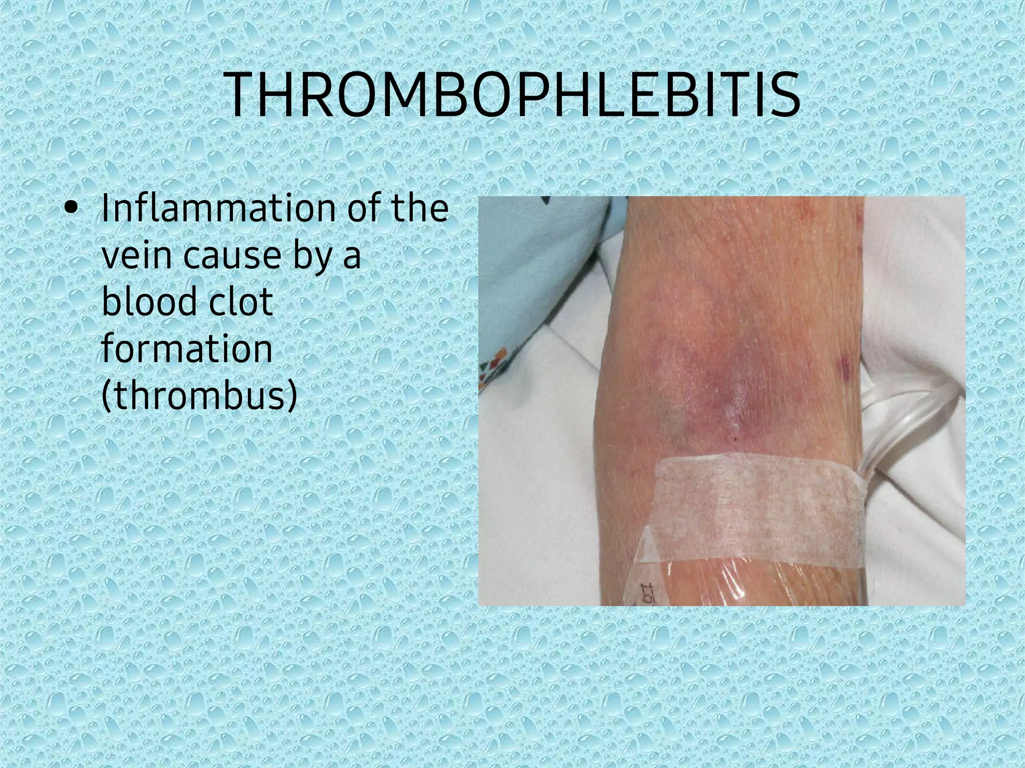
MANAGING COMPLICATIONS
OF IV THERAPY-
PROPERTY OF GESUNDHEITS
Risks Associated
with IVT
Risks
1. Needlestick
injury
2. Infectious
organism
exposure
Needlestick Injury
An AIDS patient became agitated and tried to
remove the intravenous catheters. Hospital
staff struggled to restrain the patient. During
the struggle, an IV infusion line was pulled,
exposing the connector needle. A nurse
recovered the connector needle at the end of
the IV line and attempted to reinsert it. The
patient kicked her arm, pushing the needle into
the hand of the second nurse. Three months
later, the nurse who sustained the needlestick
injury tested positive for HIV1.
Prevention:




Avoid the use of needles where safe and
effective alternatives are available.
Avoid recapping needles.
Report all needlestick and other sharps
related injuries to ensure that you
receive appropriate follow-up care.
Create/maintain a safe, comprehensive
disposal system.
Infectious Organism Exposure




Prevention:
Do proper hand hygiene.
Do not reuse tourniquets.
Wear gloves.
Cleanse insertion sites with the
recommended solutions.
•
–
–
–
–
–
–
–
The following list is a summary of
some of the rules to be observed in
the workplace:
HEPATITIS B vaccine
STANDARD PRECAUTIONS
SHARPS AND WASTE DISPOSAL
PROTECTIVE DEVICE/EQUIPMENT
GLOVES
LAUNDRY
COMMUNICATING HAZARDS
COMPLICATIONS ASSOCIATED
WITH IVT
IV SITE PLACEMENT
PHLEBITIS

-
•
-



•

•

INFLAMMATION OF THE VEIN
CAN BE CAUSED BY THE FF:
1.MECHANICAL PHLEBITIS
Occurs when:
the movement of a foreign object (iv cannula) within a
vein causes friction
device left in vein to long
The size of iv cannula is too big for the selected vein.
2. CHEMICAL PHLEBITIS
Caused by the drug or fluid infused
3. INFECTIVE PHLEBITIS
Caused by an introduction of bacteria into the vein
SIGNS AND SYMPTOMS
•
•
•
Localized redness
and swelling
Pain or burning
along the length of
the vein
Vein being hard and
cord like
PHLEBITIS SCALE
•
•
•
•
•
GRADE 0 – no symptoms
GRADE 1 – erythema at access site, with or
without pain
GRADE 2 - pain at access site, with erythema or
edema
GRADE 3 - pain at access site, with erythema or
edema , streak formation, palpable venous cord
GRADE 4 - pain at access site, with erythema or
edema , streak formation, palpable venous cord,
with purulent discharge




Nursing Management
Stop the infusion at the first sign of redness or
pain.
Apply warm, moist compresses to the affected
area.
Document your patient ’s condition and
interventions.
If indicated,insert a new catheter at a different
site,preferably on the opposite arm,using a larger
vein or a smaller device and restart the infusion




PREVENTIVE MEASURES
Use proper venipuncture technique.
Use a trusted drug reference or consult
with the pharmacist for instructions on
drug dilution,when necessary.
Monitor administration rates and inspect
the I.V. site frequently.
Change the infusion site according to
your institution policy.
THROMBOPHLEBITIS
• Inflammation of the
vein cause by a
blood clot
formation
(thrombus)
SIGNS AND SYMPTOMS
•
•
•
•
REDNESS
SWELLING
HARDENED VEIN
SEVERE DISCOMFORT
-
-
-
-
-
NURSING
INTERVENTION
Remove the device;
restart infusion at
opposite limb if
necessary
Apply warm compress
Watch for ivt related
infection
Notify physician
Document
-
PREVENTIVE MEASURES
Frequent assessment
of iv site
HEMATOMA
-
-
POSSIBLE CAUSES:
Leakage of blood into tissue
Vein punctured through ventral wall at
time of venipuncture
SIGNS AND SYMPTOMS
•
•
BRUISING AROUND
VENIPUNCTURE SITE
TENDERNESS AT
VENIPUNCTURE SITE
-
-
-
-
-
NURSING
INTERVENTIONS
Remove the device
Apply pressure and
cold compress
Recheck the bleeding
Notify the physician
Document
PREVENTIVE MEASURES
- Choose an appropriate
vein and cannula size
- Release the tourniquet
as soon as successful
insertion is achieved
INFILTRATION
-
-
-
LEAKING OF NON-VESICANT INTO THE
SURROUNDING TISSUE
CAUSES:
IMPROPER PLACEMENT OR DISLODGE OF
THE CATHETER
TIP OF THE CATHETER IS POSITIONED
NEAR THE FLEXION AREA
SIGNS AND SYMPTOMS
-
-
-
-
-
-
blanching at site
Cool skin around site
Discomfort, burning
or pain at site
Feeling of tightness
at site
Sluggish flow rate
Swelling (may extend
along entire limb





NURSING INTERVENTIONS
Stop the infusion and remove the device.
Elevate the limb to increase patient
comfort; a compress may be applied.
Check the patient’s pulse and capillary
refill time.
Check the site frequently.
Document your findings and interventions
performed.





PREVENTIVE MEASURES
Select an appropriate I.V. site, avoiding
areas of flexion areas.
Use proper venipuncture technique.
Follow your facility policy for securing, the
I.V. catheter.
Observe the I.V. site frequently.
Advise the patient to report any swelling
or tenderness at the I.V. site.
EXTRAVASATION
- LEAKING OF VESICANT DRUGS INTO
SURROUNDING TISSUE
SIGNS AND SYMPTOMS
-
-
-
- blanching, burning,
discomfort at the iv
site
Cool skin around site
swelling
Blister formation





NURSING INTERVENTIONS
Stop the I.V. flow and remove the I.V. line.
Elevate the affected extremity.
Perform frequent assessments of sensation,
motor function,and circulation of the affected
extremity.
Record the extravasation site,your patient’s
symptoms,the estimated amount of extravasated
solution, and the treatment.
Follow the manufacturer recommendations to
apply either cold or warm compress to the
affected area.





PREVENTIVE MEASURES
Avoid veins that are small or fragile, veins in areas of
flexion, veins in extremities with pre-existing edema.
Be aware of vesicant medications.
Follow your facility policy regarding vesicant
administration via a peripheral I.V.; some institutions
require that vesicants are administered via central
venous access device only.
Give vesicant last when multiple drugs are ordered.
Strictly adhere to proper venipuncture technique.
CATHETER DISLODGEMENT
Possible Causes:
- loosened tape or tubing
- confused patient attempting to
remove it
SIGNS AND SYMPTOMS
•
•
- catheter back out of
the vein
- infusate infiltrating
into tissue
NURSING
INTERVENTIONS
- remove the device,
restart infusion at
opposite limb if
necessary
- notify the physician
- document
PREVENTIVE MEASURES
- secure properly
- use armboard or splint
CIRCULATORY OVERLOAD
-
-
-
Possible Causes:
flow rate too rapid
miscalculation of fluid
requirements
roller clamp loosened
SIGNS AND SYMPTOMS
-
-
-
respiratory distress
increased blood
pressure
large positive fluid
balance ( intake is
greater than output )
-
-
-
-
-
-
NURSING
INTERVENTIONS
administer oxygen as
needed
regulate to kvo as
ordered
raise head of the bead
notify physician
administer
medications as
ordered
document
-
-
-
PREVENTIVE MEASURES
use pump controller
recheck calculations
of fluid
monitor infusion
frequently
ALLERGIC REACTION
Possible Causes:
- Administration of
drugs, iv fluids,
blood products
SIGNS AND SYMPTOMS
-
-
-
-
-
-
skin rashes
fever and chills
flushing
itchiness
respiratory distress
cardiac arrest
-
-
-
-
-
-
NURSING
INTERVENTIONS
Stop infusion
Maintain a patent
airway
Notify physician
Administer
antihistamine, anti-
inflammatory, anti-
pyretics , cortisone
as ordered
Give .2-.5ml
epinephrine
IM(lateral thigh) as
-
-
-
PREVENTIVE MEASURES
obtain the patients
allergy history
assist with test
dosing
monitor patient
during the first
15minutes of
administration of new
drug
SYSTEMIC INFECTION
-
-
POSSIBLE CAUSES:
- Failure to maintain
aseptic non touch
technique
- Immunocompromised
pateint
Prolonged in dwelling
time of device
Severe phlebitis
SIGNS AND SYMPTOMS
• Fever, chills and
body malaise for
no apparent
reason



NURSING INTERVENTIONS
Stop the infusion and notify the
prescriber.
Remove the device, and culture
the site and catheter as ordered.
Administer medications as
prescribed.





PREVENTIVE MEASURES
Perform hand hygiene, don gloves , and use
aseptic non touch technique during I.V. insertion.
Clean the site with approved skin antiseptic
before inserting I.V. catheter.
Ensure careful hand hygiene before any
contact with the infusion system or the patient.
Disinfect injection port before each use.
Follow your institutions policy for dressing
changes and changing of the solution and
administration set.
AIR EMBOLISM
-are caused when gas
bubbles enter blood
vessels.
-
-
POSSIBLE CAUSES:
EMPTY SOLUTION
CONTAINER
DISCONNECTED
TUBING
SIGNS AND SYMPTOMS
•
•
•
•
•
•
•
low blood pressure
irregular heartbeat
extreme fatigue
(tiredness) or lack of
strength
blurred vision
disorientation
cyanosis
irregular breathing



NURSING INTERVNTIONS
Discontinue the infusion.
Administer oxygen as ordered.
Place patient on left side trendelenburg position, this
position helps trap the air in the apex of the right atrium
rather than entering the right ventricle and from there
moving into the pulmonary arterial system.



TREATMENT
HYPERBARIC OXYGEN THERAPY
a non invasive procedure in which the
patient breath 100% oxygen.
the increase pressure in hyperbaric
chamber makes bubbles smaller and
helps push them back into physical
solution,while high oxygen pressure
washes out the gas from the bubble.
.
HYPERBARIC CHAMBER



Once the bubbles
are smaller or
gone, blood flow
resumes.
It allows poorly
oxygenated
tissues to receive
high levels of
oxygen.
Each treatment
session will take
approximately 1-2
hrs per session.
OCCLUSION
-
-
-
-
-
POSSIBLE CAUSES:
Blood back up in the
line
Hypercoagulable
patient
Intermittent device
not flushed
Line clamped too
long
Kink in the IV tubing
or catheter
SIGNS AND SYMPTOMS
•
•
SLUGGISH FLOW RATE
STOPPED IV FLOW
-
-
NURSING
INTERVENTIONS
Use mild flush
pressure during
injection
If unsuccessful, start
another line.
-
-
-
PREVENTIVE MEASURES
Maintain iv flow rate
Flush promptly after
intermittent piggy
back administration
Have the patient walk
with his arm folded to
his chest
VEIN IRRITATION
- caused by
Erosive
medications
or drugs
SIGNS AND SYMPTOMS
•
•
Pain during the
infusion
Red skin over the vein
during infusion
-
-
NURSING
INTERVENTIONS
Slower the flow rate
as ordered
Try using an
electronic flow device
PREVENTIVE MEASURES
- Dilute solutions before
administration
VENOUS SPASM
POSSIBLE CAUSES:
•
•
•
Trauma (from
chemical irritation or
temperature
extremes)
Entrance of the IV
cannula into the vein,
especially if the
cannula is too large
for vein site
Rapid infusate
infusion, particularly
with a small-gauge
SIGNS AND SYMPTOMS
•
•
•
•
Pain (usually sharp and severe) that
radiates from the IV site up the extremity
Redness over vein (indication of phlebitis)
Slowed infusion rate
Stopped infusion
NURSING
INTERVENTIONS
•
•
•
Once spasm is
identified, slow the
infusion rate.
Apply warm
compresses to the
site of spasm.
Discontinue the IV if
spasm continues in
spite of measures
used to stop it.
PREVENTIVE MEASURES
•
•
•
Use blood warmer
IV fluids and
medications should
be administered at
room temperature
Dilute irritating
infusates
NERVE, TENDON, OR LIGAMENT
DAMAGE
•
•
•
•
•
POSSIBLE CAUSE:
incorrect insertion and placement of the IV
cannula
Improper securing and stabilization of the
cannula and infusion line
Extravasation
Infiltration
Anatomic displacement caused by
hematoma
SIGNS AND SYMPTOMS
•
•
•
•
•
•
•
•
•
Extreme pain
Tingling
Numbness
Loss of sensation
Loss of movement
Cyanosis
Pallor
Deformity
Paralysis
•
•
•
•
NURSING
INTERVENTIONS
Stop the procedure
Avoid moving the
cannula back and
forth in the
subcutaneous tissue
in an attempt to find a
vein.
Notify the Physician
Document
•
•
PREVENTIVE MEASURES
Always observe
proper venipunctured
technique
Secure properly
SEVERED CATHETER
POSSIBLE CAUSES:
•
•
Catheter
inadvertently cut by
scissor
Reinsertion of the
needle into the
catheter
SIGNS AND SYMPTOMS
•
•
Leakage from the
catheter shaft
sheared catheter
NURSING INTERVENTIONS
•
•
•
If the broken portion
of the catheter is
visible, attempt to
retrieve if
unsuccessful notify
the physician
If the broken of the
catheter enters the
bloodstream, place a
tourniquet above the
iv site to prevent its
progression
Notify the physician
PREVENTIVE MEASURES
•
•
•
Avoid using scissors
around the iv site
Never reinsert the
needle into the
catheter
Remove
unsuccessfully
inserted catheter and
needle together
Vascular Access Device Related
Infection
•
•
•
•
Possible Causes:
Inadequate skin antisepsis prior to VAD insertion
Multiple manipulation of infusion delivery system
Patient age, condition, acuity
Inadequate care and maintenance practices
SIGNS AND SYMPTOMS
>tenderness of site
>erythema within 2 cm of
catheter-skin junction
>necrosis of skin over
reservoir of implanted port
>acute on set of fever, chills,
and hypotension
Nursing
Interventions
>Apply warm, moist
compress
>Obtain culture of purulent
exudate on form the tip of
catheter
>Apply to topical ointment to
affected area
>Notify the doctor
>Administration oral on
parenteral infective therapy
>Continue to monitor signs
& review laboratory findings
>Perform site care &
maintenance catheter is not
removed
Preventive
Measures
>Perform hand hygiene and
maintain aseptic technique
prior to placing any VAD-
related intervention.
>Disinfect needleless
connectors prior to access
>Remove VAD when no
longer needed
>Teach patients/caregivers
who will set or manage their
VAD
WHAT TO DO WHEN INFUSION
SLOWS DOWN OR STOPS
1.
2.



Assess the I.V. system to locate the problem.
Start at the insertion site. Check for infiltration,
extravasation, or phlebitis.
Check for patency. Obstruction of flow is caused
or affected by the following factors:
2.1 Patients limb is flexed; patient lying on the
side. Reposition limb to release venous pressure.
2.2 Tip of needle or cannula is against the vein
wall. Lift or pull-back the needle or cannula a
little.
2.3 Adhesive taping maybe too tight, release
every apply tapes.
•
•
•
2.4. Small cannulas or tubing may kink or fold,
gently adjust.
2.5. Local edema or poor tissue perfusion from
disease can block venous flow. Transfer I.V. line to
an unaffected site.
2.6. Presence of precipitates in solution either
from incompatibility of fluids and medications or
from infusion. Replace the entire venipuncture
device and solution. It may expose the patient to
embolism.
3. Check the clamps. Some sets have two:
the roller clamp and the side clamp. Check if both
are open or if these are properly adjusted.
4. Check the patency of the air vent;
reposition it if needed
5. Check fluid level: if empty replace
as prescribed. If solution is too cold,
it may cause venous spasm and
decrease the flow; keep room
temperature regulated. Check the
spike of the set; push it more inside
the fluid bag or adjust it.
6. Check filters: ordinary sets usually
do not have in-line filters. If it has,
follow the manufacturer’s guide
instructions. Blood transfusion filters
retain blood product debris.
•
•
•
If flow rate decreases or stops after
more than one unit has been
transfused you may have to change
the set.
7. Check tubings: if patient is lying
on it or if it is kinked or it may be
crimped with too tight roller clamps,
release and round-up the tubing to
its original shape
8. Is gauge of the needle too small?
Is fluid container too low above the
venipuncture site? Adjust it around
36-48 inches above the site.
REFERENCES
•
•
•
Association of Nursing Service
Administrators of the Philippines, Inc.
(ANSAP). 2000. Nursing Standards on
Intravenous Practice 7th EDITION.
Cahil, Matthew. I.V. Therapy made
Incredibly Easy. Springhouse Corporation,
Pennsylvania.
Dionne, Lynn. Manual of I.V. Therapeutics.
Philips, F.A., Davis Co. Philadelphia.
•
•
•
Intravenous Nursing Society, Supplement
to Journal of Intravenous Nursing, Jan./
February 1998 vol.21, Fresh Pond Square,
10 Faucett street, Cambridge, MAO 218.
Lippincott Williams and Wilkins. 2005.
JUST THE FACTS I.V. Therapy.
Nursing Journal May and July 2000.

IVT COMPLICATIONS & MANAGEMENT(1).pdf

  • 1.
    MANAGING COMPLICATIONS OF IVTHERAPY- PROPERTY OF GESUNDHEITS
  • 2.
  • 3.
  • 4.
    Needlestick Injury An AIDSpatient became agitated and tried to remove the intravenous catheters. Hospital staff struggled to restrain the patient. During the struggle, an IV infusion line was pulled, exposing the connector needle. A nurse recovered the connector needle at the end of the IV line and attempted to reinsert it. The patient kicked her arm, pushing the needle into the hand of the second nurse. Three months later, the nurse who sustained the needlestick injury tested positive for HIV1.
  • 6.
    Prevention:     Avoid the useof needles where safe and effective alternatives are available. Avoid recapping needles. Report all needlestick and other sharps related injuries to ensure that you receive appropriate follow-up care. Create/maintain a safe, comprehensive disposal system.
  • 7.
    Infectious Organism Exposure     Prevention: Doproper hand hygiene. Do not reuse tourniquets. Wear gloves. Cleanse insertion sites with the recommended solutions.
  • 8.
    • – – – – – – – The following listis a summary of some of the rules to be observed in the workplace: HEPATITIS B vaccine STANDARD PRECAUTIONS SHARPS AND WASTE DISPOSAL PROTECTIVE DEVICE/EQUIPMENT GLOVES LAUNDRY COMMUNICATING HAZARDS
  • 9.
  • 10.
  • 11.
    PHLEBITIS  - • -    •  •  INFLAMMATION OF THEVEIN CAN BE CAUSED BY THE FF: 1.MECHANICAL PHLEBITIS Occurs when: the movement of a foreign object (iv cannula) within a vein causes friction device left in vein to long The size of iv cannula is too big for the selected vein. 2. CHEMICAL PHLEBITIS Caused by the drug or fluid infused 3. INFECTIVE PHLEBITIS Caused by an introduction of bacteria into the vein
  • 12.
    SIGNS AND SYMPTOMS • • • Localizedredness and swelling Pain or burning along the length of the vein Vein being hard and cord like
  • 13.
    PHLEBITIS SCALE • • • • • GRADE 0– no symptoms GRADE 1 – erythema at access site, with or without pain GRADE 2 - pain at access site, with erythema or edema GRADE 3 - pain at access site, with erythema or edema , streak formation, palpable venous cord GRADE 4 - pain at access site, with erythema or edema , streak formation, palpable venous cord, with purulent discharge
  • 14.
        Nursing Management Stop theinfusion at the first sign of redness or pain. Apply warm, moist compresses to the affected area. Document your patient ’s condition and interventions. If indicated,insert a new catheter at a different site,preferably on the opposite arm,using a larger vein or a smaller device and restart the infusion
  • 15.
        PREVENTIVE MEASURES Use propervenipuncture technique. Use a trusted drug reference or consult with the pharmacist for instructions on drug dilution,when necessary. Monitor administration rates and inspect the I.V. site frequently. Change the infusion site according to your institution policy.
  • 16.
    THROMBOPHLEBITIS • Inflammation ofthe vein cause by a blood clot formation (thrombus)
  • 17.
  • 18.
    - - - - - NURSING INTERVENTION Remove the device; restartinfusion at opposite limb if necessary Apply warm compress Watch for ivt related infection Notify physician Document - PREVENTIVE MEASURES Frequent assessment of iv site
  • 19.
    HEMATOMA - - POSSIBLE CAUSES: Leakage ofblood into tissue Vein punctured through ventral wall at time of venipuncture
  • 20.
    SIGNS AND SYMPTOMS • • BRUISINGAROUND VENIPUNCTURE SITE TENDERNESS AT VENIPUNCTURE SITE
  • 21.
    - - - - - NURSING INTERVENTIONS Remove the device Applypressure and cold compress Recheck the bleeding Notify the physician Document PREVENTIVE MEASURES - Choose an appropriate vein and cannula size - Release the tourniquet as soon as successful insertion is achieved
  • 22.
    INFILTRATION - - - LEAKING OF NON-VESICANTINTO THE SURROUNDING TISSUE CAUSES: IMPROPER PLACEMENT OR DISLODGE OF THE CATHETER TIP OF THE CATHETER IS POSITIONED NEAR THE FLEXION AREA
  • 23.
    SIGNS AND SYMPTOMS - - - - - - blanchingat site Cool skin around site Discomfort, burning or pain at site Feeling of tightness at site Sluggish flow rate Swelling (may extend along entire limb
  • 24.
         NURSING INTERVENTIONS Stop theinfusion and remove the device. Elevate the limb to increase patient comfort; a compress may be applied. Check the patient’s pulse and capillary refill time. Check the site frequently. Document your findings and interventions performed.
  • 25.
         PREVENTIVE MEASURES Select anappropriate I.V. site, avoiding areas of flexion areas. Use proper venipuncture technique. Follow your facility policy for securing, the I.V. catheter. Observe the I.V. site frequently. Advise the patient to report any swelling or tenderness at the I.V. site.
  • 26.
    EXTRAVASATION - LEAKING OFVESICANT DRUGS INTO SURROUNDING TISSUE
  • 27.
    SIGNS AND SYMPTOMS - - - -blanching, burning, discomfort at the iv site Cool skin around site swelling Blister formation
  • 28.
         NURSING INTERVENTIONS Stop theI.V. flow and remove the I.V. line. Elevate the affected extremity. Perform frequent assessments of sensation, motor function,and circulation of the affected extremity. Record the extravasation site,your patient’s symptoms,the estimated amount of extravasated solution, and the treatment. Follow the manufacturer recommendations to apply either cold or warm compress to the affected area.
  • 29.
         PREVENTIVE MEASURES Avoid veinsthat are small or fragile, veins in areas of flexion, veins in extremities with pre-existing edema. Be aware of vesicant medications. Follow your facility policy regarding vesicant administration via a peripheral I.V.; some institutions require that vesicants are administered via central venous access device only. Give vesicant last when multiple drugs are ordered. Strictly adhere to proper venipuncture technique.
  • 30.
    CATHETER DISLODGEMENT Possible Causes: -loosened tape or tubing - confused patient attempting to remove it
  • 31.
    SIGNS AND SYMPTOMS • • -catheter back out of the vein - infusate infiltrating into tissue
  • 32.
    NURSING INTERVENTIONS - remove thedevice, restart infusion at opposite limb if necessary - notify the physician - document PREVENTIVE MEASURES - secure properly - use armboard or splint
  • 33.
    CIRCULATORY OVERLOAD - - - Possible Causes: flowrate too rapid miscalculation of fluid requirements roller clamp loosened
  • 34.
    SIGNS AND SYMPTOMS - - - respiratorydistress increased blood pressure large positive fluid balance ( intake is greater than output )
  • 35.
    - - - - - - NURSING INTERVENTIONS administer oxygen as needed regulateto kvo as ordered raise head of the bead notify physician administer medications as ordered document - - - PREVENTIVE MEASURES use pump controller recheck calculations of fluid monitor infusion frequently
  • 36.
    ALLERGIC REACTION Possible Causes: -Administration of drugs, iv fluids, blood products
  • 37.
    SIGNS AND SYMPTOMS - - - - - - skinrashes fever and chills flushing itchiness respiratory distress cardiac arrest
  • 38.
    - - - - - - NURSING INTERVENTIONS Stop infusion Maintain apatent airway Notify physician Administer antihistamine, anti- inflammatory, anti- pyretics , cortisone as ordered Give .2-.5ml epinephrine IM(lateral thigh) as - - - PREVENTIVE MEASURES obtain the patients allergy history assist with test dosing monitor patient during the first 15minutes of administration of new drug
  • 39.
    SYSTEMIC INFECTION - - POSSIBLE CAUSES: -Failure to maintain aseptic non touch technique - Immunocompromised pateint Prolonged in dwelling time of device Severe phlebitis
  • 40.
    SIGNS AND SYMPTOMS •Fever, chills and body malaise for no apparent reason
  • 41.
       NURSING INTERVENTIONS Stop theinfusion and notify the prescriber. Remove the device, and culture the site and catheter as ordered. Administer medications as prescribed.
  • 42.
         PREVENTIVE MEASURES Perform handhygiene, don gloves , and use aseptic non touch technique during I.V. insertion. Clean the site with approved skin antiseptic before inserting I.V. catheter. Ensure careful hand hygiene before any contact with the infusion system or the patient. Disinfect injection port before each use. Follow your institutions policy for dressing changes and changing of the solution and administration set.
  • 43.
    AIR EMBOLISM -are causedwhen gas bubbles enter blood vessels. - - POSSIBLE CAUSES: EMPTY SOLUTION CONTAINER DISCONNECTED TUBING
  • 44.
    SIGNS AND SYMPTOMS • • • • • • • lowblood pressure irregular heartbeat extreme fatigue (tiredness) or lack of strength blurred vision disorientation cyanosis irregular breathing
  • 45.
       NURSING INTERVNTIONS Discontinue theinfusion. Administer oxygen as ordered. Place patient on left side trendelenburg position, this position helps trap the air in the apex of the right atrium rather than entering the right ventricle and from there moving into the pulmonary arterial system.
  • 46.
       TREATMENT HYPERBARIC OXYGEN THERAPY anon invasive procedure in which the patient breath 100% oxygen. the increase pressure in hyperbaric chamber makes bubbles smaller and helps push them back into physical solution,while high oxygen pressure washes out the gas from the bubble. .
  • 47.
    HYPERBARIC CHAMBER    Once thebubbles are smaller or gone, blood flow resumes. It allows poorly oxygenated tissues to receive high levels of oxygen. Each treatment session will take approximately 1-2 hrs per session.
  • 48.
    OCCLUSION - - - - - POSSIBLE CAUSES: Blood backup in the line Hypercoagulable patient Intermittent device not flushed Line clamped too long Kink in the IV tubing or catheter
  • 49.
    SIGNS AND SYMPTOMS • • SLUGGISHFLOW RATE STOPPED IV FLOW
  • 50.
    - - NURSING INTERVENTIONS Use mild flush pressureduring injection If unsuccessful, start another line. - - - PREVENTIVE MEASURES Maintain iv flow rate Flush promptly after intermittent piggy back administration Have the patient walk with his arm folded to his chest
  • 51.
    VEIN IRRITATION - causedby Erosive medications or drugs
  • 52.
    SIGNS AND SYMPTOMS • • Painduring the infusion Red skin over the vein during infusion
  • 53.
    - - NURSING INTERVENTIONS Slower the flowrate as ordered Try using an electronic flow device PREVENTIVE MEASURES - Dilute solutions before administration
  • 54.
    VENOUS SPASM POSSIBLE CAUSES: • • • Trauma(from chemical irritation or temperature extremes) Entrance of the IV cannula into the vein, especially if the cannula is too large for vein site Rapid infusate infusion, particularly with a small-gauge
  • 55.
    SIGNS AND SYMPTOMS • • • • Pain(usually sharp and severe) that radiates from the IV site up the extremity Redness over vein (indication of phlebitis) Slowed infusion rate Stopped infusion
  • 56.
    NURSING INTERVENTIONS • • • Once spasm is identified,slow the infusion rate. Apply warm compresses to the site of spasm. Discontinue the IV if spasm continues in spite of measures used to stop it. PREVENTIVE MEASURES • • • Use blood warmer IV fluids and medications should be administered at room temperature Dilute irritating infusates
  • 57.
    NERVE, TENDON, ORLIGAMENT DAMAGE • • • • • POSSIBLE CAUSE: incorrect insertion and placement of the IV cannula Improper securing and stabilization of the cannula and infusion line Extravasation Infiltration Anatomic displacement caused by hematoma
  • 58.
    SIGNS AND SYMPTOMS • • • • • • • • • Extremepain Tingling Numbness Loss of sensation Loss of movement Cyanosis Pallor Deformity Paralysis
  • 59.
    • • • • NURSING INTERVENTIONS Stop the procedure Avoidmoving the cannula back and forth in the subcutaneous tissue in an attempt to find a vein. Notify the Physician Document • • PREVENTIVE MEASURES Always observe proper venipunctured technique Secure properly
  • 60.
    SEVERED CATHETER POSSIBLE CAUSES: • • Catheter inadvertentlycut by scissor Reinsertion of the needle into the catheter
  • 61.
    SIGNS AND SYMPTOMS • • Leakagefrom the catheter shaft sheared catheter
  • 62.
    NURSING INTERVENTIONS • • • If thebroken portion of the catheter is visible, attempt to retrieve if unsuccessful notify the physician If the broken of the catheter enters the bloodstream, place a tourniquet above the iv site to prevent its progression Notify the physician PREVENTIVE MEASURES • • • Avoid using scissors around the iv site Never reinsert the needle into the catheter Remove unsuccessfully inserted catheter and needle together
  • 63.
    Vascular Access DeviceRelated Infection • • • • Possible Causes: Inadequate skin antisepsis prior to VAD insertion Multiple manipulation of infusion delivery system Patient age, condition, acuity Inadequate care and maintenance practices
  • 64.
    SIGNS AND SYMPTOMS >tendernessof site >erythema within 2 cm of catheter-skin junction >necrosis of skin over reservoir of implanted port >acute on set of fever, chills, and hypotension
  • 65.
    Nursing Interventions >Apply warm, moist compress >Obtainculture of purulent exudate on form the tip of catheter >Apply to topical ointment to affected area >Notify the doctor >Administration oral on parenteral infective therapy >Continue to monitor signs & review laboratory findings >Perform site care & maintenance catheter is not removed Preventive Measures >Perform hand hygiene and maintain aseptic technique prior to placing any VAD- related intervention. >Disinfect needleless connectors prior to access >Remove VAD when no longer needed >Teach patients/caregivers who will set or manage their VAD
  • 66.
    WHAT TO DOWHEN INFUSION SLOWS DOWN OR STOPS
  • 67.
    1. 2.    Assess the I.V.system to locate the problem. Start at the insertion site. Check for infiltration, extravasation, or phlebitis. Check for patency. Obstruction of flow is caused or affected by the following factors: 2.1 Patients limb is flexed; patient lying on the side. Reposition limb to release venous pressure. 2.2 Tip of needle or cannula is against the vein wall. Lift or pull-back the needle or cannula a little. 2.3 Adhesive taping maybe too tight, release every apply tapes.
  • 68.
    • • • 2.4. Small cannulasor tubing may kink or fold, gently adjust. 2.5. Local edema or poor tissue perfusion from disease can block venous flow. Transfer I.V. line to an unaffected site. 2.6. Presence of precipitates in solution either from incompatibility of fluids and medications or from infusion. Replace the entire venipuncture device and solution. It may expose the patient to embolism. 3. Check the clamps. Some sets have two: the roller clamp and the side clamp. Check if both are open or if these are properly adjusted.
  • 69.
    4. Check thepatency of the air vent; reposition it if needed 5. Check fluid level: if empty replace as prescribed. If solution is too cold, it may cause venous spasm and decrease the flow; keep room temperature regulated. Check the spike of the set; push it more inside the fluid bag or adjust it. 6. Check filters: ordinary sets usually do not have in-line filters. If it has, follow the manufacturer’s guide instructions. Blood transfusion filters retain blood product debris.
  • 70.
    • • • If flow ratedecreases or stops after more than one unit has been transfused you may have to change the set. 7. Check tubings: if patient is lying on it or if it is kinked or it may be crimped with too tight roller clamps, release and round-up the tubing to its original shape 8. Is gauge of the needle too small? Is fluid container too low above the venipuncture site? Adjust it around 36-48 inches above the site.
  • 71.
    REFERENCES • • • Association of NursingService Administrators of the Philippines, Inc. (ANSAP). 2000. Nursing Standards on Intravenous Practice 7th EDITION. Cahil, Matthew. I.V. Therapy made Incredibly Easy. Springhouse Corporation, Pennsylvania. Dionne, Lynn. Manual of I.V. Therapeutics. Philips, F.A., Davis Co. Philadelphia.
  • 72.
    • • • Intravenous Nursing Society,Supplement to Journal of Intravenous Nursing, Jan./ February 1998 vol.21, Fresh Pond Square, 10 Faucett street, Cambridge, MAO 218. Lippincott Williams and Wilkins. 2005. JUST THE FACTS I.V. Therapy. Nursing Journal May and July 2000.