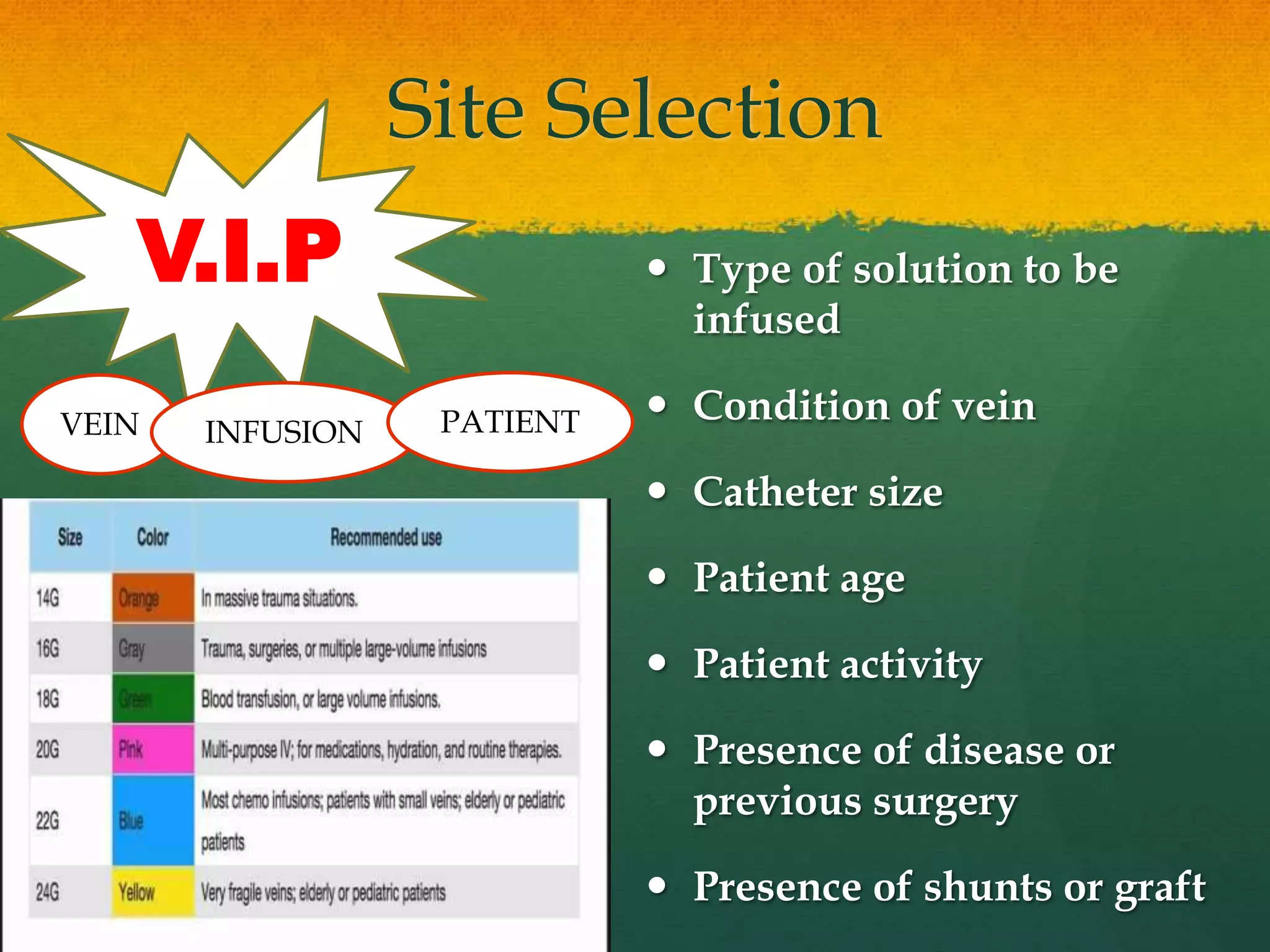
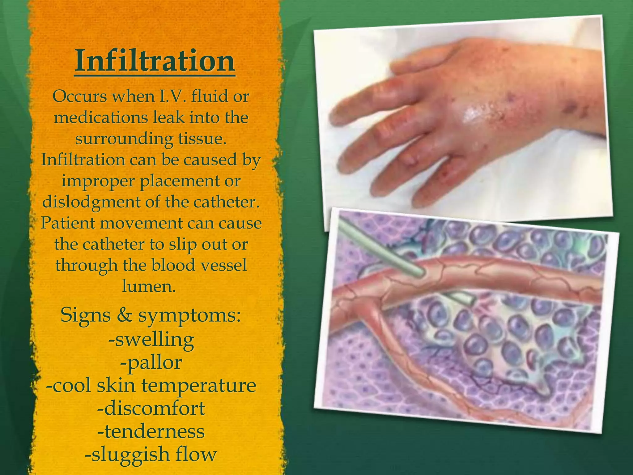
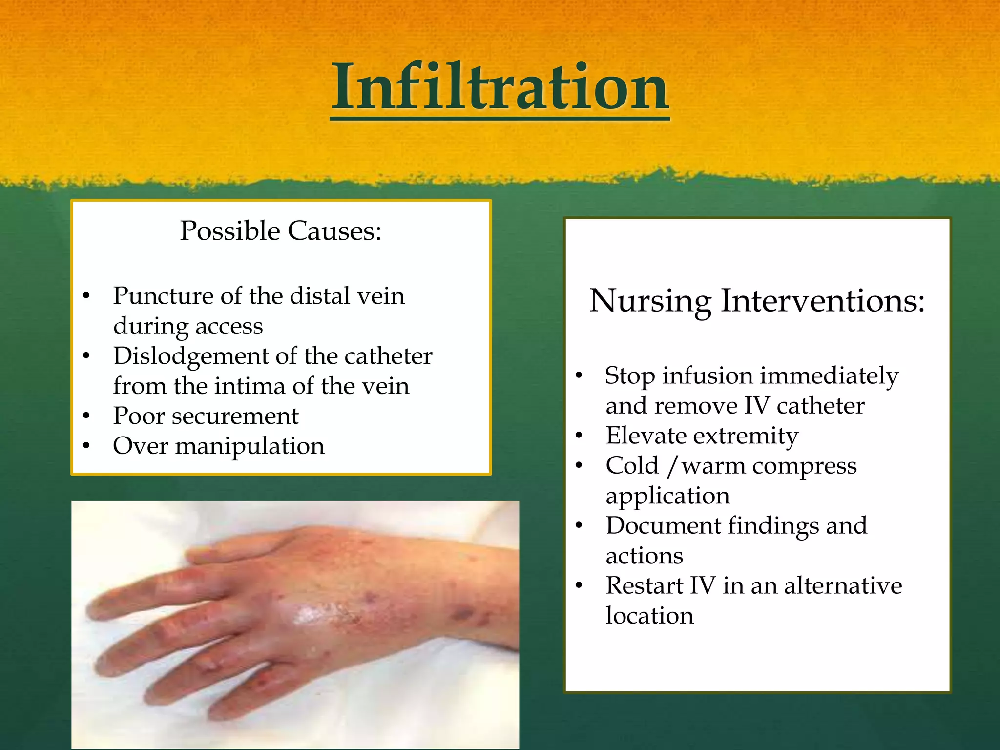
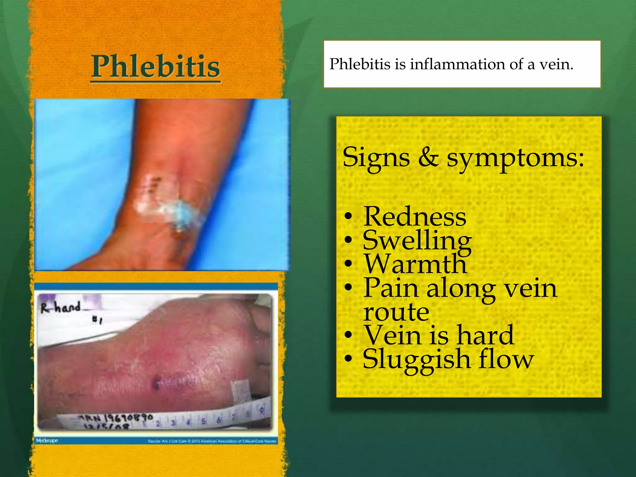
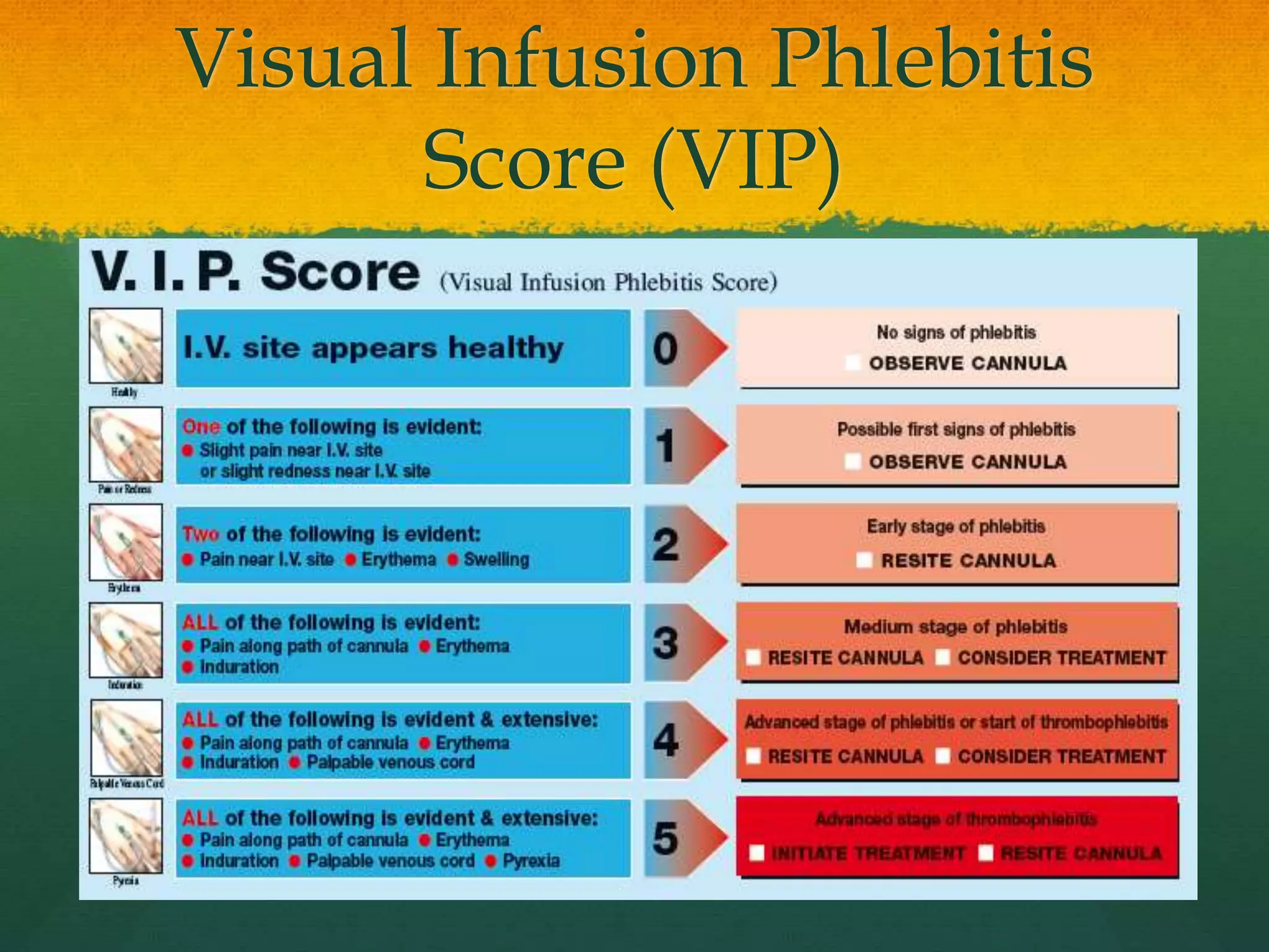
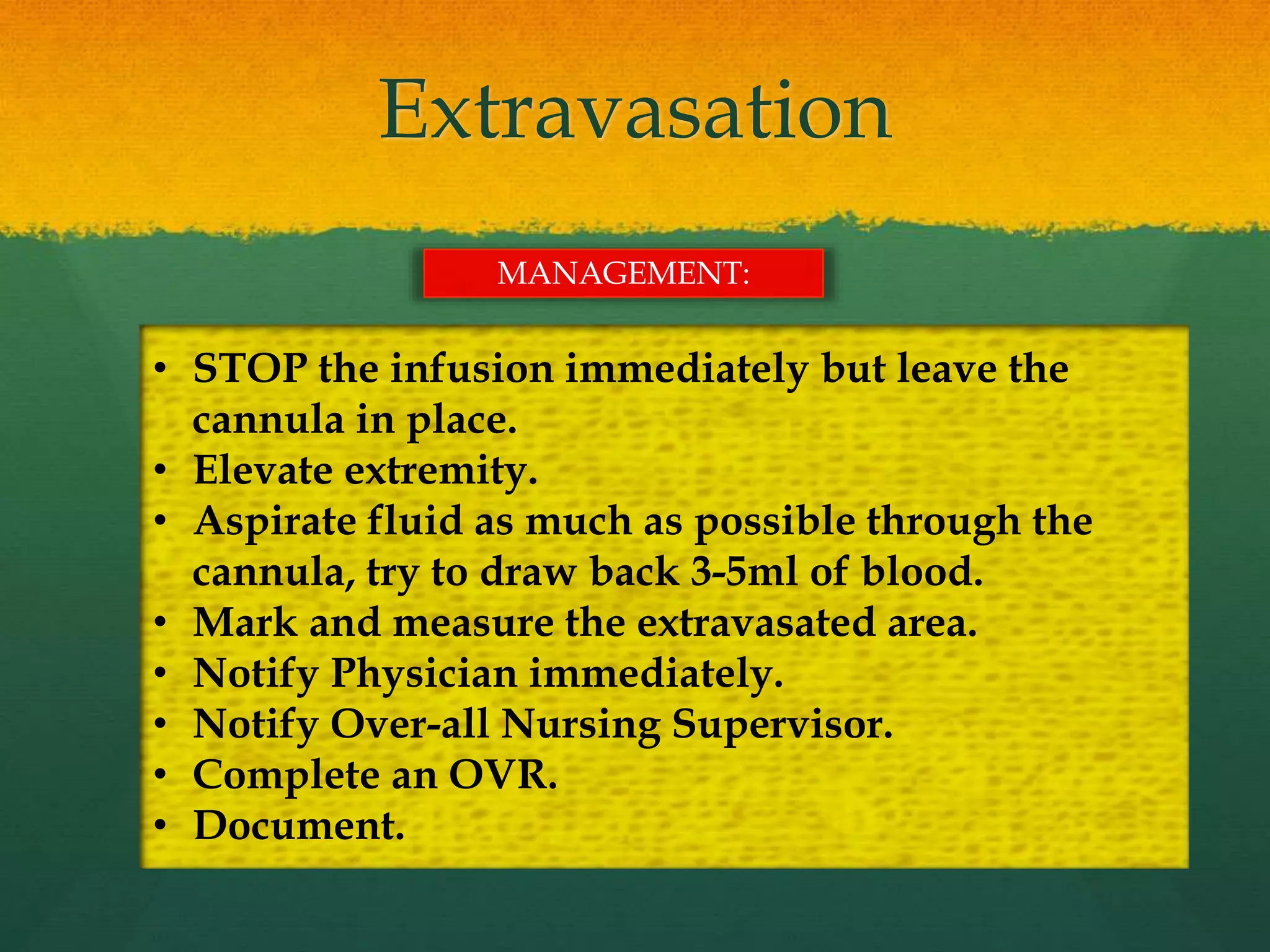
IV Therapy: Indications and
Complications
MICHELLE ANTIMANO, RN
INTRODUCTION
 It is estimated that over 85% of hospitalized patients
have an IV therapy during their stay in the hospital.
 At least 2% of medical lawsuits involve a
complication from a peripheral IV line.
Vein Anatomy
Site Selection
 Type of solution to be
infused
 Condition of vein
 Catheter size
 Patient age
 Patient activity
 Presence of disease or
previous surgery
 Presence of shunts or graft
V.I.P
VEIN INFUSION PATIENT
Vein dilation techniques
 Tourniquet.
 Gravity.
 Fist clenching
 Tapping vein
 Warm compresses
 Blood pressure cuff
 Multiple tourniquet technique
Indications
Fluid and electrolyte
maintenance, restoration
and replacement
To establish a lifeline for
rapidly needed
medications
Blood and blood products
administration
Medication and nutrition
administration
Complications
Local Complications Systemic Complications
Occur at the
insertion site of
an IV device or
close at an IV
site
Assessing and
monitoring are
the key
components to
early
intervention.
Occur within the
vascular system,
remote from the
IV site
Can be life
threatening.
Infiltration
Occurs when I.V. fluid or
medications leak into the
surrounding tissue.
Infiltration can be caused by
improper placement or
dislodgment of the catheter.
Patient movement can cause
the catheter to slip out or
through the blood vessel
lumen.
Signs & symptoms:
-swelling
-pallor
-cool skin temperature
-discomfort
-tenderness
-sluggish flow
Infiltration
Possible Causes:
• Puncture of the distal vein
during access
• Dislodgement of the catheter
from the intima of the vein
• Poor securement
• Over manipulation
Nursing Interventions:
• Stop infusion immediately
and remove IV catheter
• Elevate extremity
• Cold /warm compress
application
• Document findings and
actions
• Restart IV in an alternative
location
Phlebitis Phlebitis is inflammation of a vein.
Signs & symptoms:
• Redness
• Swelling
• Warmth
• Pain along vein
route
• Vein is hard
• Sluggish flow
Mechanical Phlebitis
Possible causes:
• Cannula too large for
vein
• Cannula inserted near
a joint creating a piston
motion against vein
wall when patients
moves
• Inadequate dressing
and securement
Chemical Phlebitis
Possible causes:
• Infusion of Alkaline
solutions
• Infusion of
hyper/hypotonic
solutions
• Speed and method
of infusion delivery
Bacterial Phlebitis
Possible causes:
• Break in aseptic technique
during insertion or
routine care
• Inadequate skin
preparation
• Use of contaminated
supplies, solutions or
medications
• Cannula in situ passed
date of expiry
Management:
• Remove IV cannula and reinsert in new location
• Apply warm compress
• Notify Physician
• Notify Over-all Nursing Supervisor
• Document assessments and actions
Visual Infusion Phlebitis
Score (VIP)
Hematoma
Hematoma occur when blood leaks into the extravascular space.
Possible causes:
• Related to
venipuncture
technique
• Trauma to the vein
during insertion
• Discontinuing IV
without applying
adequate pressure
• Signs and
symptoms:
• Discoloration or
bruising of the
skin around site
• Site swelling and
discomfort
• Inability to
advance or flush
IV line
Management:
• Remove venipuncture device.
• Apply pressure and warm soaks to
• affected area and recheck for bleeding
• Document patient’s condition and your
interventions
Extravasation
Inadvertent administration of a vesicant solution into surrounding tissue.
Signs and symptoms:
• Swelling
• Burning or pain at the
insertion site
• Slow or stopped infusion
Vesicant- is a fluid or
medication that causes
formation of blisters,
with subsequent
sloughing of tissues
occurring from the tissue
necrosis.
Examples of Vesicants
 Vancomycin
 Nafcillin
 Calcium Chloride
 Potassium Chloride
 Sodium Chloride
 Calcium Gluconate
 Dobutamine
 Diazepam
 Dopamine
 Norepinephrine
 Phenytoin
 Promethazine
 Propofol
 Vasopressin
 Radiologic contrast agents
 Chemotherapeutic agents
Extravasation
• STOP the infusion immediately but leave the
cannula in place.
• Elevate extremity.
• Aspirate fluid as much as possible through the
cannula, try to draw back 3-5ml of blood.
• Mark and measure the extravasated area.
• Notify Physician immediately.
• Notify Over-all Nursing Supervisor.
• Complete an OVR.
• Document.
MANAGEMENT:
Air Embolism
An air embolism takes place when a bubbles of air introduced into the circulating blood.
Occurs most frequently with central venous
devices
Signs and symptoms:
• Dyspnea, cyanosis, hypotension
• Weak, rapid pulse
• Chest pain
Loss of consciousness
Management:
• Immediate corrective action for suspected Air
embolism includes:
• Stop infusion by clamping the line
• Place patient on left trendelenburg position.
• Administer oxygen.
• Notify your immediate supervisor and
physician immediately.
• Document assessments and interventions.
Fluid Overload
An excess of fluid disrupting homeostasis caused by an infusion at a rate greater
than the patient’s system is able to accommodate.
Signs and symptoms:
• Tachypnea, dyspnea
• Tachycardia
• Hypertension
• Raised CVP measurement and
distended neck veins
Management:
• Stop the infusion.
• Notify physician.
• Administer treatment as ordered.
• Document assessments and actions.
Septicemia
Systemic infection can occur as a result of phlebitis, poor securement
of venipuncture, prolonged dwell time of catheter and failure to
maintain aseptic technique during insertion or site care.
Signs and symptoms:
• Redness, swelling and
pain at site
• Fever, chills and malaise
• Tachycardia
Management:
• Notify the physician.
• Monitor vital signs.
• Administer medications as
prescribed.
• Culture the site and the
device.
IV Therapy

IV Therapy

  • 1.
    IV Therapy: Indicationsand Complications MICHELLE ANTIMANO, RN
  • 2.
    INTRODUCTION  It isestimated that over 85% of hospitalized patients have an IV therapy during their stay in the hospital.  At least 2% of medical lawsuits involve a complication from a peripheral IV line.
  • 3.
  • 4.
    Site Selection  Typeof solution to be infused  Condition of vein  Catheter size  Patient age  Patient activity  Presence of disease or previous surgery  Presence of shunts or graft V.I.P VEIN INFUSION PATIENT
  • 5.
    Vein dilation techniques Tourniquet.  Gravity.  Fist clenching  Tapping vein  Warm compresses  Blood pressure cuff  Multiple tourniquet technique
  • 6.
    Indications Fluid and electrolyte maintenance,restoration and replacement To establish a lifeline for rapidly needed medications Blood and blood products administration Medication and nutrition administration
  • 7.
    Complications Local Complications SystemicComplications Occur at the insertion site of an IV device or close at an IV site Assessing and monitoring are the key components to early intervention. Occur within the vascular system, remote from the IV site Can be life threatening.
  • 8.
    Infiltration Occurs when I.V.fluid or medications leak into the surrounding tissue. Infiltration can be caused by improper placement or dislodgment of the catheter. Patient movement can cause the catheter to slip out or through the blood vessel lumen. Signs & symptoms: -swelling -pallor -cool skin temperature -discomfort -tenderness -sluggish flow
  • 9.
    Infiltration Possible Causes: • Punctureof the distal vein during access • Dislodgement of the catheter from the intima of the vein • Poor securement • Over manipulation Nursing Interventions: • Stop infusion immediately and remove IV catheter • Elevate extremity • Cold /warm compress application • Document findings and actions • Restart IV in an alternative location
  • 10.
    Phlebitis Phlebitis isinflammation of a vein. Signs & symptoms: • Redness • Swelling • Warmth • Pain along vein route • Vein is hard • Sluggish flow
  • 11.
    Mechanical Phlebitis Possible causes: •Cannula too large for vein • Cannula inserted near a joint creating a piston motion against vein wall when patients moves • Inadequate dressing and securement Chemical Phlebitis Possible causes: • Infusion of Alkaline solutions • Infusion of hyper/hypotonic solutions • Speed and method of infusion delivery Bacterial Phlebitis Possible causes: • Break in aseptic technique during insertion or routine care • Inadequate skin preparation • Use of contaminated supplies, solutions or medications • Cannula in situ passed date of expiry Management: • Remove IV cannula and reinsert in new location • Apply warm compress • Notify Physician • Notify Over-all Nursing Supervisor • Document assessments and actions
  • 12.
  • 13.
    Hematoma Hematoma occur whenblood leaks into the extravascular space. Possible causes: • Related to venipuncture technique • Trauma to the vein during insertion • Discontinuing IV without applying adequate pressure • Signs and symptoms: • Discoloration or bruising of the skin around site • Site swelling and discomfort • Inability to advance or flush IV line Management: • Remove venipuncture device. • Apply pressure and warm soaks to • affected area and recheck for bleeding • Document patient’s condition and your interventions
  • 14.
    Extravasation Inadvertent administration ofa vesicant solution into surrounding tissue. Signs and symptoms: • Swelling • Burning or pain at the insertion site • Slow or stopped infusion Vesicant- is a fluid or medication that causes formation of blisters, with subsequent sloughing of tissues occurring from the tissue necrosis.
  • 15.
    Examples of Vesicants Vancomycin  Nafcillin  Calcium Chloride  Potassium Chloride  Sodium Chloride  Calcium Gluconate  Dobutamine  Diazepam  Dopamine  Norepinephrine  Phenytoin  Promethazine  Propofol  Vasopressin  Radiologic contrast agents  Chemotherapeutic agents
  • 16.
    Extravasation • STOP theinfusion immediately but leave the cannula in place. • Elevate extremity. • Aspirate fluid as much as possible through the cannula, try to draw back 3-5ml of blood. • Mark and measure the extravasated area. • Notify Physician immediately. • Notify Over-all Nursing Supervisor. • Complete an OVR. • Document. MANAGEMENT:
  • 17.
    Air Embolism An airembolism takes place when a bubbles of air introduced into the circulating blood. Occurs most frequently with central venous devices Signs and symptoms: • Dyspnea, cyanosis, hypotension • Weak, rapid pulse • Chest pain Loss of consciousness Management: • Immediate corrective action for suspected Air embolism includes: • Stop infusion by clamping the line • Place patient on left trendelenburg position. • Administer oxygen. • Notify your immediate supervisor and physician immediately. • Document assessments and interventions.
  • 18.
    Fluid Overload An excessof fluid disrupting homeostasis caused by an infusion at a rate greater than the patient’s system is able to accommodate. Signs and symptoms: • Tachypnea, dyspnea • Tachycardia • Hypertension • Raised CVP measurement and distended neck veins Management: • Stop the infusion. • Notify physician. • Administer treatment as ordered. • Document assessments and actions.
  • 19.
    Septicemia Systemic infection canoccur as a result of phlebitis, poor securement of venipuncture, prolonged dwell time of catheter and failure to maintain aseptic technique during insertion or site care. Signs and symptoms: • Redness, swelling and pain at site • Fever, chills and malaise • Tachycardia Management: • Notify the physician. • Monitor vital signs. • Administer medications as prescribed. • Culture the site and the device.

Editor's Notes

  • #2 Salamalaikom everyone! The purpose of this lecture is to provide a brief overview of IVT, including care and potential complications.
  • #3 IVT is very common practice , particularly with patients in acute care settings. Is estimated that over 85% of hospitalized patients have an IVT at some time during their stay, and that at least 2% of medical lawsuits involve a complication from a peripheral IV line. It is important that nurses understand the theory behind initiating and maintaining an IV, including familiarity with anatomy, selection of equipment assessment and prevention of potential complications.
  • #4 It is important for nurses to become familiar with the anatomy of blood vessels and blood flow, specially with regards to the venous system and the administration of IV therapy. Understanding the anatomy of vein will help to facilitate appropriate decisions about the placement and maintenance of an IV catheter. Each vein is composed of 3 layers: The innermost layer is called tunica intima-it is composed of elastic membrane lining and smooth endothelium, the muscular middle layer is tunica media, it is composed of smooth muscles and elastic fibers and the outermost part is called tunica tunica adventitia or externa and composed of connective tissues. Veins also contain valves that provide foothold for the blood as it travels against gravity. Venous valves and the muscles of the leg contract to helpprevent backflow from occuring and facilitate the flow back to the heart.
  • #5 IN SELECTING THE BEST SITE FOR VENIPUNCTURE, REMEMBER THE VIP. VEIN, INFUSION, PATIENT. VEIN- FOR THE VEIN CONSIDER ITS LOCATION, CONDITION AND PHYSICAL PATH ALONG ITS ENSURITY. FOR THE INFUSION, CONSIDER THE PURPOSE AND DURATION. FOR THE PATIENT COSIDER THE DEGREE OF COOPERATION AND COMPLIANCE AND PREFERENCE. THE INFUSION NURSES SOCIETY STANDARD AND PRACTICE STATES THAT THE CATHETER SHOULD BE THE SMALLEST SIZE AND SHORTEST LINK THAT WOULD ACCOMMODATE THE PRESCRIBE THERAPY There are several factors you need to consider before initiating venipunctures: Type of solution to be infused. Hypertonic solutions and medications are irritating to vein Condition of vein choose a vein that soft, straight and bouncy. Avoid veins that are near to previously infected sites. Catheter size the choice of cannula should be of the smallest gauge that will accommodate the prescribed therapy. Patient age. Elderly and children need additional time for assessment and management of insertion. Presence of disease or previous surgery Patients with vascular disease or dehydration may have limited venous access. If a patient has a condition causing poor vascular return (mastectomy, stroke) he affected side must be avoided. Do not use the arm or hand that has a patent graft or shunt for dialysis.
  • #6 Use the techniques below to dilate the vein: Torniquet Latex or nonlatex used most frequently. Placed 6–8 inches above the venipuncture site. If BP high, move farther from venipuncture site. If BP low, move as close as possible without risking site contamination. Gravity. Position the extremity lower than the heart. Fist clenching. Instruct patient to open and close his/her fist. Tapping vein. Using thumb and second finger, flick the vein; this releases histamines beneath the skin and causes dilation. Do not slap the vein. Warm compresses. 10 minutes maximum. Bp cuff. Inflate to 30 mmHg; great for fragile veins. Multiple tourniquet technique. Use 2 to 3 latex tourniquets; apply one high on arm and leave for 2 minutes; apply second at mid arm below antecubital fossa; collateral veins should appear; apply third if needed.
  • #8 As with any invasive procedure, there are potential complications that can occur with IVT these complications will be reviewed , including assessment and management. As with any invasive procedure, there are potential complications that can occur with IVT these complications will be reviewed , including assessment and management.
  • #9 Infiltration is the inadvertent leakage of nonvesicant solution into the surrounding tissue.
  • #10 OCCURS WHEN IV FLUID LEAKS INTO SORROUNDING TISSUES. Causes of Infiltration include improper insertion of IV cath, Catheter dislodgement occurs when the catheter backs out of the vein. The IV solution may infiltrate and the catheter will be backed out further then normal from the vein and the tape may be loose. Treatment includes treating the infiltration, if any, dressing the old site and restarting the IV.
  • #11 Most frequent complication of peripheral iv. If it is associated with clot formation as well, it is known as thrombophlebitis. Phlebitis is the inflammation of a vein. If it is associated with clot formation as well it is known as thrombophlebitis. Signs and symptoms of phlebitis include redness, swelling, pain, and edema at the insertion site and/or along the vein. It is often caused by poor blood flow around the venipuncture device, friction from catheter movement in the vein, clotting at the catheter tip (thrombophlebitis, or a medication or fluid with a high or low pH or high osmolarity. Treatment includes removal of the catheter, and application of warm soaks. Thrombosis occurs when the platelets adhere to the tunica intima of the vein due to vessel injury during venipuncture. The vein will appear painful, red and swollen. The IV infusate will not run quickly. If thrombosis occurs, you will need to remove the IV catheter and restart the IV in the opposite arm if at all possible. Warm soaks can also be applied.
  • #13 VIP score tool for assessment of the early signs of phlebitis.>2 score means positive for phlebitis VIP score tool for assessment of the early signs of phlebitis.>2 score means positive for phlebitis
  • #14 Hematomas occur when blood leaks into the extravascular space. The patient will have tenderness at the site, a bruise may be evident at the site, and the infusion will not flow. The IV catheter must be removed and restarted elsewhere. Additionally, you should apply pressure until the bleeding stops and warms soaks to aid in the absorption of the blood. Hematomas occur when blood leaks into the extravascular space. The patient will have tenderness at the site, a bruise may be evident at the site, and the infusion will not flow. The IV catheter must be removed and restarted elsewhere. Additionally, you should apply pressure until the bleeding stops and warms soaks to aid in the absorption of the blood.
  • #15 The patient may need referral to plastic surgery.
  • #16 Each vesicant may have a corresponding antidote, collaborate with pharmacist to determine.
  • #18 Air embolism occurs when the solution container runs empty and the added container pushes air down the line into the patient. The patient will experience respiratory distress, unequal breath sounds, a weak pulse, increased central venous pressure, decreased blood pressure, and loss of consciousness. If air embolism is suspected, discontinue the infusion, place the patient in Trendelenburg, administer oxygen, and notify the physician.
  • #19 Circulatory overload can occur when the IV roller clamp is loosened and the infusate is allowed to run into the vein quickly. The patient may be anxious, experience respiratory distress, crackles in the lung bases, increased blood pressure and neck engorgement. Treatment includes raising the head of the bed, administering oxygen and IV furosimide as ordered, and prompt notification of the physician.
  • #20 Systemic infection or bacteremia can occur as a result of phlebitis, poor taping that allows the venipuncture device to move in and out of the vein, prolonged dwell time of the catheter, and failure to maintain aseptic technique during insertion or site care. The patient may experience malaise, fever, and chills. Treatment includes contacting the physician, culturing the site and device, administering antibiotics and hemodynamic support.