ANATOMY
Venous Drainage ofUpper Limbs
• Superficial venous system- skin and superficial fascia
• Deep venous system- deeper fascia, muscles and bones
4.
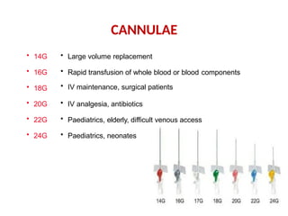
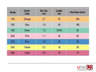
CANNULAE
• 14G
• 16G
•18G
• 20G
• 22G
• 24G
• Large volume replacement
• Rapid transfusion of whole blood or blood components
• IV maintenance, surgical patients
• IV analgesia, antibiotics
• Paediatrics, elderly, difficult venous access
• Paediatrics, neonates
6.
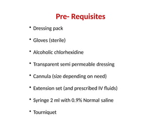
Pre- Requisites
• Dressingpack
• Gloves (sterile)
• Alcoholic chlorhexidine
• Transparent semi permeable dressing
• Cannula (size depending on need)
• Extension set (and prescribed IV fluids)
• Syringe 2 ml with 0.9% Normal saline
• Tourniquet
7.
• Check patientfor allergies to medications, cleansing
fluids & dressings
• Assess the dominant/non-dominant side and check
the veins for suitability
• Provide a clear explanation of the procedure
ASSESSMENT
8.
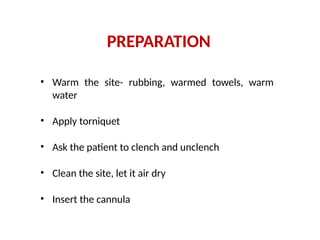
• Warm thesite- rubbing, warmed towels, warm
water
• Apply torniquet
• Ask the patient to clench and unclench
• Clean the site, let it air dry
• Insert the cannula
PREPARATION
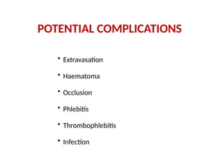
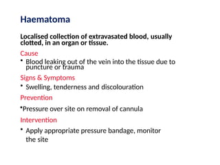
Haematoma
Localised collection ofextravasated blood, usually
clotted, in an organ or tissue.
Cause
• Blood leaking out of the vein into the tissue due to
puncture or trauma
Signs & Symptoms
• Swelling, tenderness and discolouration
Prevention
•Pressure over site on removal of cannula
Intervention
• Apply appropriate pressure bandage, monitor
the site
11.
Phlebitis
Inflammation of thevein
Cause
• Poor aseptic technique
• High osmolarity I.V. infusions or
drugs
• Trauma to the vein during
insertion/incorrect cannula gauge
• Prolonged use of the same site
Signs & Symptoms
• Tenderness, redness, heat and
oedema
• Advanced-induration, palpable
venous cord
Intervention
• Remove cannula
• Apply warm compress
• Observe for signs of
infection
• If phlebitis is advanced
antibiotics may be
required
12.
Occlusion
Cause
• Cannula notflushed- fibrin formation in or
around the tip of cannula
• Kinking of the cannula
Signs & Symptoms
• Slowing or cessation of fluid
• Blood in the line
Intervention
• Check for kinks in cannula
• Raise IV higher
• Flush
• Remove cannula
13.
Thrombophlebitis
Inflammation and formationof a thrombus
in the vein
Causes
• Injury to the vein
• Infection
• Chemical irritation
•Prolonged use of the same vein
Signs & Symptoms
• Tenderness/redness
• Heat/oedema
• Cordlike appearance of the
vein
• Slowing of the IV infusion
Intervention
• Remove cannula
• Observe for signs of
infection
14.
Infection
Pathogen in thetissue
surrounding the I.V. site.
Cause
• Lack of asepsis
•Prolonged use of the same site
Signs & Symptoms
• Tenderness and swelling
• Erythema/purulent drainage
Intervention
• Remove cannula
• Antibiotics may
be required