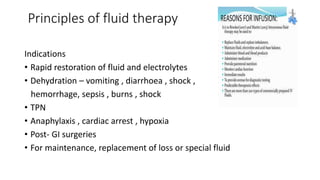
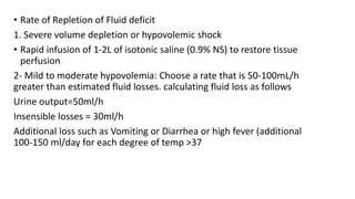
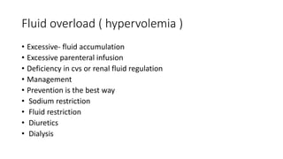
IV FLUIDS
DR. ANIRUDH AGRAWAL
DNB RESIDENT
GENERAL SURGERY
TOTAL BODY WATER
• APPROX 60% OF BODY WEIGHT (30l)
• VARIES WITH AGE , GENDER AND BODY WEIGHT
BODY WATER COMPARTMENT
• Intracellular volume- 2/3 of tbw (20 l)
• Extracellular volume – 1/3 of tbw (10l)
- intravascular – plasma volume (1/4) (2.5l)
- Extravascular – interstitial fluid and others (3/4)(7.5l)
Extra cellular fluid
1. interstitial compartment
• Small narrow space- between tissues or parts of an organ
• Interstitial fluid +
• excessive fluid accumulates – edema develops
2. Intra vascular compartment
• blood +
3. Third space
• Peritoneal cavity
• Pleural cavity
• Small amount of fluid present normally
Water- essential for cell life
• Minimum obligatory daily water input
• 500ml- ingested water
• 800 ml – water content in food
• 300 ml – water from oxidation
• Minimum obligatory daily water output
• 500 ml- urine
• 500 ml – skin
• 400 ml – respiratory tract
• 200 ml – stool
• Average – input and output – 2.4 l/day
↑ Requirement of water in-
• Fever
• Burns
• Sweating
• Surgical drains
• GI loss thorugh vomiting and diarrohea
• Sweating
• Tachyponea
• Polyuria
Fluid therapy
• Osmolality – amount of solute dissolved in a solvent like water
measured in weight ( kg )
• Osmolarity - amount of solute dissolved in a solvent like water
measured in volume (l)
• Normal plasma osmolality – 275-295 mosm/kg
Principles of fluid therapy
Indications
• Rapid restoration of fluid and electrolytes
• Dehydration – vomiting , diarrhoea , shock ,
hemorrhage, sepsis , burns , shock
• TPN
• Anaphylaxis , cardiac arrest , hypoxia
• Post- GI surgeries
• For maintenance, replacement of loss or special fluid
Types of fluid
1. Colloid
• Solutions –large molecules – don’t pass cell membrane
• Remain in intra vascular compartment
• Expand intravascular volume
• Draw fluid from extra vascular spaces via oncotic pressure
2. Crystalloid
• Solutions –small molecules – easily pass cell membrane
• Increase fluid volume – interstitial + intra vascular
• Divided into- isotonic
hypotonic
hypertonic
Isotonic fluids
• concentration of the particles (solutes)=to that of plasma
• So it doesn't move into cells and remains within the extracellular
compartment thus intravascular volume.
Isotonic solutions include:
• 0.9% sodium chloride (0.9% NaCl)
• lactated Ringer's solution
• 5% dextrose in water (D5W)
• Ringer's solution
A. 0.9% sodium chloride (Normal Saline)
• 0.9% NaCl- 154-Na+ K+ Ca+2 Mg+2 Cl -154 Dextrose mOsm/L-308
• Normal saline - % Nacl similar to concentration of na+ and cl- in intra vascular space
• Used in –
• to treat low extracellular fluid, as in fluid volume deficit from- Hemorrhage
• Severe vomiting or diarrhea
• Heavy drainage fromGl suction, fistulas, or wounds
• Shock
• Mild hyponatremia
• Metabolic acidosis (such as diabetic ketoacidosis)
• It's the fluid of choice for resuscitation efforts.
• it's the only fluid used with administration of blood products
• Used cautiously- cardiac or renal patient( volume overload0
B. Ringer’s lactate or Hartmann solution
• Na -130 , k-4, Ca - 3 ,Cl-109 mosm/l -273
• most physiologically adaptable fluid - electrolyte content - body's
blood serum and plasma.
• choice for first-line fluid resuscitation- burn injuries or trauma
• Acute blood loss- third space fluid shift
• LR- metabolized- liver –lactate to bicarbonate ( metabolic acidosis )
• Cautious use – renal impairement( K)
Liver disease
ph> 7.5
C. Dextrose 5%
• Dextrose – 50 gm/l mosm/l-278
• Isotonic – when metabolized – become hypotonic – fluid shift into cells
• Provides free water – expand intra + extra cellular compartment
• 170 calories/l – no electrolytes
• Do not use – renal failure – fluid overload
cardiac failure
ICP - Cerebral edema
Mix with blood – hemolyze
Not used for resuscitation – won’t remain in intravascular space
Early postoperative period - ADH secretion due to surgical stress
Precaution in usage of isotonic fluids
• Hypovolemia to hypervolemia
• Signs and symptoms of hypervolemia
• hypertension
• bounding pulse
• pulmonary crackles
• peripheral edema
• dyspnea
• shortness of breath
• jugular venous distention (JVD)•
• Monitor intake and output
• Elevate the head of bed at 35 to 45 degrees, unless contraindicated.
• If edema is present, elevate the patient‘s legs
• signs and symptoms of continued hypovolemia, including:
• urine output of less than 0.5 mL/kg /hour
• poor skin turgor
• Tachycardia
• weak, thready pulse
• hypotension
Hypotonic fluids
• hypotonic solutions - concentration of solutes (electrolytes)
• osmolality < 250 mOsm/L.
• Hypotonic crystalloid solutions lowers the serum osmolality within
the vascular space, causing fluid to shift from the intravascular space
to both the intracellular and interstitial spaces.
• These solutions will hydrate cells, although their use may deplete
fluid within the circulatory system.
Types of hypotonic fluids
• 0.45% sodium chloride (0.45% NaCl),
• 0.33% sodium chloride
• 0.2% sodium chloride
• 2.5% dextrose inwater
• Hypotonic fluids are used in conditions causing intracellular
dehydration
• fluid needs to be shifted into the cell- Hypernatremia
Diabetic ketoacidosis
Hyperosmolar hyperglycemic state.
Hypotonic fluids
• Precaution – ICP - Cerebral edema
Liver disease
Trauma
Burns
• depletion of intravascular fluid volume- hypovolemia and
hypotension- Cardiovascular collapse
• Monitor patients for signs and symptoms of fluid deficit
Hypertonic solutions
• Solution - higher tonicity or solute concentration.
• osmolarity of 375 mOsm/L or higher
• The osmotic pressure gradient draws water out of the intracellular
space, increasing extracellular fluid volume- volume expander
Types and indications
• 3% sodium chloride (3% NaCl): severe hyponatremia.
cerebral edema
• 2- 5% Dextrose with normal saline (D5NS): replaces sodium, chloride
and some calories
• Precautions-
• Intra vascular volume overload – pulmonary edema
Colloid solutions
• expand the intravascular volume by drawing fluid from the interstitial
spaces into the intravascular compartment through their higher
oncotic pressure.
• the same effect as hypertonic crystalloids solutions but it requires
administration of less total volume and have a longer duration of
action because the molecules remain within the intravascular space
longer.
• Its effect can last for several days if capillary wall linings are intact
and working properly.
Colloid solutions
• Examples-
• 1- 5% albumin (Human albumin solution)- most commonly used
• - It contains plasma protein fractions obtained from human plasma and works to rapidly expand
the plasma volume used for
• volume expansion
• moderate protein replacement
• achievement of hemodynamic stability in shock states
• Contraindications- severe anemia
Heart failure
ACEI – stop for 24 hours – atypical reaction- flushing and hypotension
2. Hydroxyethalstarches
• contain – Na and Cl – hemodynamic volume replacement and burns
• Less expensive
• Precautions – risk for fluid volume overload
• Monitor for signs and symptoms of hypervolemia
• Monitor input /output
• Interfere with platelet function – bleeding time
• Anaphylactoid reactions
Components of fluid therapy
• Maintenance therapy- replaces normal ongoing losses( peri-operatively or
on ventilator)- 4-2-1 rule (weight +40 )
• 4ml/kg/hr for first 10 kg
• 2ml/kg/hr for next 10 kg
• 1 ml/kg/hr for any kgs above that
• Fluid Resuscitation- corrects any existing water and electrolyte deficits.(
hypovolemic shock )
• Parameters- BP
Urine output
JVP
Urine Na concentration
• Rate of Repletion of Fluid deficit
1. Severe volume depletion or hypovolemic shock
• Rapid infusion of 1-2L of isotonic saline (0.9% NS) to restore tissue
perfusion
2- Mild to moderate hypovolemia: Choose a rate that is 50-100mL/h
greater than estimated fluid losses. calculating fluid loss as follows
Urine output=50ml/h
Insensible losses = 30ml/h
Additional loss such as Vomiting or Diarrhea or high fever (additional
100-150 ml/day for each degree of temp >37
Fluid overload ( hypervolemia )
• Excessive- fluid accumulation
• Excessive parenteral infusion
• Deficiency in cvs or renal fluid regulation
• Management
• Prevention is the best way
• Sodium restriction
• Fluid restriction
• Diuretics
• Dialysis

IV FLUIDS, TYPES AND CLASSIFICATION pptx

  • 1.
    IV FLUIDS DR. ANIRUDHAGRAWAL DNB RESIDENT GENERAL SURGERY
  • 2.
    TOTAL BODY WATER •APPROX 60% OF BODY WEIGHT (30l) • VARIES WITH AGE , GENDER AND BODY WEIGHT
  • 3.
    BODY WATER COMPARTMENT •Intracellular volume- 2/3 of tbw (20 l) • Extracellular volume – 1/3 of tbw (10l) - intravascular – plasma volume (1/4) (2.5l) - Extravascular – interstitial fluid and others (3/4)(7.5l)
  • 4.
    Extra cellular fluid 1.interstitial compartment • Small narrow space- between tissues or parts of an organ • Interstitial fluid + • excessive fluid accumulates – edema develops 2. Intra vascular compartment • blood + 3. Third space • Peritoneal cavity • Pleural cavity • Small amount of fluid present normally
  • 5.
    Water- essential forcell life • Minimum obligatory daily water input • 500ml- ingested water • 800 ml – water content in food • 300 ml – water from oxidation • Minimum obligatory daily water output • 500 ml- urine • 500 ml – skin • 400 ml – respiratory tract • 200 ml – stool • Average – input and output – 2.4 l/day
  • 6.
    ↑ Requirement ofwater in- • Fever • Burns • Sweating • Surgical drains • GI loss thorugh vomiting and diarrohea • Sweating • Tachyponea • Polyuria
  • 7.
    Fluid therapy • Osmolality– amount of solute dissolved in a solvent like water measured in weight ( kg ) • Osmolarity - amount of solute dissolved in a solvent like water measured in volume (l) • Normal plasma osmolality – 275-295 mosm/kg
  • 8.
    Principles of fluidtherapy Indications • Rapid restoration of fluid and electrolytes • Dehydration – vomiting , diarrhoea , shock , hemorrhage, sepsis , burns , shock • TPN • Anaphylaxis , cardiac arrest , hypoxia • Post- GI surgeries • For maintenance, replacement of loss or special fluid
  • 9.
    Types of fluid 1.Colloid • Solutions –large molecules – don’t pass cell membrane • Remain in intra vascular compartment • Expand intravascular volume • Draw fluid from extra vascular spaces via oncotic pressure 2. Crystalloid • Solutions –small molecules – easily pass cell membrane • Increase fluid volume – interstitial + intra vascular • Divided into- isotonic hypotonic hypertonic
  • 10.
    Isotonic fluids • concentrationof the particles (solutes)=to that of plasma • So it doesn't move into cells and remains within the extracellular compartment thus intravascular volume. Isotonic solutions include: • 0.9% sodium chloride (0.9% NaCl) • lactated Ringer's solution • 5% dextrose in water (D5W) • Ringer's solution
  • 11.
    A. 0.9% sodiumchloride (Normal Saline) • 0.9% NaCl- 154-Na+ K+ Ca+2 Mg+2 Cl -154 Dextrose mOsm/L-308 • Normal saline - % Nacl similar to concentration of na+ and cl- in intra vascular space • Used in – • to treat low extracellular fluid, as in fluid volume deficit from- Hemorrhage • Severe vomiting or diarrhea • Heavy drainage fromGl suction, fistulas, or wounds • Shock • Mild hyponatremia • Metabolic acidosis (such as diabetic ketoacidosis) • It's the fluid of choice for resuscitation efforts. • it's the only fluid used with administration of blood products • Used cautiously- cardiac or renal patient( volume overload0
  • 12.
    B. Ringer’s lactateor Hartmann solution • Na -130 , k-4, Ca - 3 ,Cl-109 mosm/l -273 • most physiologically adaptable fluid - electrolyte content - body's blood serum and plasma. • choice for first-line fluid resuscitation- burn injuries or trauma • Acute blood loss- third space fluid shift • LR- metabolized- liver –lactate to bicarbonate ( metabolic acidosis ) • Cautious use – renal impairement( K) Liver disease ph> 7.5
  • 13.
    C. Dextrose 5% •Dextrose – 50 gm/l mosm/l-278 • Isotonic – when metabolized – become hypotonic – fluid shift into cells • Provides free water – expand intra + extra cellular compartment • 170 calories/l – no electrolytes • Do not use – renal failure – fluid overload cardiac failure ICP - Cerebral edema Mix with blood – hemolyze Not used for resuscitation – won’t remain in intravascular space Early postoperative period - ADH secretion due to surgical stress
  • 14.
    Precaution in usageof isotonic fluids • Hypovolemia to hypervolemia • Signs and symptoms of hypervolemia • hypertension • bounding pulse • pulmonary crackles • peripheral edema • dyspnea • shortness of breath • jugular venous distention (JVD)• • Monitor intake and output • Elevate the head of bed at 35 to 45 degrees, unless contraindicated. • If edema is present, elevate the patient‘s legs
  • 15.
    • signs andsymptoms of continued hypovolemia, including: • urine output of less than 0.5 mL/kg /hour • poor skin turgor • Tachycardia • weak, thready pulse • hypotension
  • 16.
    Hypotonic fluids • hypotonicsolutions - concentration of solutes (electrolytes) • osmolality < 250 mOsm/L. • Hypotonic crystalloid solutions lowers the serum osmolality within the vascular space, causing fluid to shift from the intravascular space to both the intracellular and interstitial spaces. • These solutions will hydrate cells, although their use may deplete fluid within the circulatory system.
  • 17.
    Types of hypotonicfluids • 0.45% sodium chloride (0.45% NaCl), • 0.33% sodium chloride • 0.2% sodium chloride • 2.5% dextrose inwater • Hypotonic fluids are used in conditions causing intracellular dehydration • fluid needs to be shifted into the cell- Hypernatremia Diabetic ketoacidosis Hyperosmolar hyperglycemic state.
  • 18.
    Hypotonic fluids • Precaution– ICP - Cerebral edema Liver disease Trauma Burns • depletion of intravascular fluid volume- hypovolemia and hypotension- Cardiovascular collapse • Monitor patients for signs and symptoms of fluid deficit
  • 19.
    Hypertonic solutions • Solution- higher tonicity or solute concentration. • osmolarity of 375 mOsm/L or higher • The osmotic pressure gradient draws water out of the intracellular space, increasing extracellular fluid volume- volume expander
  • 20.
    Types and indications •3% sodium chloride (3% NaCl): severe hyponatremia. cerebral edema • 2- 5% Dextrose with normal saline (D5NS): replaces sodium, chloride and some calories • Precautions- • Intra vascular volume overload – pulmonary edema
  • 21.
    Colloid solutions • expandthe intravascular volume by drawing fluid from the interstitial spaces into the intravascular compartment through their higher oncotic pressure. • the same effect as hypertonic crystalloids solutions but it requires administration of less total volume and have a longer duration of action because the molecules remain within the intravascular space longer. • Its effect can last for several days if capillary wall linings are intact and working properly.
  • 22.
    Colloid solutions • Examples- •1- 5% albumin (Human albumin solution)- most commonly used • - It contains plasma protein fractions obtained from human plasma and works to rapidly expand the plasma volume used for • volume expansion • moderate protein replacement • achievement of hemodynamic stability in shock states • Contraindications- severe anemia Heart failure ACEI – stop for 24 hours – atypical reaction- flushing and hypotension 2. Hydroxyethalstarches • contain – Na and Cl – hemodynamic volume replacement and burns • Less expensive
  • 23.
    • Precautions –risk for fluid volume overload • Monitor for signs and symptoms of hypervolemia • Monitor input /output • Interfere with platelet function – bleeding time • Anaphylactoid reactions
  • 24.
    Components of fluidtherapy • Maintenance therapy- replaces normal ongoing losses( peri-operatively or on ventilator)- 4-2-1 rule (weight +40 ) • 4ml/kg/hr for first 10 kg • 2ml/kg/hr for next 10 kg • 1 ml/kg/hr for any kgs above that • Fluid Resuscitation- corrects any existing water and electrolyte deficits.( hypovolemic shock ) • Parameters- BP Urine output JVP Urine Na concentration
  • 25.
    • Rate ofRepletion of Fluid deficit 1. Severe volume depletion or hypovolemic shock • Rapid infusion of 1-2L of isotonic saline (0.9% NS) to restore tissue perfusion 2- Mild to moderate hypovolemia: Choose a rate that is 50-100mL/h greater than estimated fluid losses. calculating fluid loss as follows Urine output=50ml/h Insensible losses = 30ml/h Additional loss such as Vomiting or Diarrhea or high fever (additional 100-150 ml/day for each degree of temp >37
  • 26.
    Fluid overload (hypervolemia ) • Excessive- fluid accumulation • Excessive parenteral infusion • Deficiency in cvs or renal fluid regulation • Management • Prevention is the best way • Sodium restriction • Fluid restriction • Diuretics • Dialysis