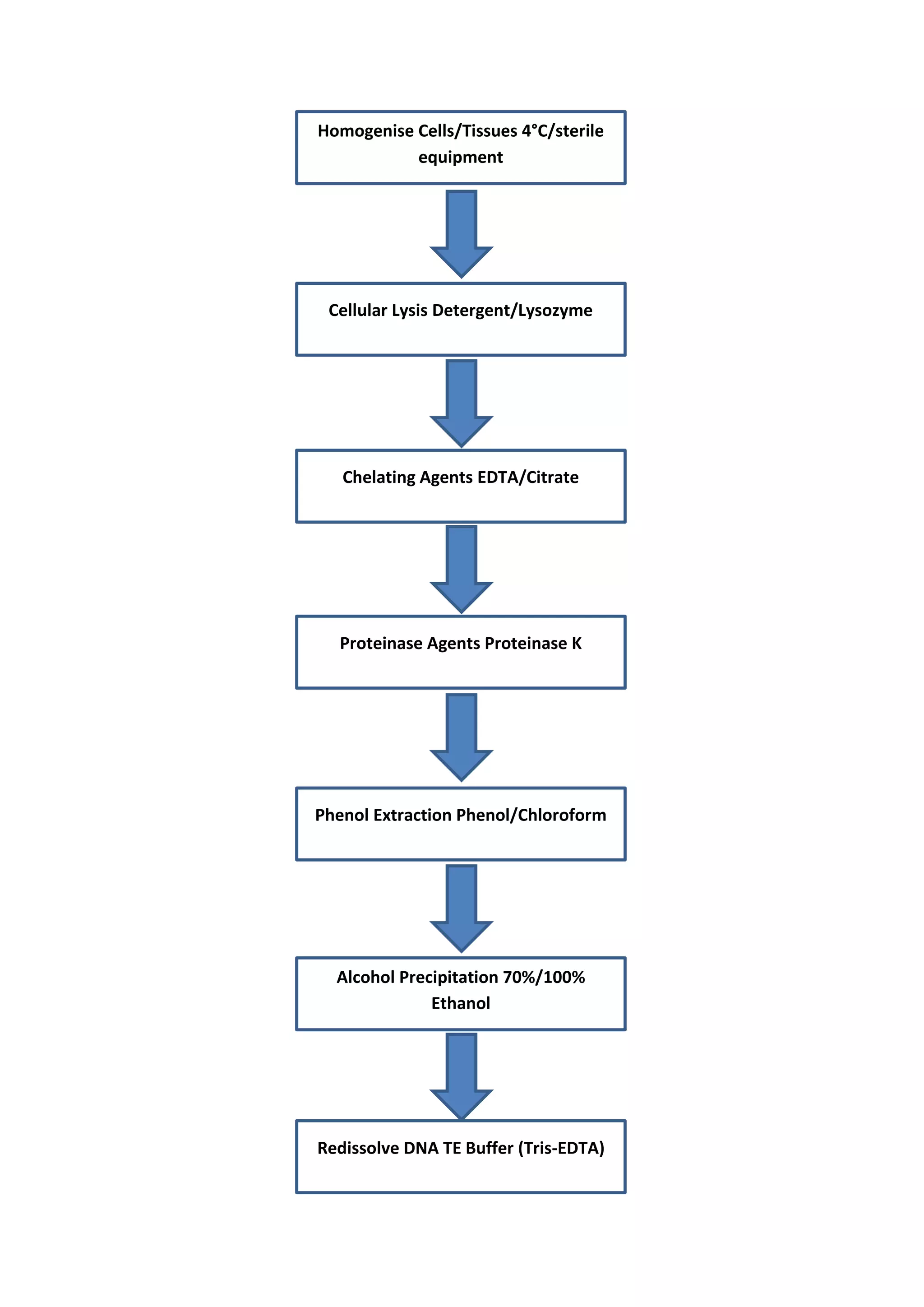
Isolation of DNA
The use of DNA for analysis or manipulation usually requires that it is
isolated and purified to a certain extent. DNA is recovered from cells by the
gentlest possible method of cell rupture to prevent the DNA from
fragmenting by mechanical shearing. This is usually in the presence of
EDTA which chelates the Mg2+ ions needed for enzymes that degrade DNA
termed DNase.
Ideally, cell walls, if present, should be digested enzymatically (e.g. cellulase
treatment of plants cell or lysozyme treatment of bacteria), and the cell
membrane should be solubilised using detergent. If physical disruption is
necessary, it should be kept to a minimum, and should involve cutting or
squashing of cells, rather than the use of shear forces.
Cell disruption (and most subsequent steps) should be performed at 4 °C,
using glassware and solutions that have been autoclaved to destroy DNase
activity.
After release of nucleic acids from the cells, RNA can be removed by
treatment with ribonuclease (RNase) that has been heat-treated to inactivate
any DNase contaminants; RNase is relatively stable to heat as a result of its
disulphide bonds, which ensure rapid renaturation of the molecule on
cooling.
The other major contaminant, protein, is removed by shaking the solution
gently with water-saturated phenol, or with a phenol/chloroform mixture,
either of which will denature proteins but not nucleic acids.
Centrifugation of the emulsion formed by this mixing produces a lower,
organic phase, separated from the upper, aqueous phase by an interface of
denatured protein. The aqueous solution is recovered and deproteinised
repeatedly, until no more material is seen at the interface.
Finally, the deproteinised DNA preparation is mixed with two volumes of
absolute ethanol, and the DNA allowed to precipitate out of solution in a
freezer.
After centrifugation, the DNA pellet is redissolved in a buffer containing
EDTA to inactivate any DNases present. This solution can be stored at 4 °C
for at least a month. DNA solutions can be stored frozen although repeated
freezing and thawing tends to damage long DNA molecules by shearing. The
procedure described above is suitable for total cellular DNA.
If the DNA from a specific organelle, it is best to isolate the organelle before
extracting its DNA, since the recovery of a particular type of DNA from a
mixture is usually rather difficult. Where a high degree of purity is required
DNA may be subjected to density gradient ultracentrifugation through
caesium chloride which is particularly useful for the preparation of plasmid
DNA. A flow chart of DNA extraction is indicated in Fig. 1.
Homogenise Cells/Tissues 4°C/sterile
equipment
Cellular Lysis Detergent/Lysozyme
Chelating Agents EDTA/Citrate
Proteinase Agents Proteinase K
Alcohol Precipitation 70%/100%
Ethanol
Phenol Extraction Phenol/Chloroform
Redissolve DNA TE Buffer (Tris-EDTA)
It is possible to check the integrity of the DNA by agarose gel electrophoresis
and determine the concentration of the DNA by using the fact that 1
absorbance unit equates to 50 μg ml-l of DNA and so:
50 x A260 = concentration of DNA sample (μgml-1)
Contaminants may also be identified by scanning UV spectrophotometry
from 200nm to 300nm. A ratio of 260nm : 280nm of approximately 1.8
indicates that the sample is free of protein contamination, which absorbs
strongly at 280nm.
Isolation of RNA
The methods used for RNA isolation are very similar to those described
above for DNA; however, RNA molecules are relatively short, and therefore
less easily damaged by shearing, so cell disruption can be rather more
vigorous.
RNA is, however, very vulnerable to digestion by RNases which are present
endogenously in various concentrations in certain cell types and
exogenously on fingers. Gloves should therefore be worn, and a strong
detergent should be included in the isolation medium to immediately
denature any RNases.
Subsequent deproteinisation should be particularly rigorous, since RNA is
often tightly associated with proteins.
DNase treatment can be used to remove DNA, and RNA can be precipitated
by ethanol. One reagent in particular which is commonly used in RNA
extraction is guanadinium thiocyanate which is both a strong inhibitor of
RNase and a protein denaturant.
A flow chart of RNA extraction is indicated in Fig. 2.
Treat Reagents Treat with RNase inhibitors e.g.
diethylpyrocarbonate (DEPC)
Homogenise Cells/Tissues 4°C/treated reagents
Cellular Lysis Detergent/Lysozyme
Proteinase Agents
Proteinase K
Alcohol Precipitation 70%/100% Ethanol
Phenol Extraction Phenol/Chloroform
Redissolve RNA
RNA solvents Guanadinium
thiocyanate
It is possible to check the integrity of an RNA extract by analysing it by agarose gel
electrophoresis. The most abundant RNA species, the rRNA molecules 23S and 16S
for prokaryotes and 18S and 28S for eukaryotes, appear as discrete bands on the
agarose gel and thus indicate that the other RNA components are likely to be
intact. This is usually carried out under denaturing conditions to prevent
secondary structure formation in the RNA. The concentration of the RNA may be
estimated by using UV spectrophotometry. At 260nm 1 absorbance unit equates to
40μgml-1 of RNA and therefore:
40 X A260 = concentration of RNA sample (μgml-1)
Contaminants may also be identified in the same way as that for DNA by scanning
UV spectrophotometry; however, in the case of RNA a 260nm : 280nm ratio of
approximately 2 would be expected for a sample containing no protein.
In many cases it is desirable to isolate eukaryotic mRNA which constitutes only 2–
5% of cellular RNA
from a mixture of
total RNA molecules.
This may be carried
out by affinity
chromatography on
oligo(dT)-cellulose
columns.
At high salt
concentrations, the
mRNA containing
poly(A) tails binds to
the complementary
oligo(dT) molecules of
the affinity column,
and so mRNA will be
retained; all other
RNA molecules can
be washed through
the column by
further high salt
solution. Finally, the
bound mRNA can be
eluted using a low
concentration of salt
(Fig. 3).
Nucleic acid species
may also be
subfractionated by more physical means such as electrophoretic or
chromatographic separations based on differences in nucleic acid fragment sizes or
physicochemical characteristics. Nanodrop spectrophotometer systems have also
aided the analysis of nucleic acids in recent years in allowing the full spectrum of
information whilst requiring only a very small (microlitre) sample volume.

Isolation of dna

  • 1.
    Isolation of DNA Theuse of DNA for analysis or manipulation usually requires that it is isolated and purified to a certain extent. DNA is recovered from cells by the gentlest possible method of cell rupture to prevent the DNA from fragmenting by mechanical shearing. This is usually in the presence of EDTA which chelates the Mg2+ ions needed for enzymes that degrade DNA termed DNase. Ideally, cell walls, if present, should be digested enzymatically (e.g. cellulase treatment of plants cell or lysozyme treatment of bacteria), and the cell membrane should be solubilised using detergent. If physical disruption is necessary, it should be kept to a minimum, and should involve cutting or squashing of cells, rather than the use of shear forces. Cell disruption (and most subsequent steps) should be performed at 4 °C, using glassware and solutions that have been autoclaved to destroy DNase activity. After release of nucleic acids from the cells, RNA can be removed by treatment with ribonuclease (RNase) that has been heat-treated to inactivate any DNase contaminants; RNase is relatively stable to heat as a result of its disulphide bonds, which ensure rapid renaturation of the molecule on cooling. The other major contaminant, protein, is removed by shaking the solution gently with water-saturated phenol, or with a phenol/chloroform mixture, either of which will denature proteins but not nucleic acids. Centrifugation of the emulsion formed by this mixing produces a lower, organic phase, separated from the upper, aqueous phase by an interface of denatured protein. The aqueous solution is recovered and deproteinised repeatedly, until no more material is seen at the interface. Finally, the deproteinised DNA preparation is mixed with two volumes of absolute ethanol, and the DNA allowed to precipitate out of solution in a freezer. After centrifugation, the DNA pellet is redissolved in a buffer containing EDTA to inactivate any DNases present. This solution can be stored at 4 °C for at least a month. DNA solutions can be stored frozen although repeated freezing and thawing tends to damage long DNA molecules by shearing. The procedure described above is suitable for total cellular DNA. If the DNA from a specific organelle, it is best to isolate the organelle before extracting its DNA, since the recovery of a particular type of DNA from a mixture is usually rather difficult. Where a high degree of purity is required DNA may be subjected to density gradient ultracentrifugation through caesium chloride which is particularly useful for the preparation of plasmid DNA. A flow chart of DNA extraction is indicated in Fig. 1.
  • 2.
    Homogenise Cells/Tissues 4°C/sterile equipment CellularLysis Detergent/Lysozyme Chelating Agents EDTA/Citrate Proteinase Agents Proteinase K Alcohol Precipitation 70%/100% Ethanol Phenol Extraction Phenol/Chloroform Redissolve DNA TE Buffer (Tris-EDTA)
  • 3.
    It is possibleto check the integrity of the DNA by agarose gel electrophoresis and determine the concentration of the DNA by using the fact that 1 absorbance unit equates to 50 μg ml-l of DNA and so: 50 x A260 = concentration of DNA sample (μgml-1) Contaminants may also be identified by scanning UV spectrophotometry from 200nm to 300nm. A ratio of 260nm : 280nm of approximately 1.8 indicates that the sample is free of protein contamination, which absorbs strongly at 280nm. Isolation of RNA The methods used for RNA isolation are very similar to those described above for DNA; however, RNA molecules are relatively short, and therefore less easily damaged by shearing, so cell disruption can be rather more vigorous. RNA is, however, very vulnerable to digestion by RNases which are present endogenously in various concentrations in certain cell types and exogenously on fingers. Gloves should therefore be worn, and a strong detergent should be included in the isolation medium to immediately denature any RNases. Subsequent deproteinisation should be particularly rigorous, since RNA is often tightly associated with proteins. DNase treatment can be used to remove DNA, and RNA can be precipitated by ethanol. One reagent in particular which is commonly used in RNA extraction is guanadinium thiocyanate which is both a strong inhibitor of RNase and a protein denaturant.
  • 4.
    A flow chartof RNA extraction is indicated in Fig. 2. Treat Reagents Treat with RNase inhibitors e.g. diethylpyrocarbonate (DEPC) Homogenise Cells/Tissues 4°C/treated reagents Cellular Lysis Detergent/Lysozyme Proteinase Agents Proteinase K Alcohol Precipitation 70%/100% Ethanol Phenol Extraction Phenol/Chloroform Redissolve RNA RNA solvents Guanadinium thiocyanate
  • 5.
    It is possibleto check the integrity of an RNA extract by analysing it by agarose gel electrophoresis. The most abundant RNA species, the rRNA molecules 23S and 16S for prokaryotes and 18S and 28S for eukaryotes, appear as discrete bands on the agarose gel and thus indicate that the other RNA components are likely to be intact. This is usually carried out under denaturing conditions to prevent secondary structure formation in the RNA. The concentration of the RNA may be estimated by using UV spectrophotometry. At 260nm 1 absorbance unit equates to 40μgml-1 of RNA and therefore: 40 X A260 = concentration of RNA sample (μgml-1) Contaminants may also be identified in the same way as that for DNA by scanning UV spectrophotometry; however, in the case of RNA a 260nm : 280nm ratio of approximately 2 would be expected for a sample containing no protein. In many cases it is desirable to isolate eukaryotic mRNA which constitutes only 2– 5% of cellular RNA from a mixture of total RNA molecules. This may be carried out by affinity chromatography on oligo(dT)-cellulose columns. At high salt concentrations, the mRNA containing poly(A) tails binds to the complementary oligo(dT) molecules of the affinity column, and so mRNA will be retained; all other RNA molecules can be washed through the column by further high salt solution. Finally, the bound mRNA can be eluted using a low concentration of salt (Fig. 3). Nucleic acid species may also be subfractionated by more physical means such as electrophoretic or chromatographic separations based on differences in nucleic acid fragment sizes or physicochemical characteristics. Nanodrop spectrophotometer systems have also aided the analysis of nucleic acids in recent years in allowing the full spectrum of information whilst requiring only a very small (microlitre) sample volume.