Downloaded 185 times
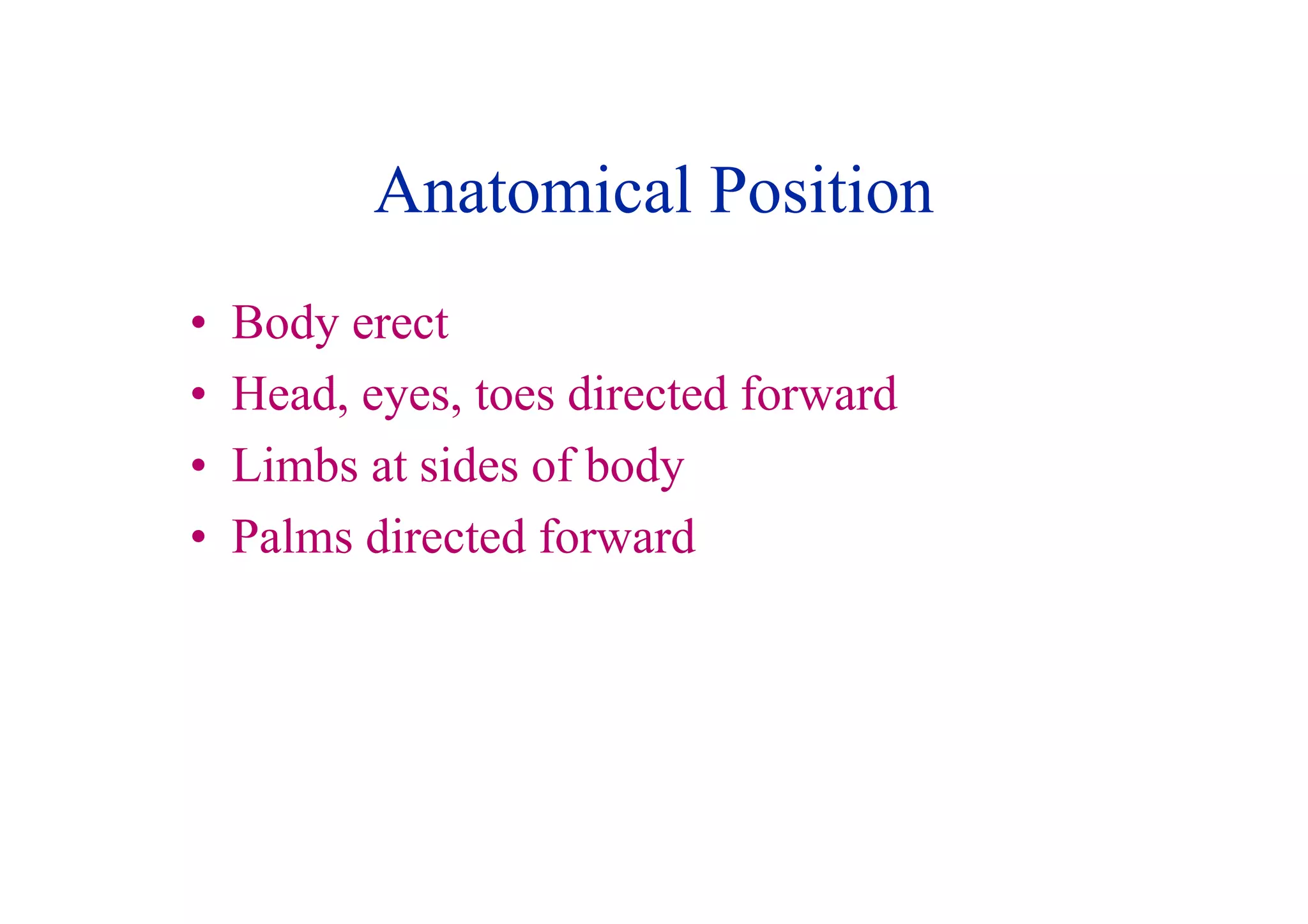
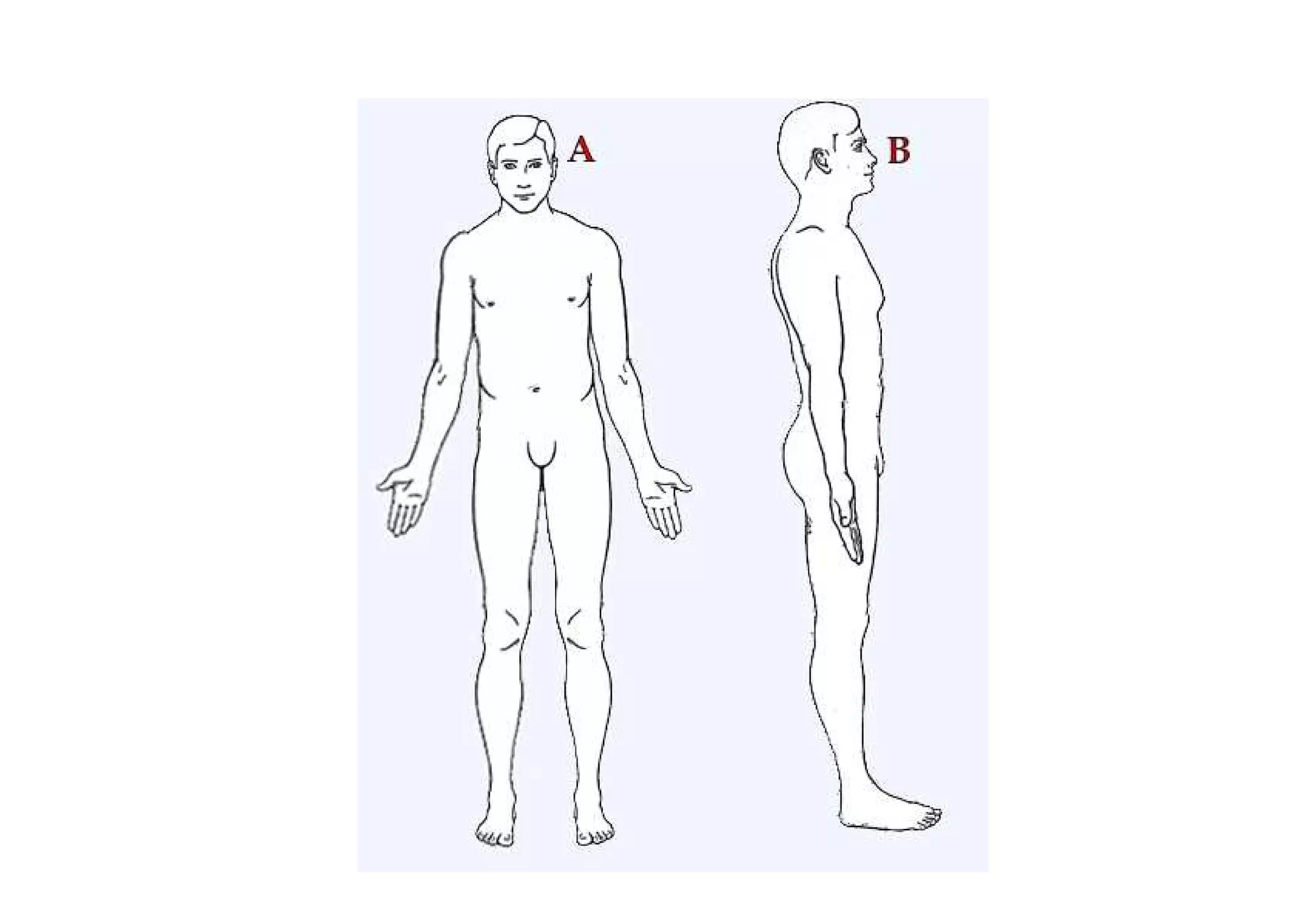
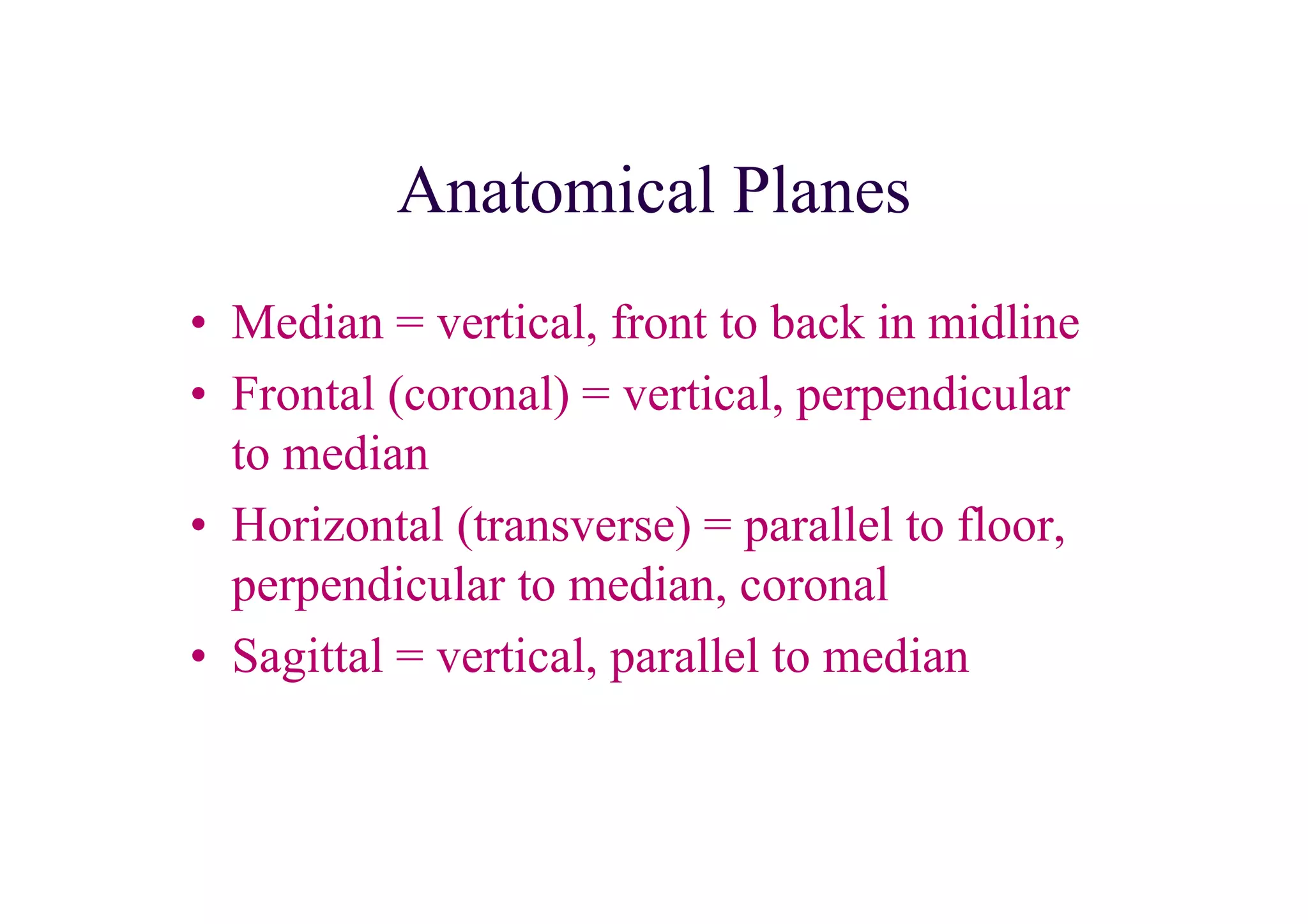
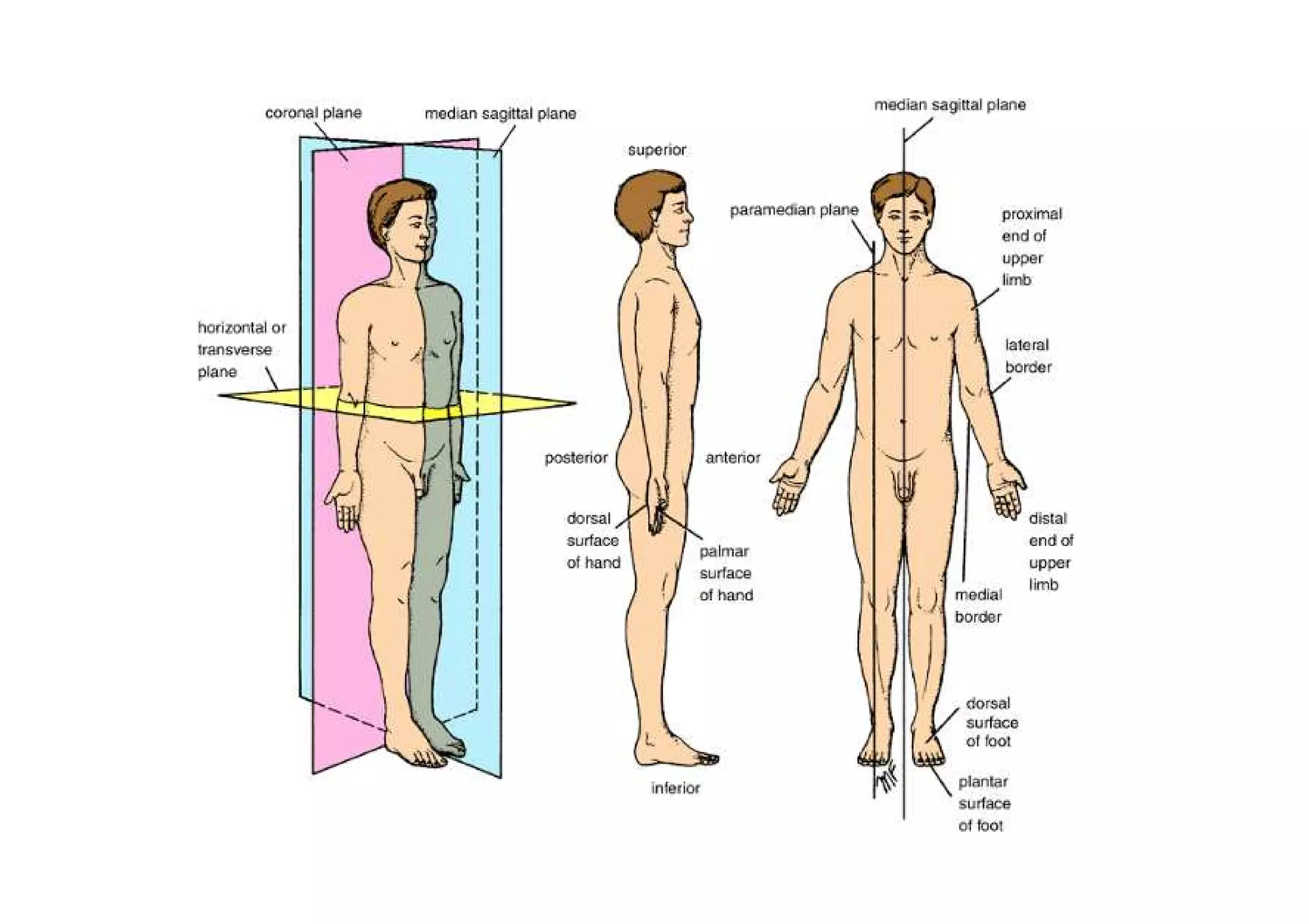
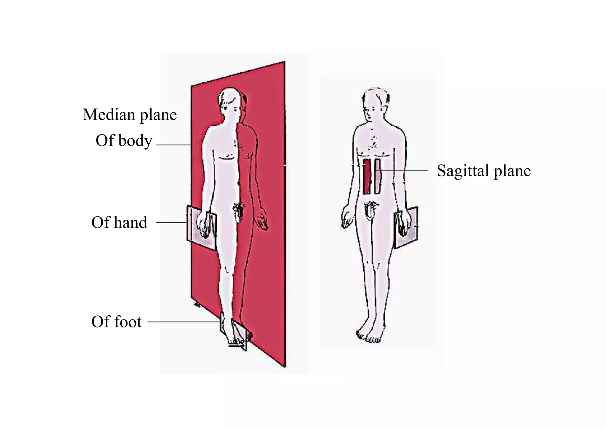
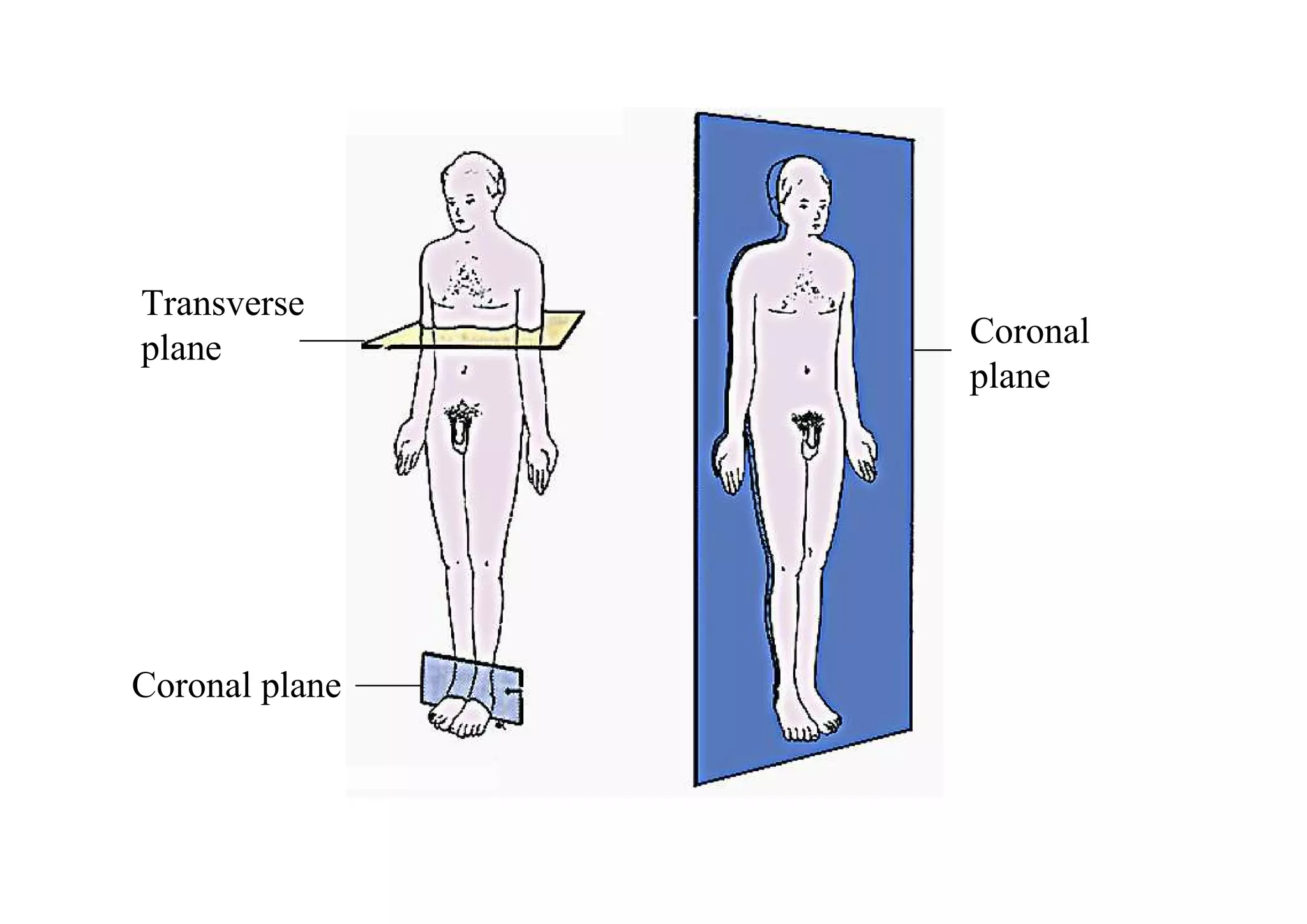
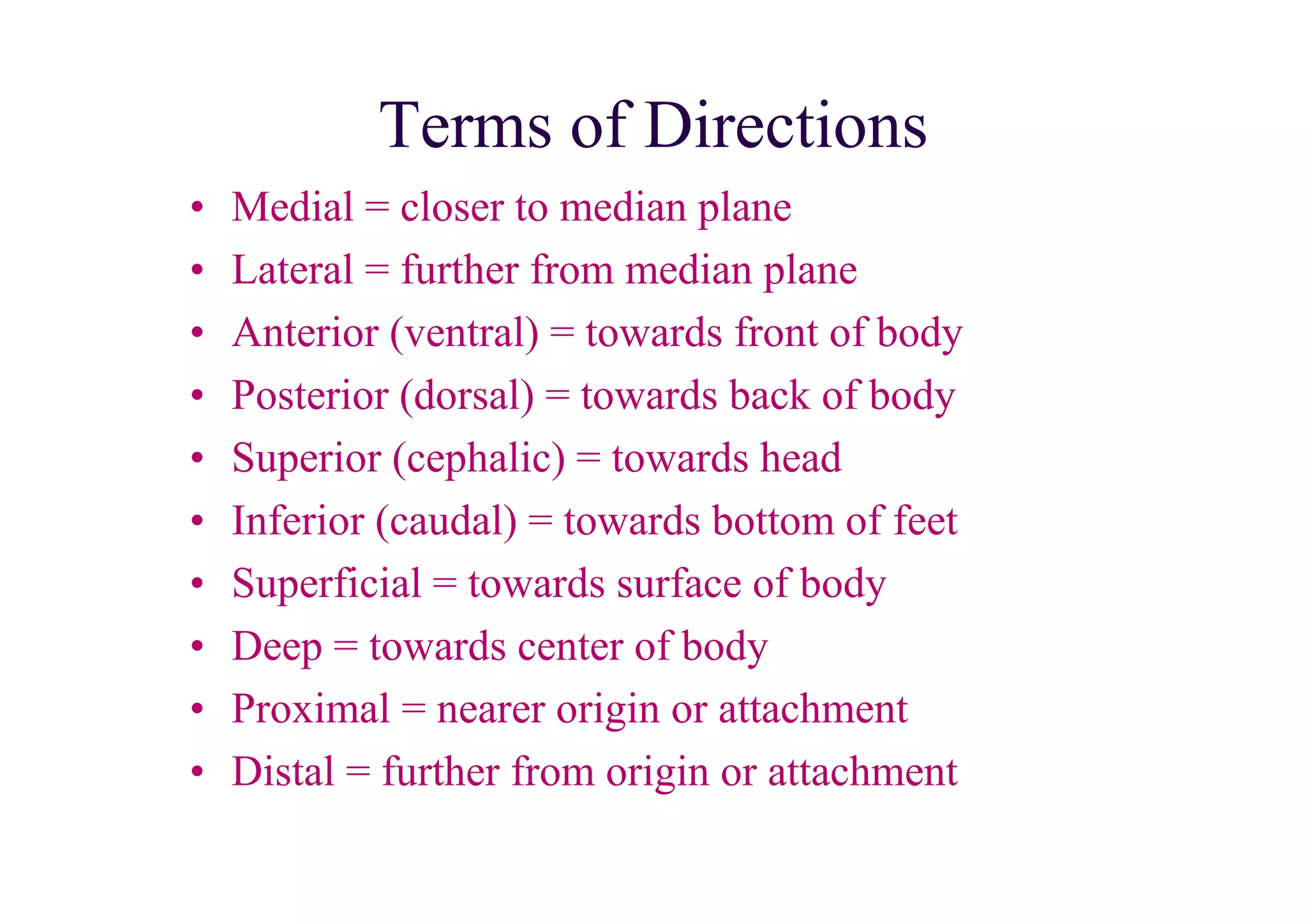

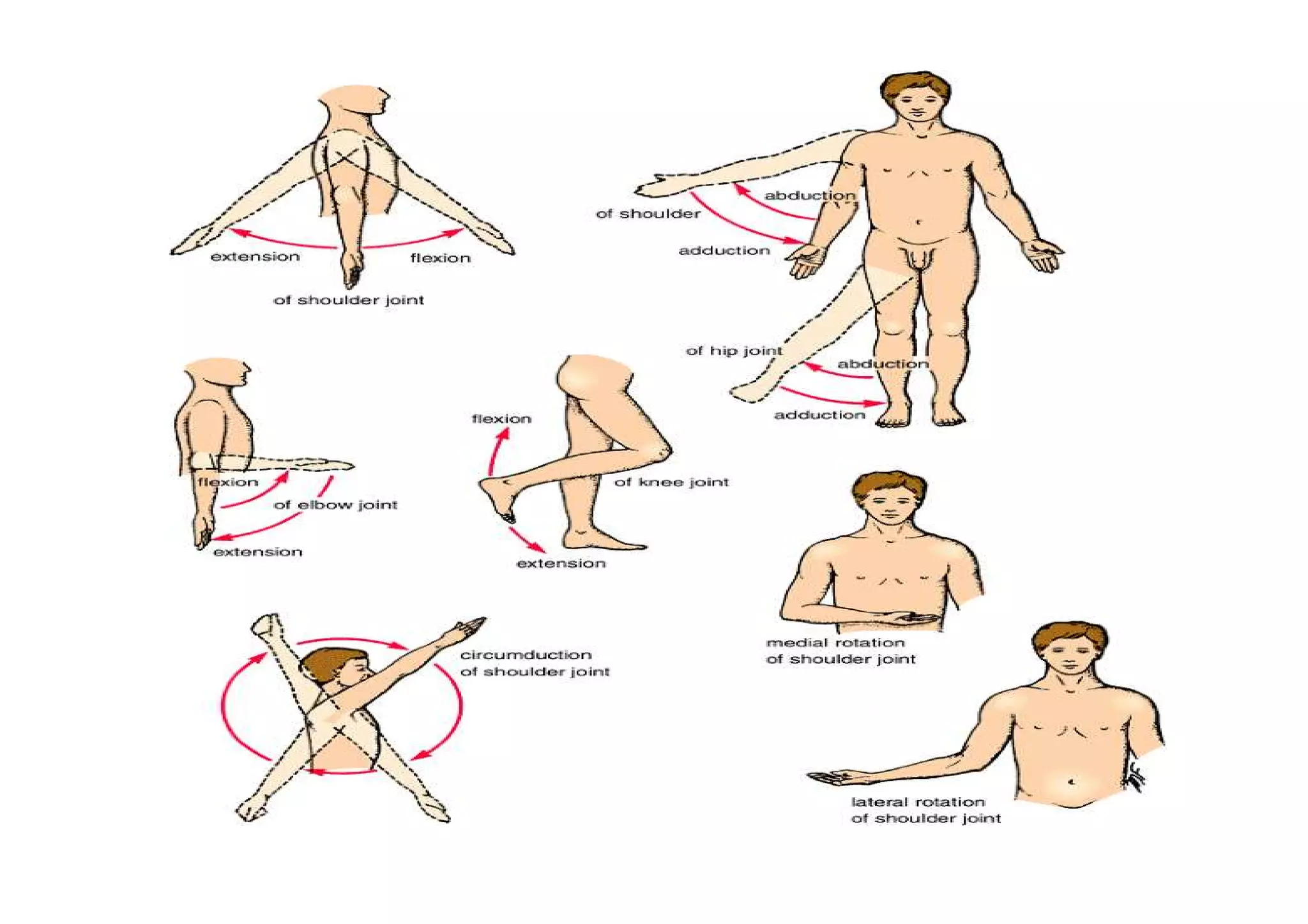
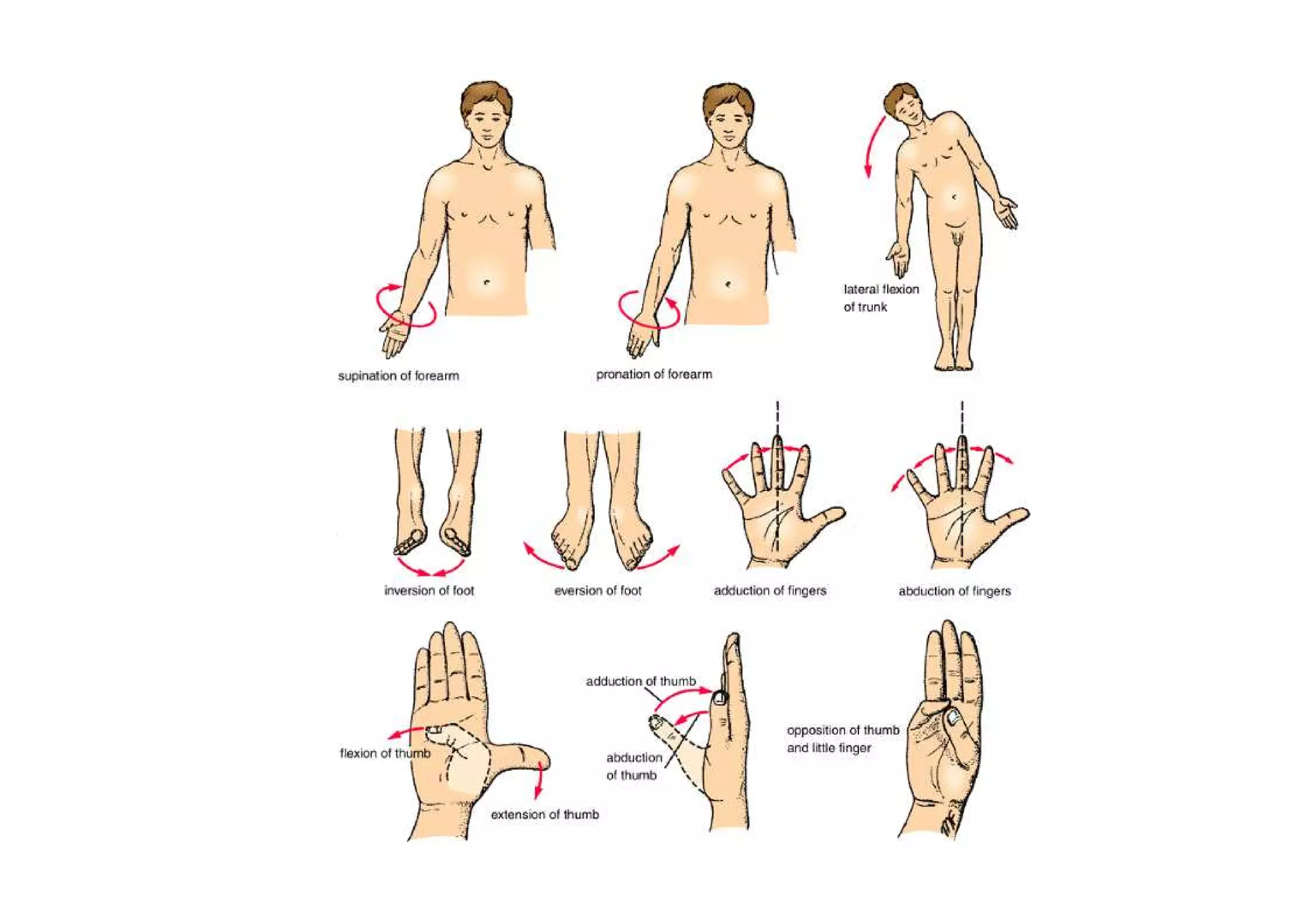
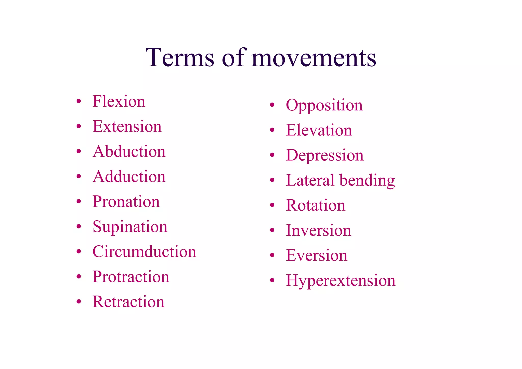
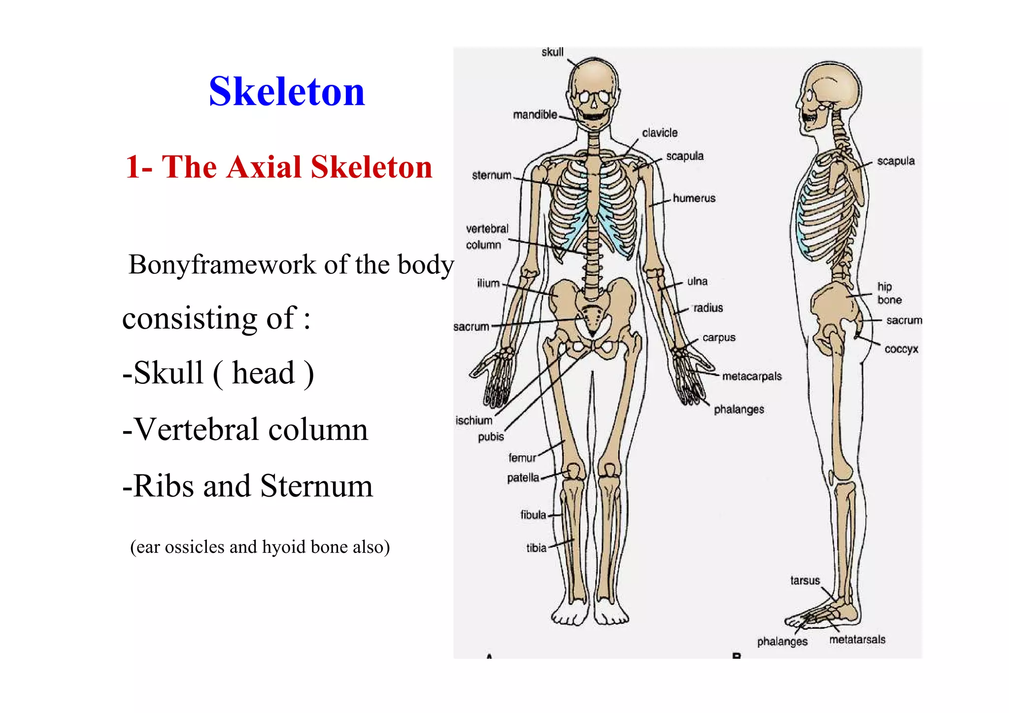

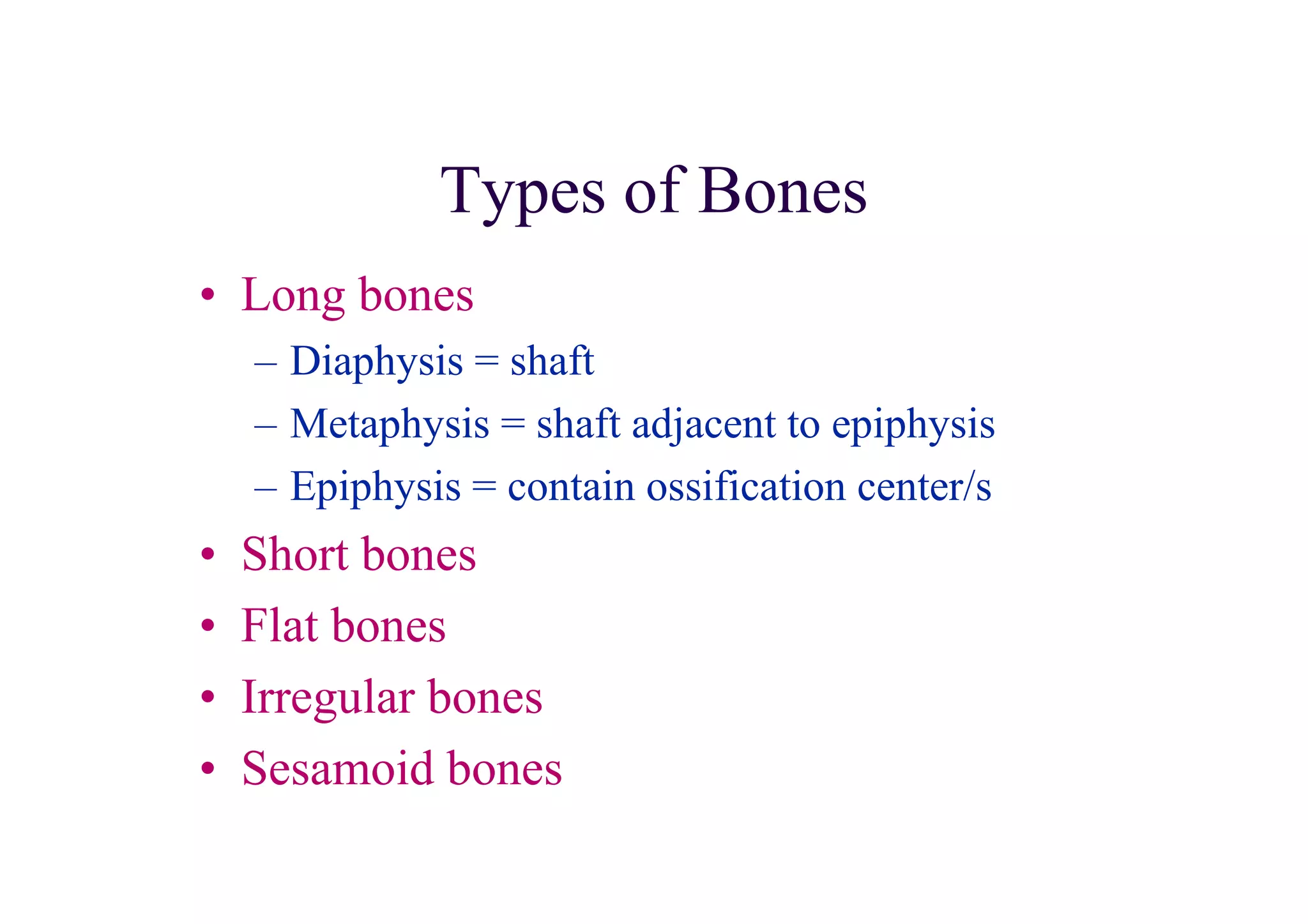
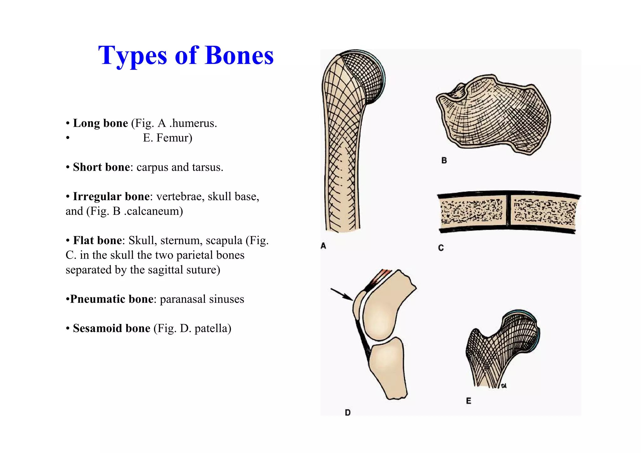
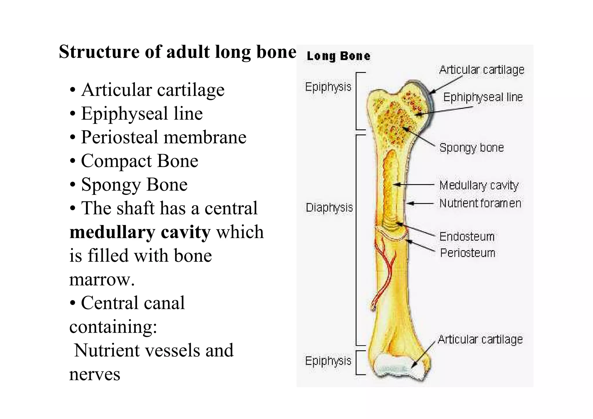
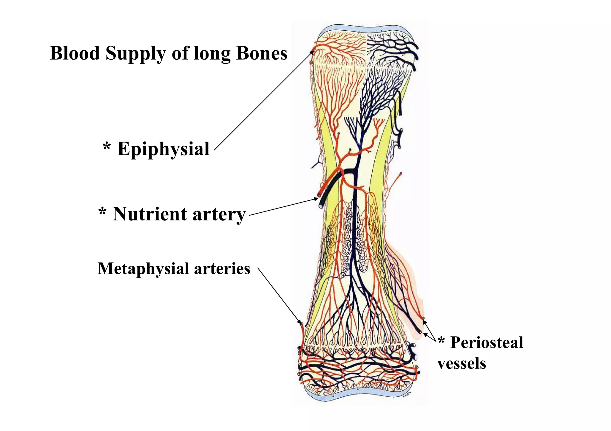
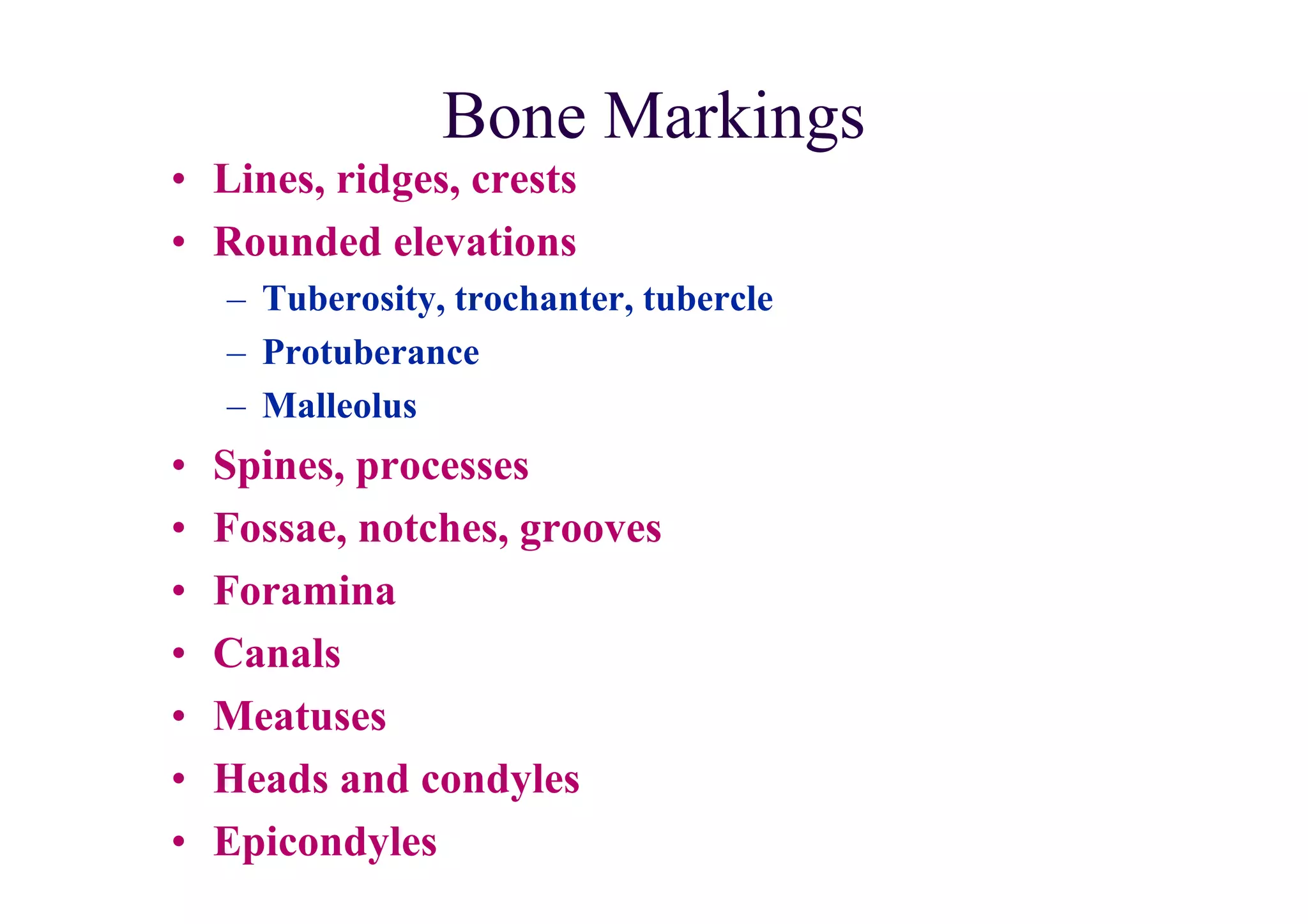

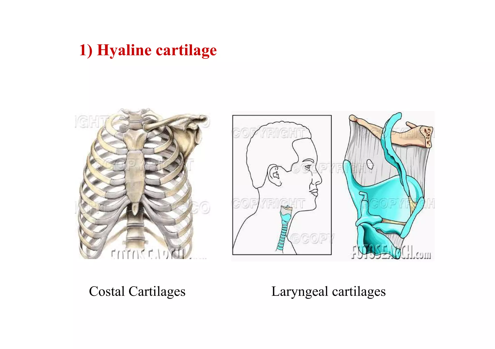
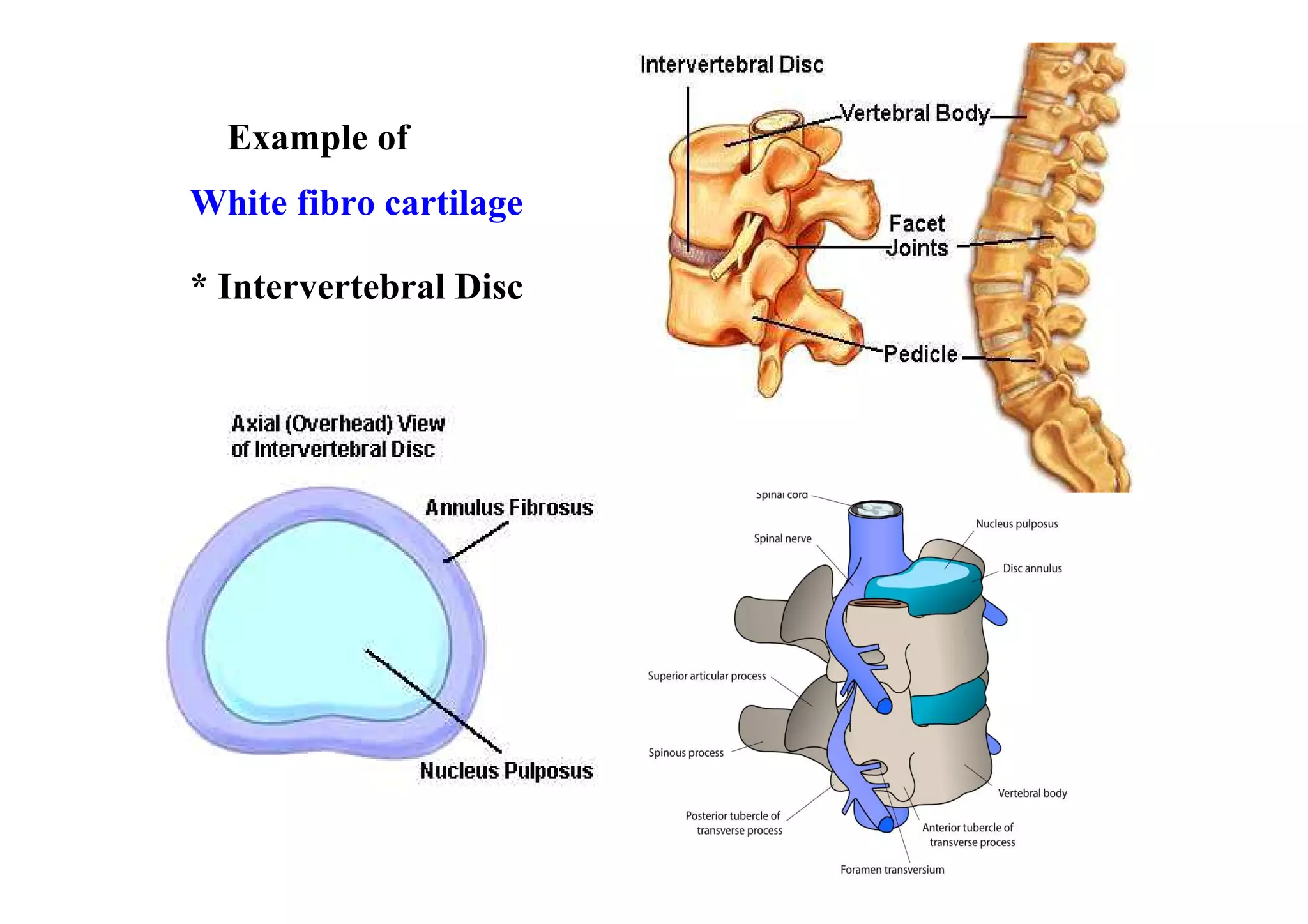
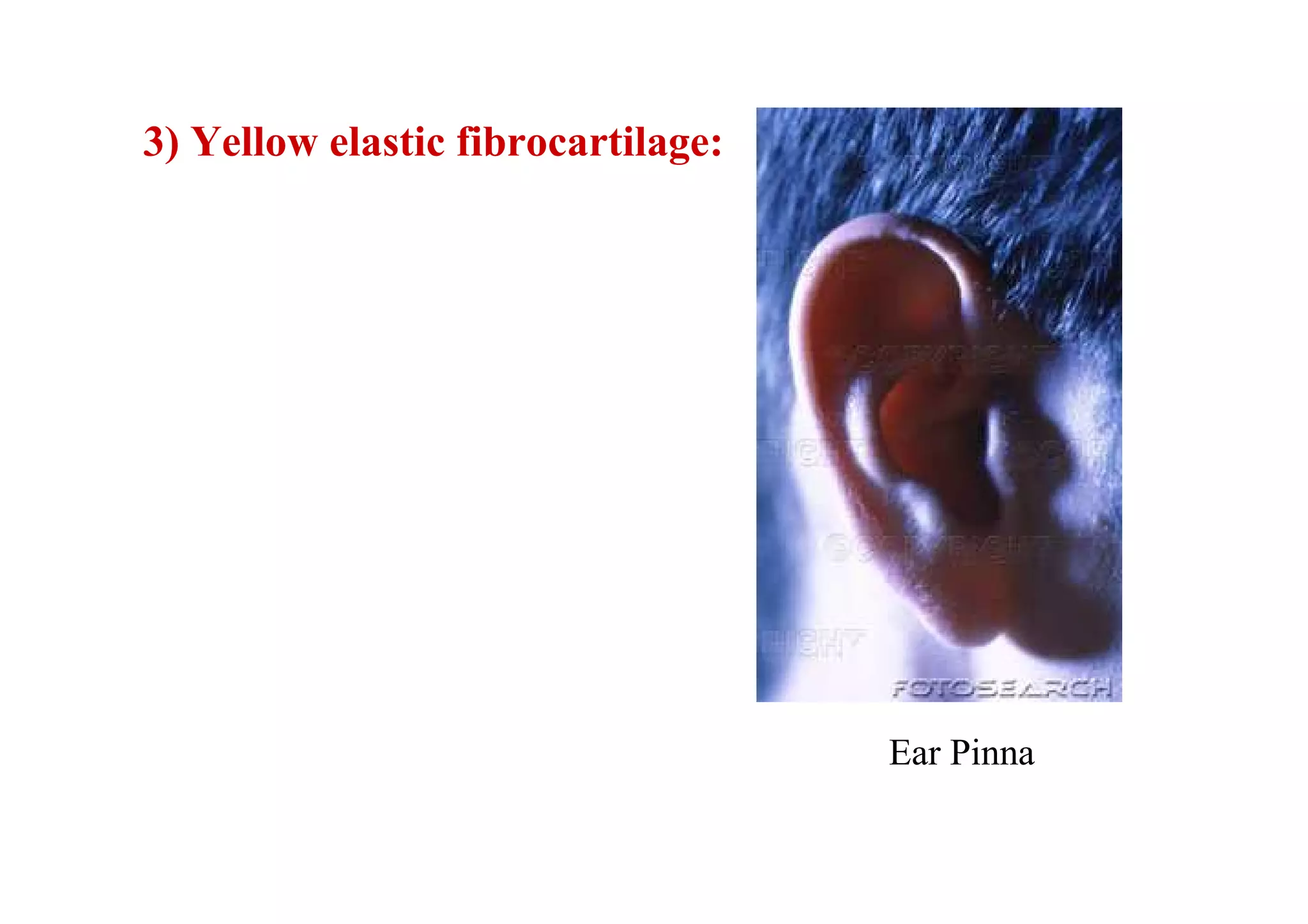


This document provides an introduction to anatomy by defining key terms and concepts. It discusses the different disciplines of anatomy, including macroscopic, microscopic, developmental, and neuroanatomy. It also describes the anatomical position, anatomical planes, and terms used to describe directions in the body. The document outlines the major components of the skeletal system, including different bone types. It provides examples of bone markings and functions. Finally, it introduces the different types of cartilage in the body.


![10. triangles of neck, tmj & applied anatomy[1]](https://cdn.slidesharecdn.com/ss_thumbnails/10-trianglesofnecktmjappliedanatomy1-100604084704-phpapp01-thumbnail.jpg?width=640&height=640&fit=bounds)


