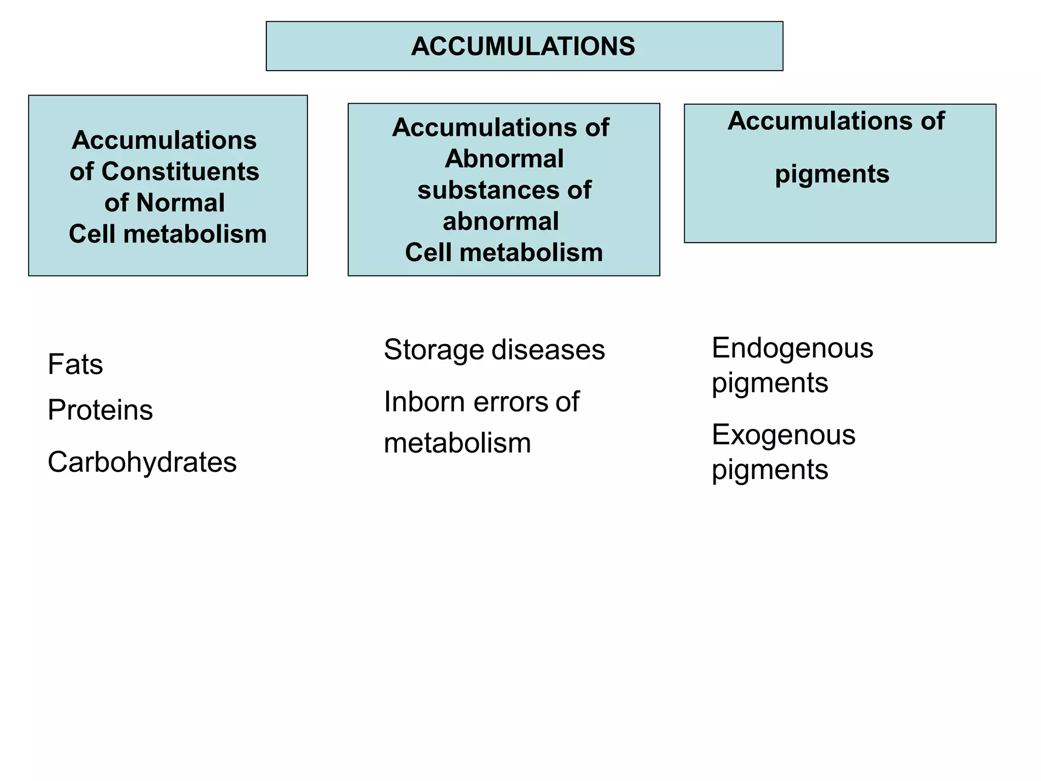
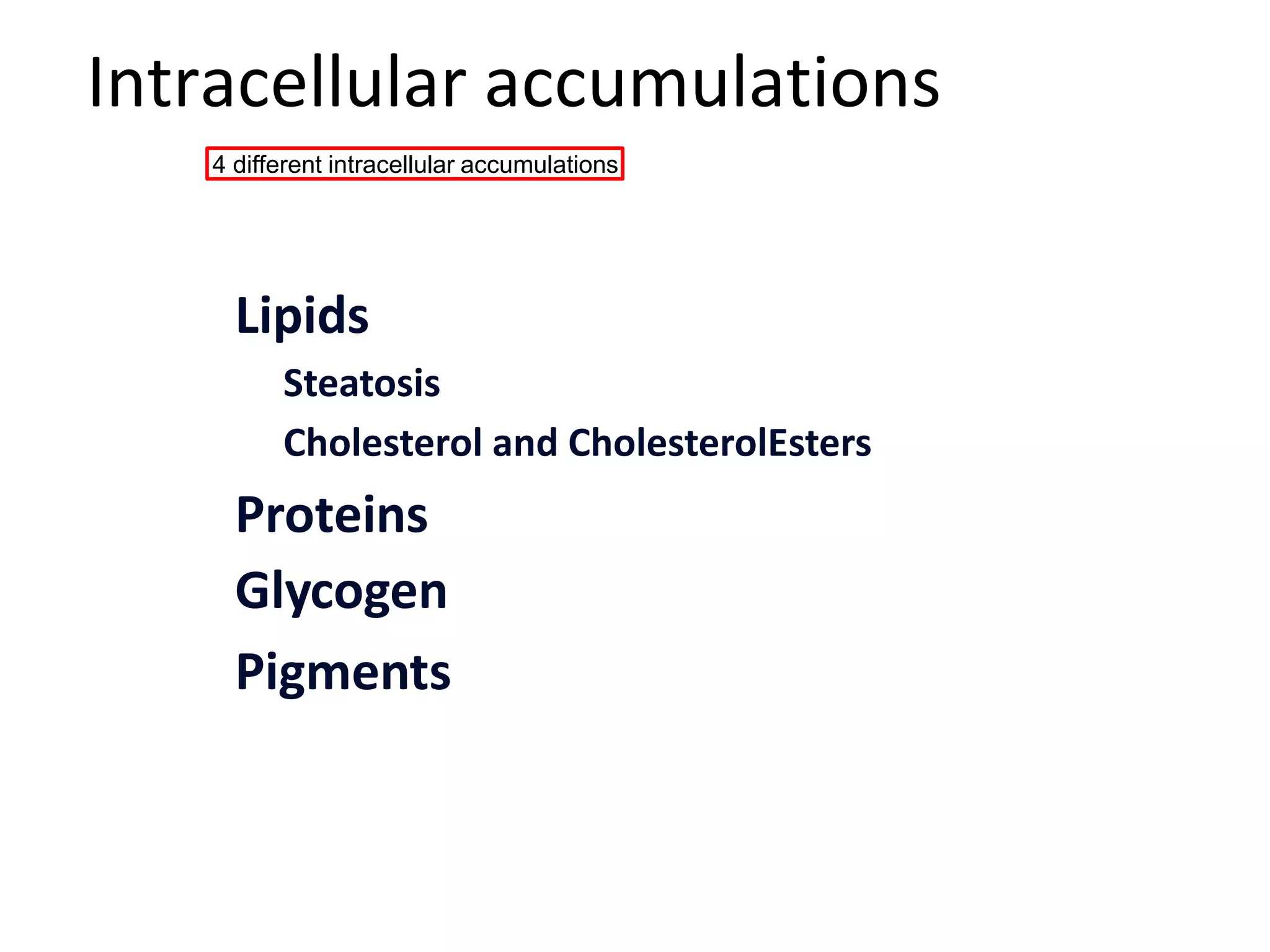
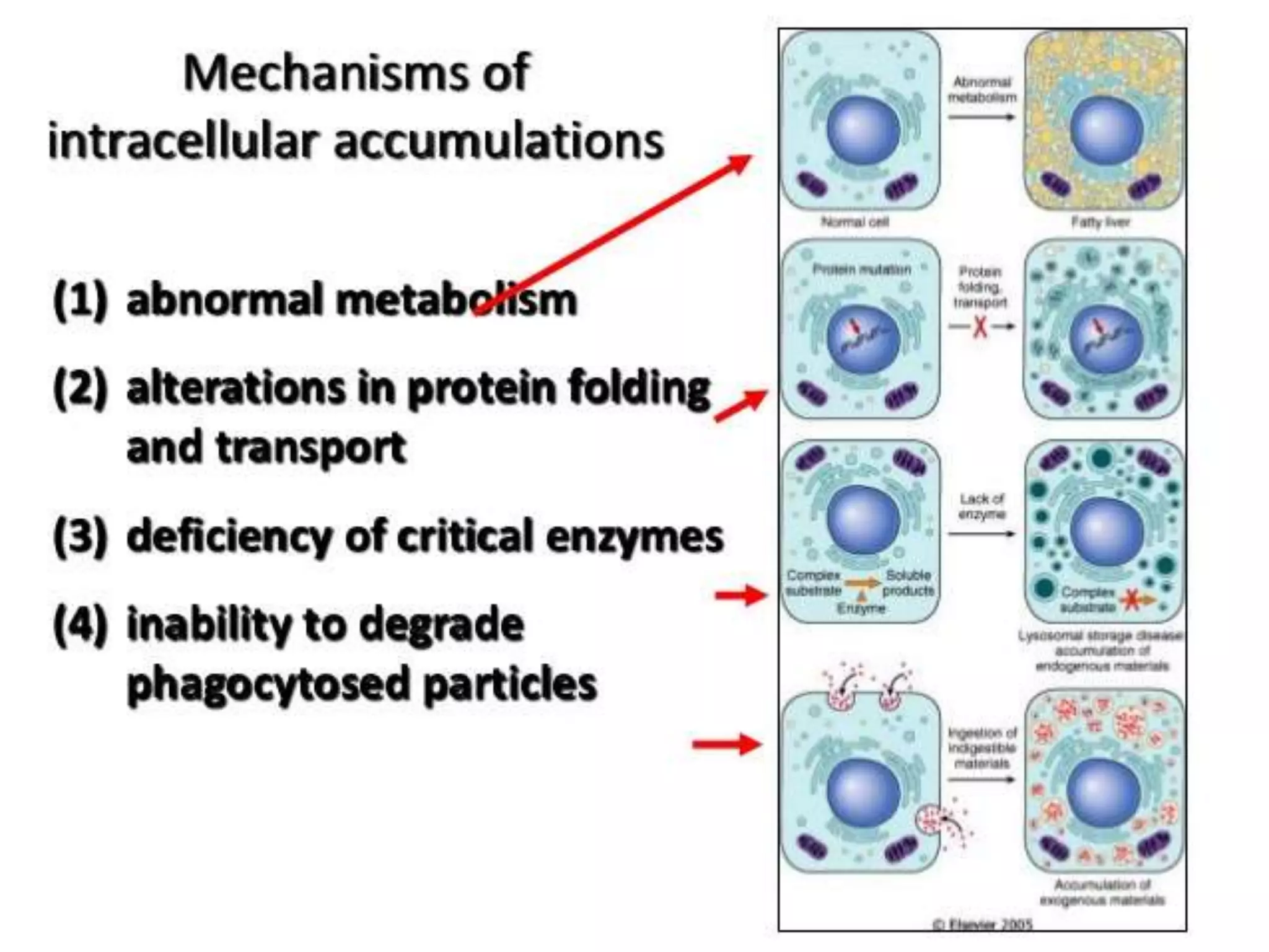
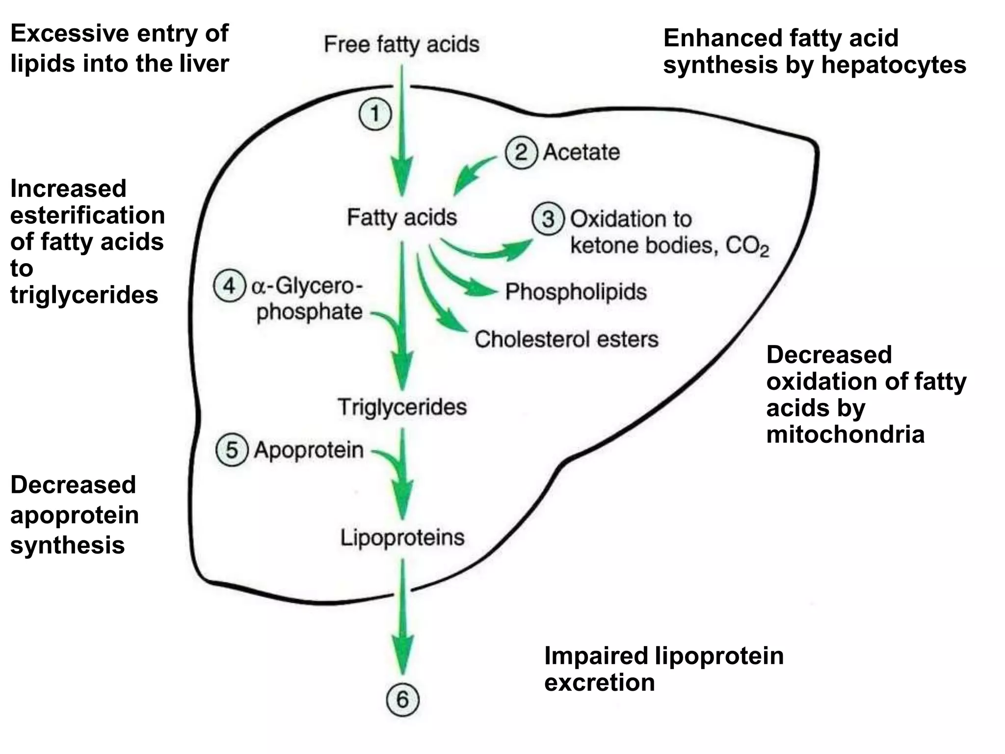
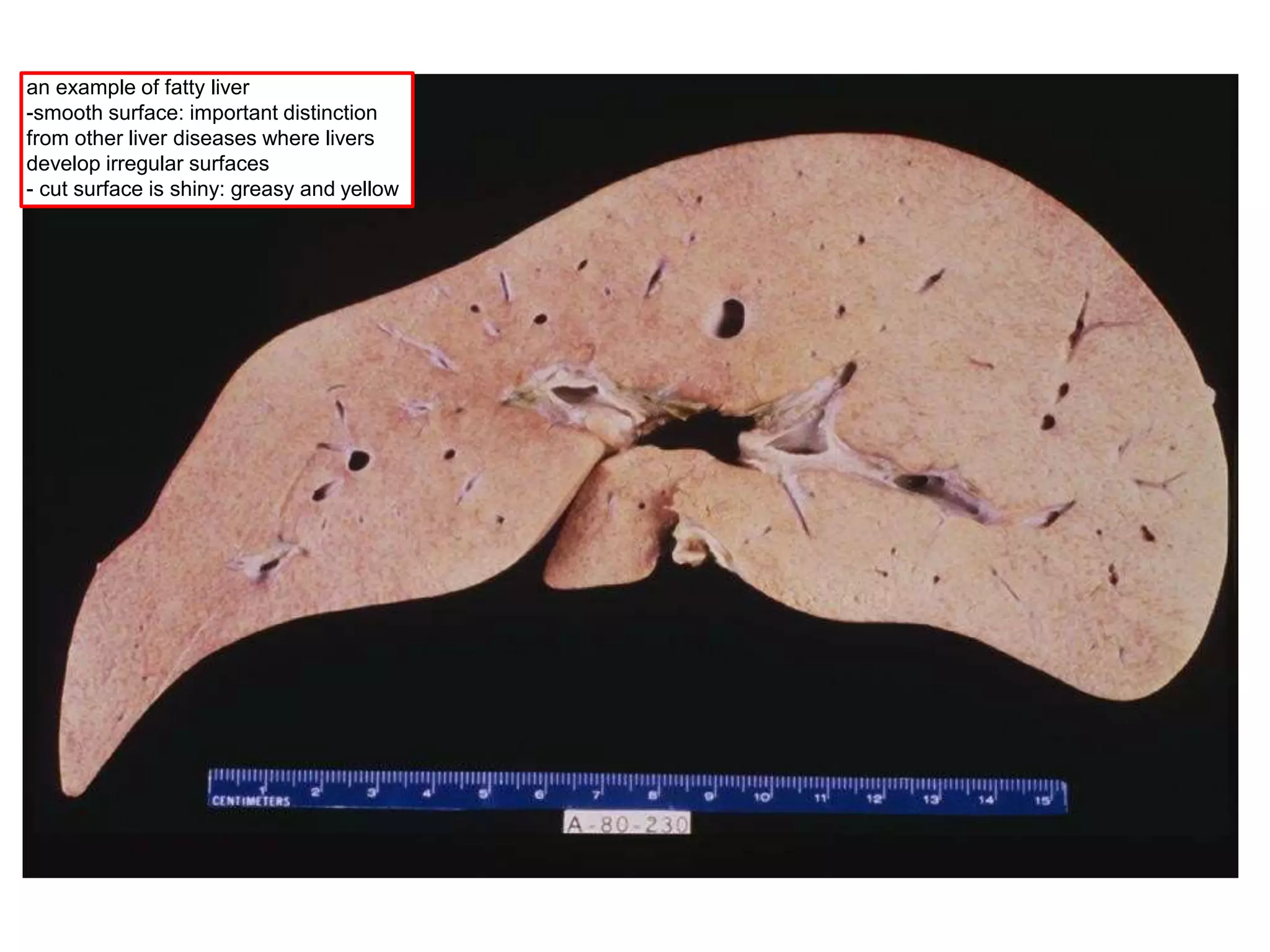
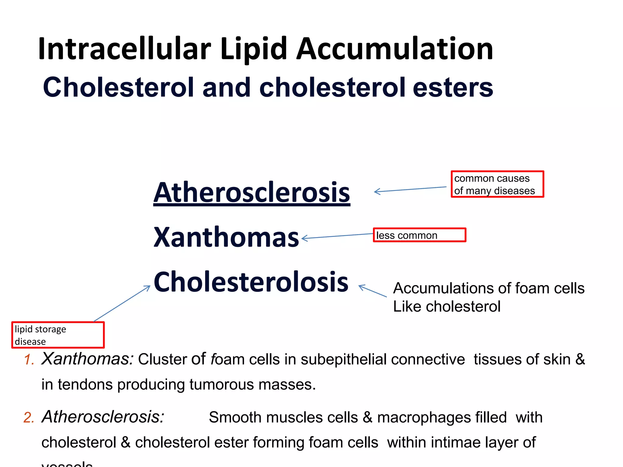
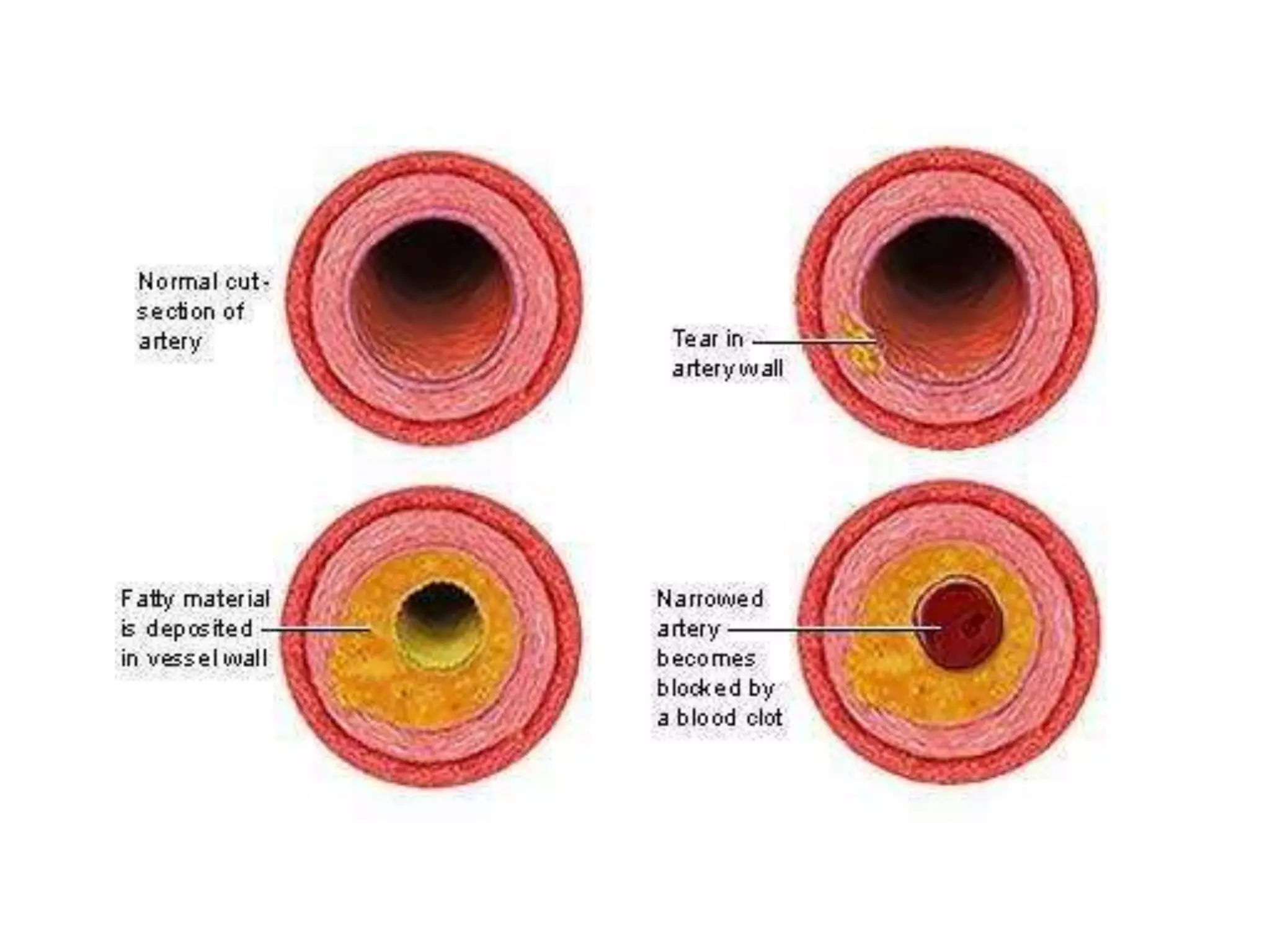
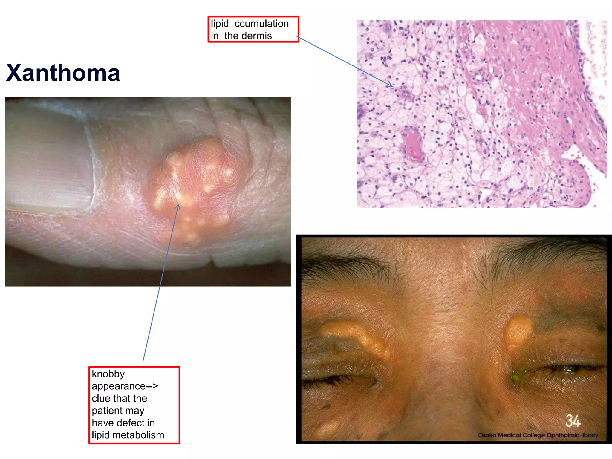
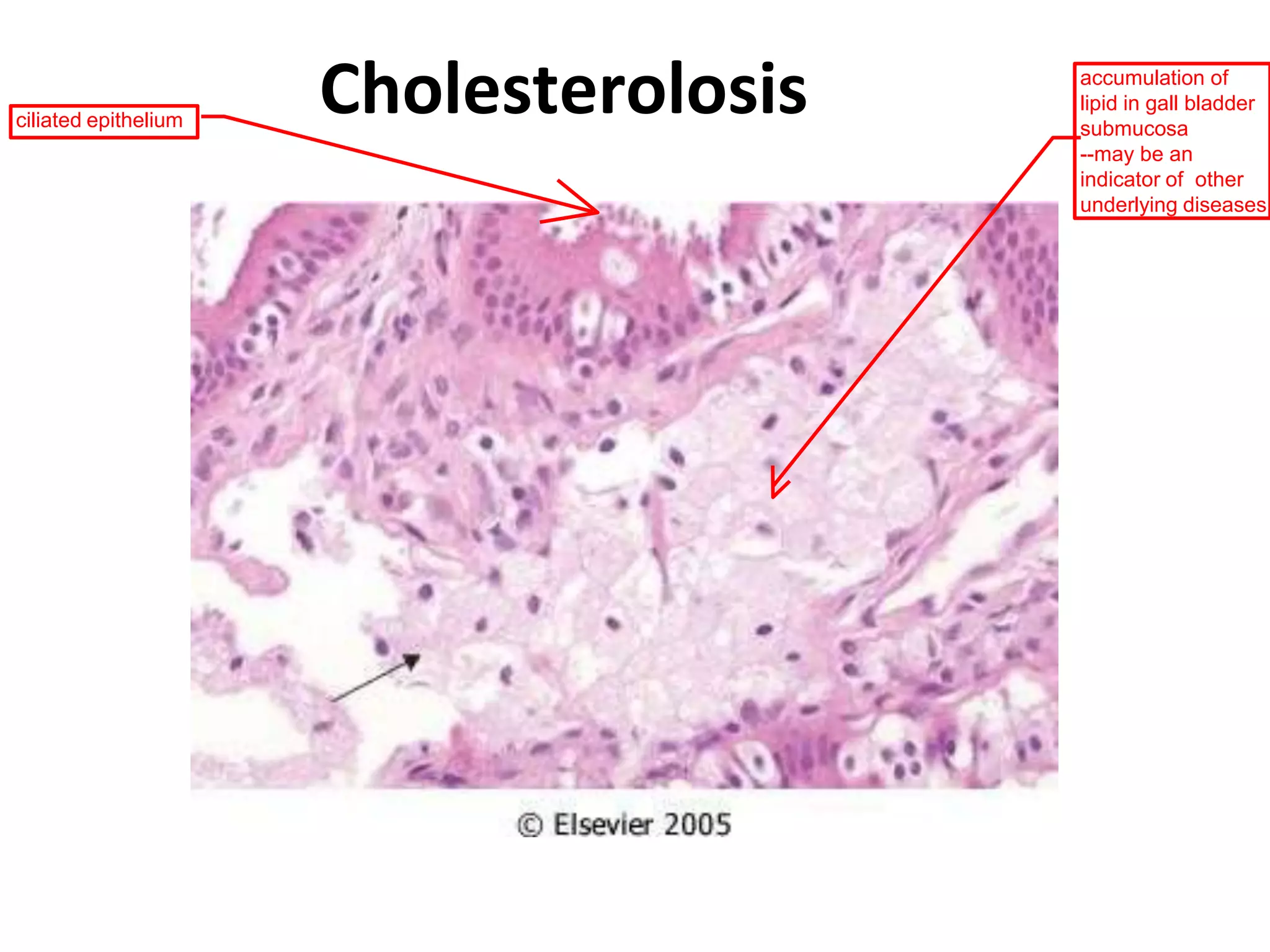
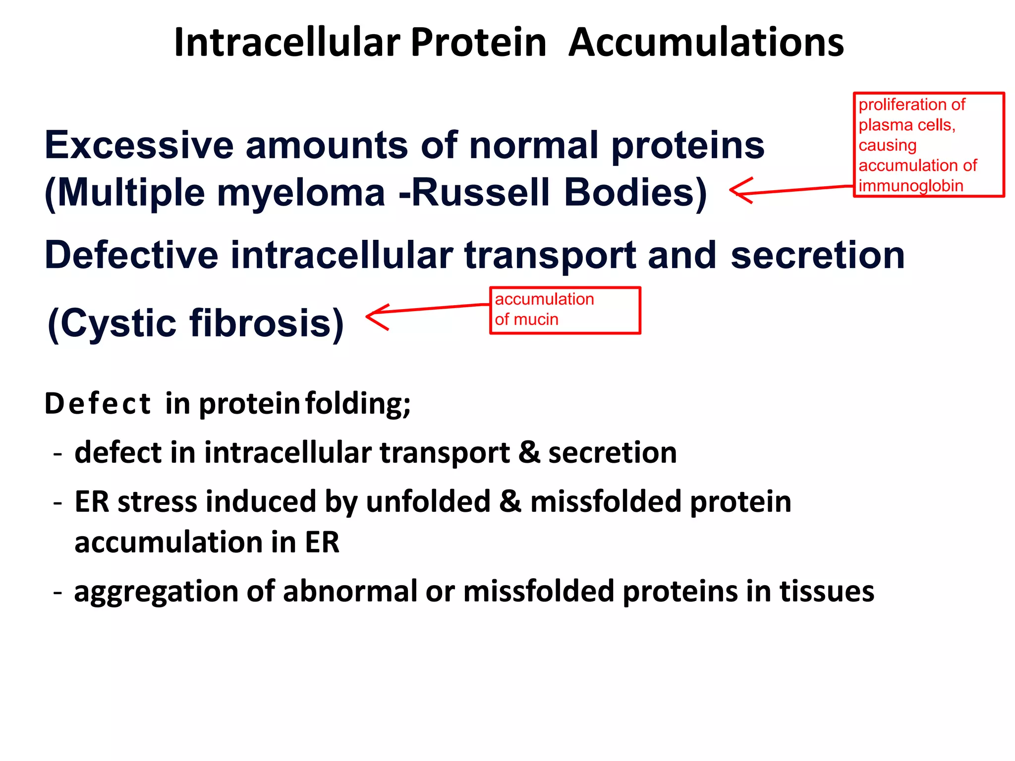
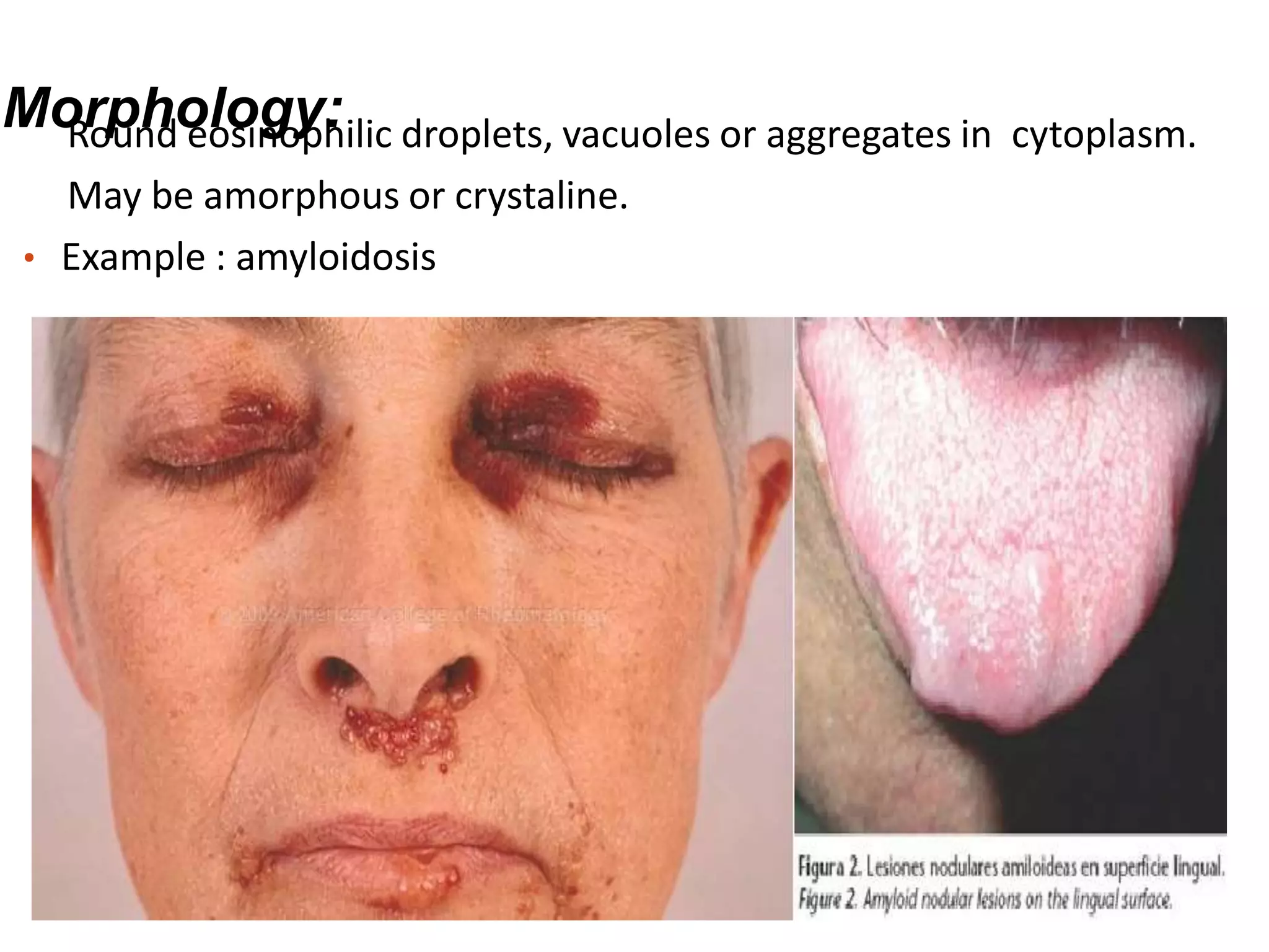
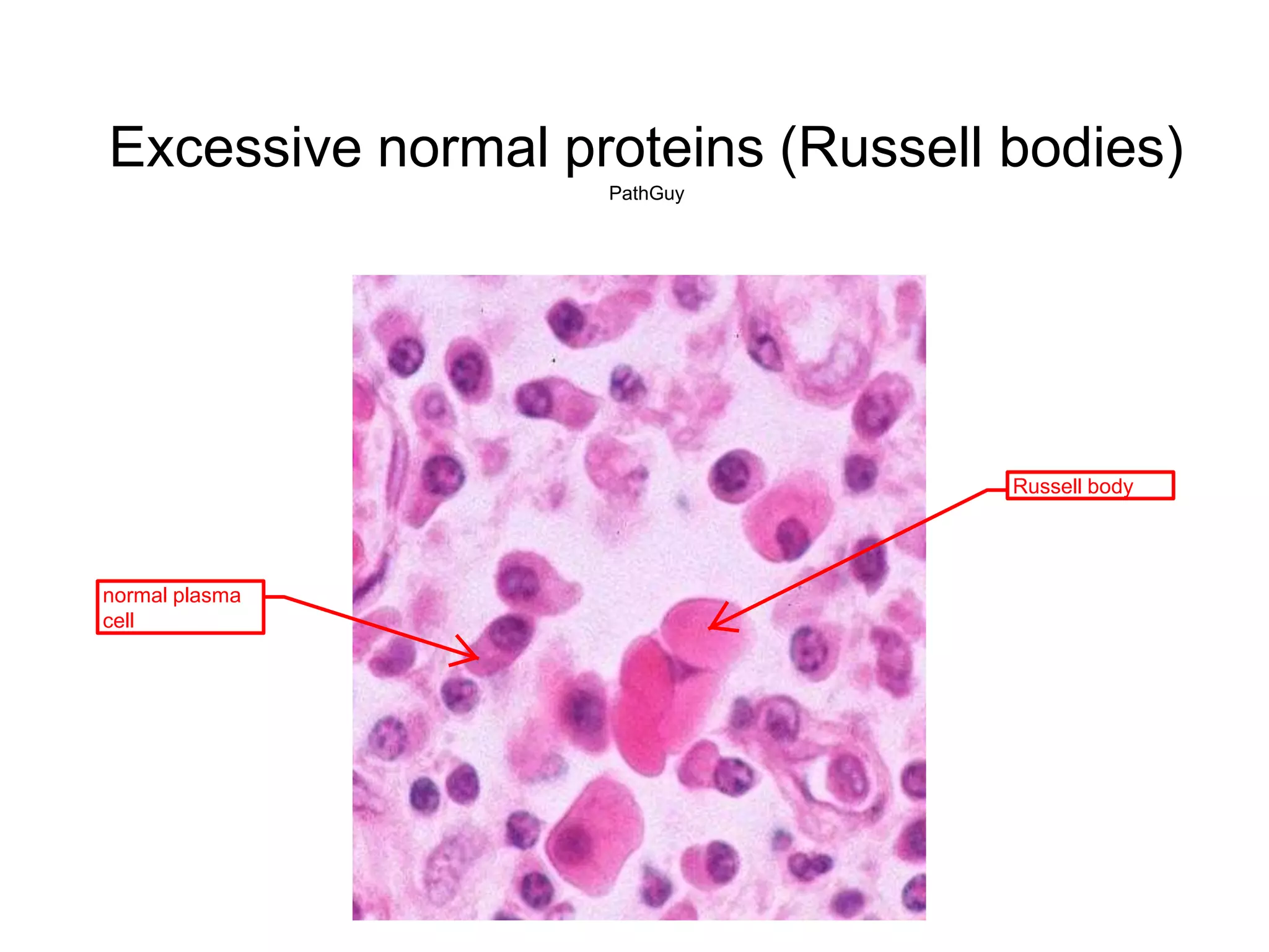
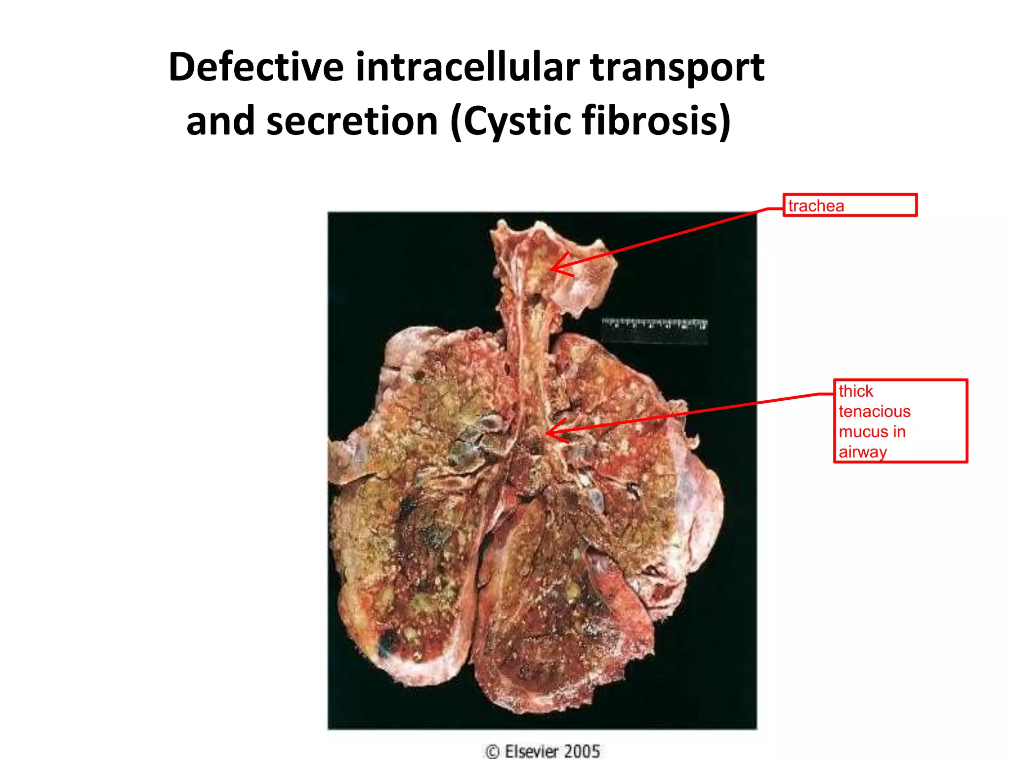
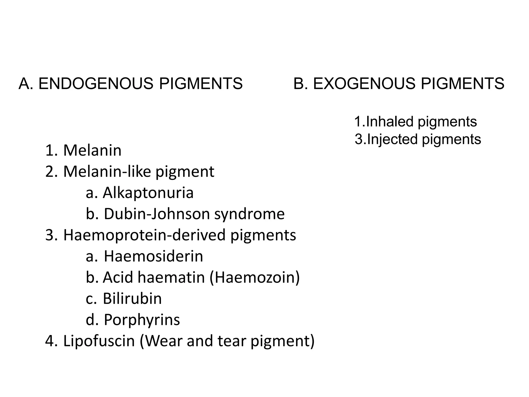
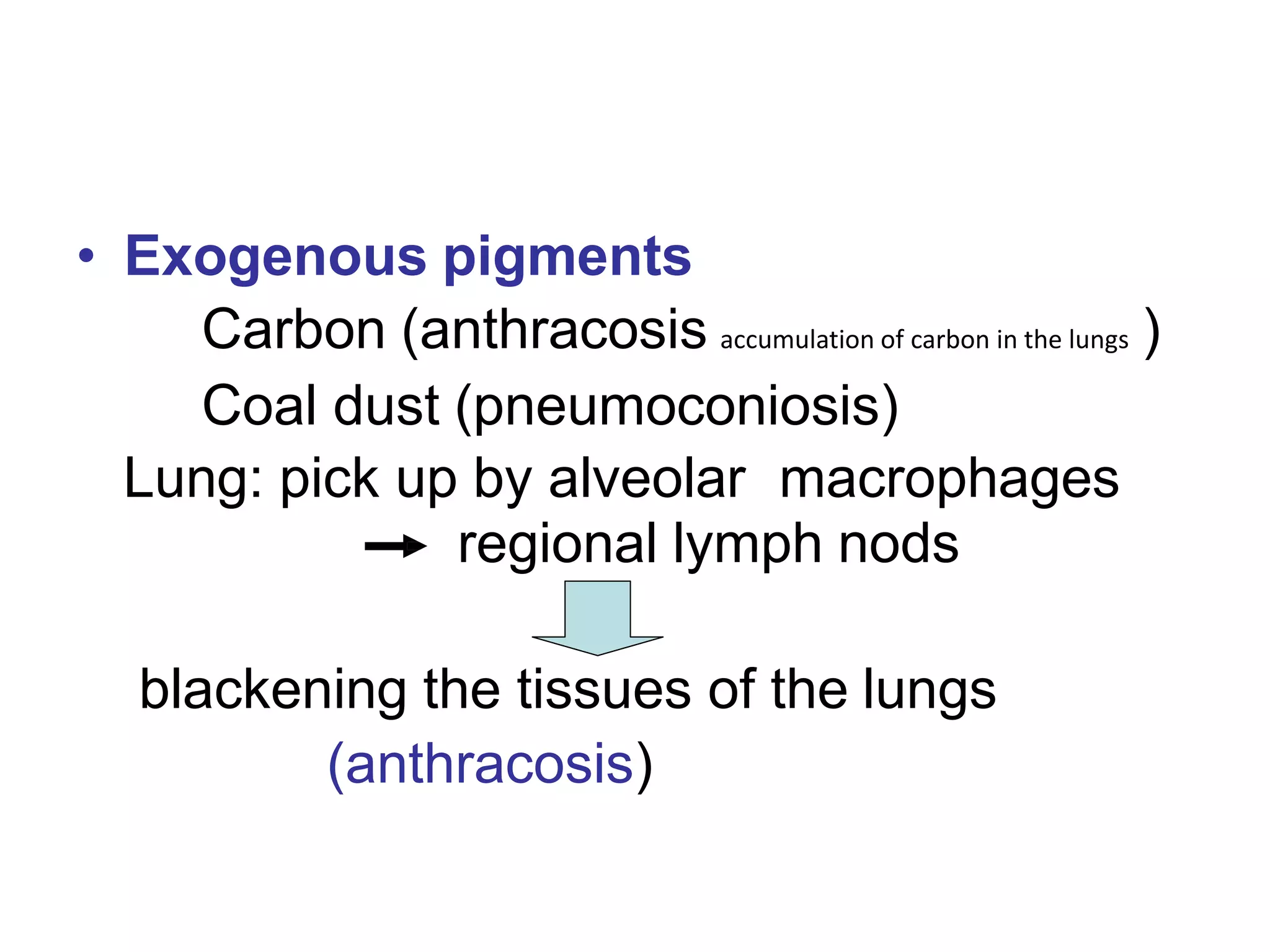
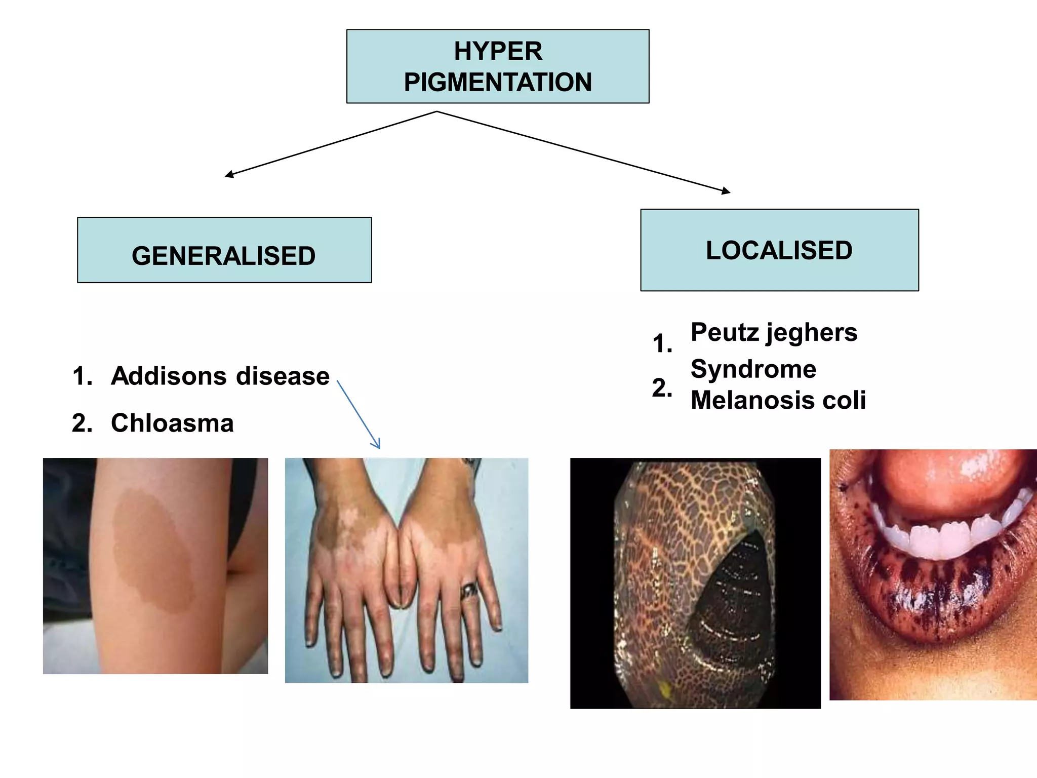
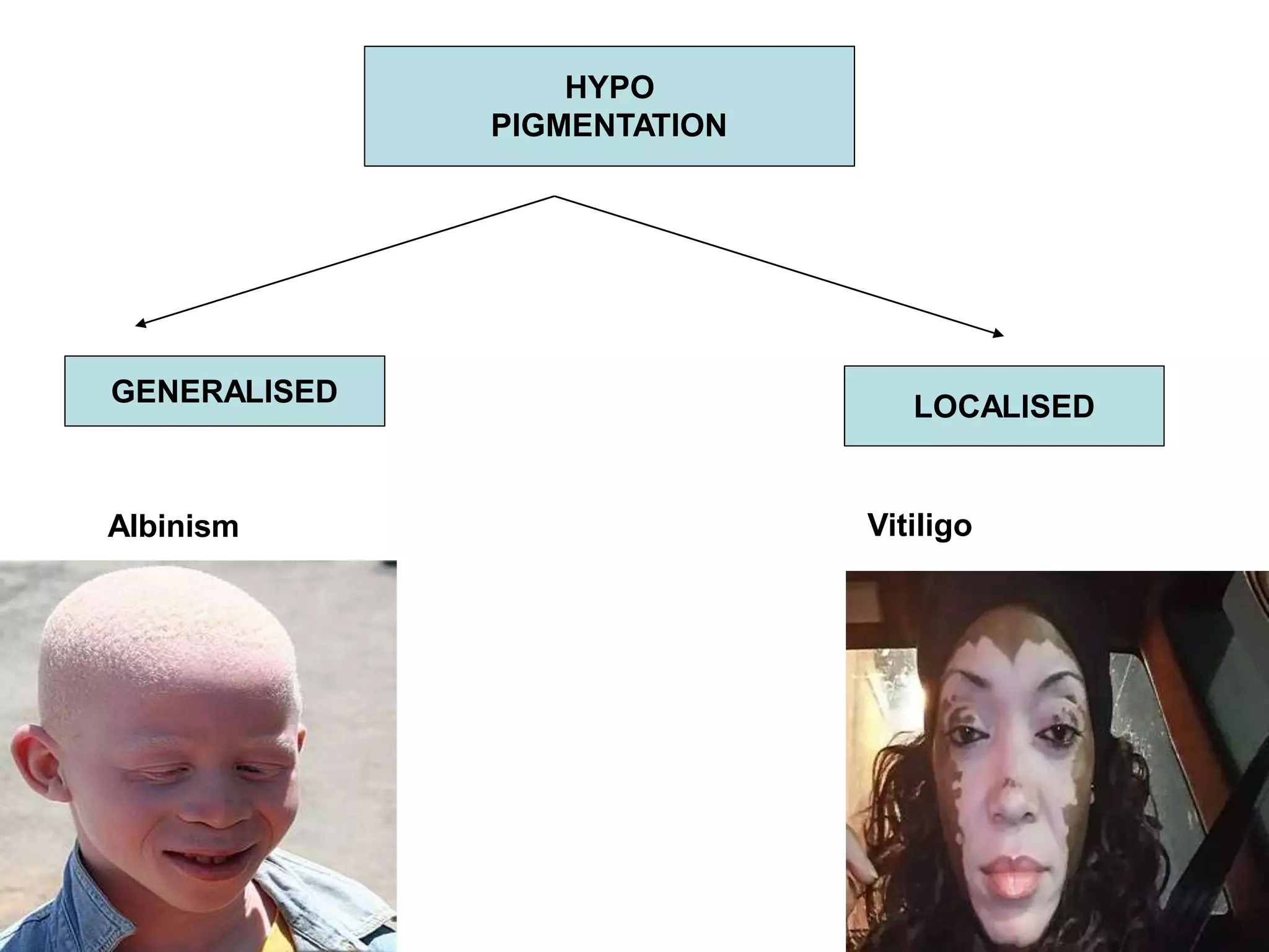
PRESENTATIONON:
Intracellular Accumulation
Presented by: Mohammad Muztaba
Asst. Professor(Jr.)
Department of Pharmacology Bhavdiya institute sibar sohawal
Ayodhya
Intracellular accumulations
• Manifestation of metabolic derangement
• Accumulation may be transient & reversible or
permanent. Effects: range from harmless to toxic
• Three categories
Accumulations
of Constituents
of Normal
Cell metabolism
Accumulations of
Abnormal
substances of
abnormal
Cell metabolism
Accumulations of
pigments
Fats
Proteins
Carbohydrates
Storage diseases
Inborn errors of
metabolism
Endogenous
pigments
Exogenous
pigments
ACCUMULATIONS
Intracellular accumulations
4 different intracellular accumulations
Lipids
Steatosis
Cholesterol and CholesterolEsters
Proteins
Glycogen
Pigments
FATTY CHANGE (Steatosis)
• Fatty metamorphosis
• Intracellular accumulation of neutral
fat(triglycerides) within the parenchymal
cells.
• Common in liver
• Can occur in heart, skeletal muscle and
kidneys.
• Causes of fatty liver
• - ALCHOL ABUSE (alcohol dependence)
• - DIABETES MELLITUS
• OBESITY (Obesity is a condition in which a person has excess
body fat)
• - PROTIEN MALNUTRION (starvation suffering or death caused
by lack of food.)
• -DRUGS/TOXINS
• -ANOXIA
• -PREGNANCY
Excessive entry of
lipids into the liver
Enhanced fatty acid
synthesis by hepatocytes
Decreased
oxidation of fatty
acids by
mitochondria
Increased
esterification
of fatty acids
to
triglycerides
Decreased
apoprotein
synthesis
Impaired lipoprotein
excretion
an example of fatty liver
-smooth surface: important distinction
from other liver diseases where livers
develop irregular surfaces
- cut surface is shiny: greasy and yellow
Other Lipid Accumulations
• Cholesterol and cholesterol esters
– In atherosclerosis, cholesterol accumulates in
smooth muscle cells and macrophages in the
intima of arteries
– In hereditary hyperlipemia, cholesterol
accumulates in macrophages, usually under
the skin, forming tumor-like structures known
as xanthomas
Intracellular Lipid Accumulation
Cholesterol and cholesterol esters
Atherosclerosis
Xanthomas
Cholesterolosis
common causes
of many diseases
less common
lipid storage
disease
1. Xanthomas: Cluster of foam cells in subepithelial connective tissues of skin &
in tendons producing tumorous masses.
2. Atherosclerosis: Smooth muscles cells & macrophages filled with
cholesterol & cholesterol ester forming foam cells within intimae layer of
Accumulations of foam cells
Like cholesterol
Xanthoma
lipid ccumulation
in the dermis
knobby
appearance-->
clue that the
patient may
have defect in
lipid metabolism
Cholesterolosisciliated epithelium
accumulation of
lipid in gall bladder
submucosa
--may be an
indicator of other
underlying diseases
Intracellular Protein Accumulations
Defective intracellular transport and secretion
Excessive amounts of normal proteins
(Multiple myeloma -Russell Bodies)
accumulation
of mucin
(Cystic fibrosis)
proliferation of
plasma cells,
causing
accumulation of
immunoglobin
Defect in proteinfolding;
- defect in intracellular transport & secretion
- ER stress induced by unfolded & missfolded protein
accumulation in ER
- aggregation of abnormal or missfolded proteins in tissues
Morphology:Round eosinophilic droplets, vacuoles or aggregates in cytoplasm.
May be amorphous or crystaline.
• Example : amyloidosis
Excessive normal proteins (Russell bodies)
PathGuy
normal plasma
cell
Russell body
Defective intracellular transport
and secretion (Cystic fibrosis)
trachea
thick
tenacious
mucus in
airway
Intra cellular accumulations
GLYCOGEN
Abnormal metabolism of glucose or glycogen result in
excessive intracellular accumulation of glycogen
Example… uncontrolled diabetes mellitus (main example
of abnormal glucose metabolism
Intra cellular accumulations
Pigments
DEFINITION
• Pigments are coloured substances, some of
which are normal constituents of cells (e.g.,
melanin), whereas others are abnormal and
accumulate in cells only under special
circumstances
• Pigments can be exogenous, coming from
outside the body,
• or endogenous, synthesized within the
body itself.
A. ENDOGENOUS PIGMENTS B. EXOGENOUS PIGMENTS
1.Inhaled pigments
3.Injected pigments
1. Melanin
2. Melanin-like pigment
a. Alkaptonuria
b. Dubin-Johnson syndrome
3. Haemoprotein-derived pigments
a. Haemosiderin
b. Acid haematin (Haemozoin)
c. Bilirubin
d. Porphyrins
4. Lipofuscin (Wear and tear pigment)
Exogenous pigments
Exogenous pigments are the pigments
introduced into the body from outside such as
by inhalation, ingestion or inoculation.
• Exogenous pigments
Carbon (anthracosis accumulation of carbon in the lungs )
Coal dust (pneumoconiosis)
Lung: pick up by alveolar macrophages
regional lymph nods
blackening the tissues of the lungs
(anthracosis)
Here is anthracotic pigment
in macrophages in a hilar
lymph node
The black streaks seen
between lobules of lung
beneath the pleural
surface are due to
accumulation of
anthracotic pigment.
Injected Pigments (Tattooing)
Pigments like India ink, cinnabar and carbon are introduced into
the dermis in the process of tattooing where the pigment is taken
up by macrophages and lies permanently in the connective tissue.
The examples of injected pigments are
Prolonged use of ointments containing mercury, and tattooing by
pricking the skin with dyes.
Lipofuschin
• fine yellow-brown pigment granules
composed of lipid-containing residues
of liposomal digestion.
• found in the liver, kidney, heart muscle,
retina, adrenals, nerve cells, and
ganglion cells
Melanin
The pigment that gives human skin, hair, and eyes their
color.
Melanin is produced by cells called melanocytes.
Melanin is group of natural pigments found in most
organisms.
Melanin is produced through a multistage chemical
process known as melanogenesis,
where the oxidation of the amino acid tyrosine, is
followed by polymerization
DISORDERS OF
MELANIN
PIGMENTATION
HYPER
PIGMENTATION
HYPO
PIGMENTATION
HYPER
PIGMENTATION
GENERALISED
1.
1. Addisons disease 2.
2. Chloasma
LOCALISED
Peutz jeghers
Syndrome
Melanosis coli
HYPO
PIGMENTATION
GENERALISED
Albinism
LOCALISED
Vitiligo
Hemosiderin
• Hemoglobin-derived, golden yellow-to-
brown, granular or crystalline pigment
• Major storage forms of iron
• Hemosiderin or haemosiderin is an iron-
storage complex. It is only found within cells
(as opposed to circulating in blood) and
appears to be a complex of ferritin,(Ferritin is a
universal intracellular protein that stores iron ) denatured
ferritin and other material. The iron within
deposits of hemosiderin is very poorly
available to supply iron when needed.
Excess of iron
Iron pigment appears as a coarse, golden, granular pigment lying
within the cell's cytoplasm

Intracell accumulation final

  • 1.
    PRESENTATIONON: Intracellular Accumulation Presented by:Mohammad Muztaba Asst. Professor(Jr.) Department of Pharmacology Bhavdiya institute sibar sohawal Ayodhya
  • 2.
    Intracellular accumulations • Manifestationof metabolic derangement • Accumulation may be transient & reversible or permanent. Effects: range from harmless to toxic • Three categories
  • 3.
    Accumulations of Constituents of Normal Cellmetabolism Accumulations of Abnormal substances of abnormal Cell metabolism Accumulations of pigments Fats Proteins Carbohydrates Storage diseases Inborn errors of metabolism Endogenous pigments Exogenous pigments ACCUMULATIONS
  • 4.
    Intracellular accumulations 4 differentintracellular accumulations Lipids Steatosis Cholesterol and CholesterolEsters Proteins Glycogen Pigments
  • 6.
    FATTY CHANGE (Steatosis) •Fatty metamorphosis • Intracellular accumulation of neutral fat(triglycerides) within the parenchymal cells. • Common in liver • Can occur in heart, skeletal muscle and kidneys.
  • 7.
    • Causes offatty liver • - ALCHOL ABUSE (alcohol dependence) • - DIABETES MELLITUS • OBESITY (Obesity is a condition in which a person has excess body fat) • - PROTIEN MALNUTRION (starvation suffering or death caused by lack of food.) • -DRUGS/TOXINS • -ANOXIA • -PREGNANCY
  • 8.
    Excessive entry of lipidsinto the liver Enhanced fatty acid synthesis by hepatocytes Decreased oxidation of fatty acids by mitochondria Increased esterification of fatty acids to triglycerides Decreased apoprotein synthesis Impaired lipoprotein excretion
  • 9.
    an example offatty liver -smooth surface: important distinction from other liver diseases where livers develop irregular surfaces - cut surface is shiny: greasy and yellow
  • 10.
    Other Lipid Accumulations •Cholesterol and cholesterol esters – In atherosclerosis, cholesterol accumulates in smooth muscle cells and macrophages in the intima of arteries – In hereditary hyperlipemia, cholesterol accumulates in macrophages, usually under the skin, forming tumor-like structures known as xanthomas
  • 11.
    Intracellular Lipid Accumulation Cholesteroland cholesterol esters Atherosclerosis Xanthomas Cholesterolosis common causes of many diseases less common lipid storage disease 1. Xanthomas: Cluster of foam cells in subepithelial connective tissues of skin & in tendons producing tumorous masses. 2. Atherosclerosis: Smooth muscles cells & macrophages filled with cholesterol & cholesterol ester forming foam cells within intimae layer of Accumulations of foam cells Like cholesterol
  • 13.
    Xanthoma lipid ccumulation in thedermis knobby appearance--> clue that the patient may have defect in lipid metabolism
  • 14.
    Cholesterolosisciliated epithelium accumulation of lipidin gall bladder submucosa --may be an indicator of other underlying diseases
  • 15.
    Intracellular Protein Accumulations Defectiveintracellular transport and secretion Excessive amounts of normal proteins (Multiple myeloma -Russell Bodies) accumulation of mucin (Cystic fibrosis) proliferation of plasma cells, causing accumulation of immunoglobin Defect in proteinfolding; - defect in intracellular transport & secretion - ER stress induced by unfolded & missfolded protein accumulation in ER - aggregation of abnormal or missfolded proteins in tissues
  • 16.
    Morphology:Round eosinophilic droplets,vacuoles or aggregates in cytoplasm. May be amorphous or crystaline. • Example : amyloidosis
  • 17.
    Excessive normal proteins(Russell bodies) PathGuy normal plasma cell Russell body
  • 18.
    Defective intracellular transport andsecretion (Cystic fibrosis) trachea thick tenacious mucus in airway
  • 19.
    Intra cellular accumulations GLYCOGEN Abnormalmetabolism of glucose or glycogen result in excessive intracellular accumulation of glycogen Example… uncontrolled diabetes mellitus (main example of abnormal glucose metabolism
  • 20.
  • 21.
    DEFINITION • Pigments arecoloured substances, some of which are normal constituents of cells (e.g., melanin), whereas others are abnormal and accumulate in cells only under special circumstances • Pigments can be exogenous, coming from outside the body, • or endogenous, synthesized within the body itself.
  • 22.
    A. ENDOGENOUS PIGMENTSB. EXOGENOUS PIGMENTS 1.Inhaled pigments 3.Injected pigments 1. Melanin 2. Melanin-like pigment a. Alkaptonuria b. Dubin-Johnson syndrome 3. Haemoprotein-derived pigments a. Haemosiderin b. Acid haematin (Haemozoin) c. Bilirubin d. Porphyrins 4. Lipofuscin (Wear and tear pigment)
  • 23.
    Exogenous pigments Exogenous pigmentsare the pigments introduced into the body from outside such as by inhalation, ingestion or inoculation.
  • 24.
    • Exogenous pigments Carbon(anthracosis accumulation of carbon in the lungs ) Coal dust (pneumoconiosis) Lung: pick up by alveolar macrophages regional lymph nods blackening the tissues of the lungs (anthracosis)
  • 25.
    Here is anthracoticpigment in macrophages in a hilar lymph node The black streaks seen between lobules of lung beneath the pleural surface are due to accumulation of anthracotic pigment.
  • 26.
    Injected Pigments (Tattooing) Pigmentslike India ink, cinnabar and carbon are introduced into the dermis in the process of tattooing where the pigment is taken up by macrophages and lies permanently in the connective tissue. The examples of injected pigments are Prolonged use of ointments containing mercury, and tattooing by pricking the skin with dyes.
  • 27.
    Lipofuschin • fine yellow-brownpigment granules composed of lipid-containing residues of liposomal digestion. • found in the liver, kidney, heart muscle, retina, adrenals, nerve cells, and ganglion cells
  • 30.
    Melanin The pigment thatgives human skin, hair, and eyes their color. Melanin is produced by cells called melanocytes. Melanin is group of natural pigments found in most organisms. Melanin is produced through a multistage chemical process known as melanogenesis, where the oxidation of the amino acid tyrosine, is followed by polymerization
  • 31.
  • 32.
    HYPER PIGMENTATION GENERALISED 1. 1. Addisons disease2. 2. Chloasma LOCALISED Peutz jeghers Syndrome Melanosis coli
  • 33.
  • 34.
    Hemosiderin • Hemoglobin-derived, goldenyellow-to- brown, granular or crystalline pigment • Major storage forms of iron • Hemosiderin or haemosiderin is an iron- storage complex. It is only found within cells (as opposed to circulating in blood) and appears to be a complex of ferritin,(Ferritin is a universal intracellular protein that stores iron ) denatured ferritin and other material. The iron within deposits of hemosiderin is very poorly available to supply iron when needed. Excess of iron
  • 35.
    Iron pigment appearsas a coarse, golden, granular pigment lying within the cell's cytoplasm