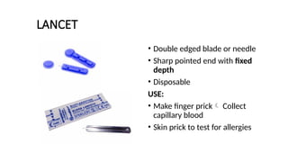
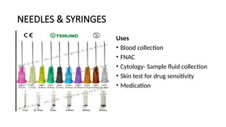
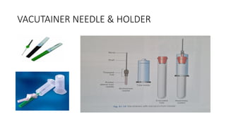
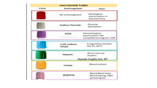
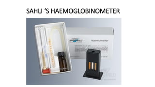
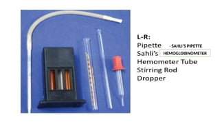
INSTRUMENT
S
HEMATOLOGY INSTRUMENTS
LANCET
• Double edged blade or needle
• Sharp pointed end with fixed
depth
• Disposable
USE:
• Make finger prick Collect
capillary blood
• Skin prick to test for allergies
NEEDLES & SYRINGES
Uses
• Blood collection
• FNAC
• Cytology- Sample fluid collection
• Skin test for drug sensitivity
• Medication
VACUTAINER NEEDLE & HOLDER
Osmotic fragility test, IPT
SAHLI ‘S HAEMOGLOBINOMETER
- SAHLI’S PIPETTE
HEMOGLOBINOMETER
• Instruments
– Sahli’s hemoglobinometer- standard brown glass
blocks mounted on a comparator
– Sahli’s hemoglobinometer tube- Graduated tube 2-22 G
%,10-140%
– Sahli’s pipette- Special pipette to measure out 20
microlitre of blood
• Reagents
– N/10 HCl
– Distilled water
Sahli’s method / Acid hematin method
Principle
• Hb + N/10 HCl Acid hematin (brown colour)
• Brown colour compared with standard brown glass
reference blocks
• Intensity of brown colour α amount of acid hematin
α amount of Hb in the blood sample
Sahli’s pipette
AMBER COLOURED BOTTLE
• Protect light sensitive chemicals
• N/10 HCl
HAEMOCYTOMETER
1. RBC PIPETTE
Uses
• RBC count
• Platelet count
• WBC count when total count
is very high
FEATURES
• Markings – 0.5,1 & 101
• Red bead
• Blood is drawn upto 0.5 mark & diluent upto 101 mark
• Dilution – 1:200
• Diluting fluid – Hayem’s fluid
• Mercuric chloride (0.25g)- Fixes, preserves and prevents the growth of
bacteria
• Sodium chloride (0.5 g)- iso – osmolarity
• Sodium sulfate (2.5 g)- Prevent clumping of RBC
• Distilled water – 100 ml
2. WBC PIPETTE
• Uses .
• Manual WBC count
• Absolute eosinophil count
• Sperm count
• Any fluid – Synovial fluid
count ,CSF
FEATURES …
• White glass bead in the bulb
• Markings – 0.5 , 1 & 11
• Blood is drawn upto 0.5 mark & diluent upto
11 mark.
• Dilution 1: 20
• Diluting fluid – Turk’s fluid
• Glacial acetic acid (2ml)- Lyses RBCs
• Gentian violet(1% sol,1 drp)- Stains WBC
nuclei
• Distilled water – 100 ml
RBC PIPETTE WBC PIPETTE
Red bead White BEAD
Larger bulb size Bulb size smaller
Lumen size Smaller Lumen size larger
Markings – 0.5,1 &101 Markings -0.5, 1 & 11
Dilution 1:200 Dilution 1:20
Improved NEUBAUER counting chamber
Other Hemocytometer
• Old Neubauer chamber
• Fuchs- Rosenthal counting
chamber
• Thoma chamber
• Burker chamber
• Louis-Charles Malassez
device
• Entire ruled area – square of 3
mm side
• 9 large squares – 1mm sides
• Central square – subdivided
into 400 smaller squares ,
0.050mm (1/20) side
• Each 5th line double line
• Smallest squares – 25 groups
of 16 squares each
• Depth of squares on placing
cover slip – 0.1 mm
16 large squares in central area 25 large squares in central area
Triple lines are widely spaced Triple lines are closely spaced
Separating lines are dull Separating lines are vivid and clear
THICK COVERSLIP
PROPERTIES
• Smooth surface
• Even thickness- 0.3mm, 0.4mm,
0.5mm
• Fluid depth of 0.1mm
• Sizes- 16x22mm, 22x23mm
CAPILLARY TUBES
•Uses
• To determine whole blood
coagulation time
•Procedure
– Capillary blood from finger prick is
collected into glass capillary tubes
– Small sections of tube are
broken off at timed intervals
– Time when blood strings out
between broken surface is
taken as clotting time.
PACKED CELL VOLUME (PCV) OR HAEMATOCRIT
• PCV is defined as ratio of volume of RBCs to that of
whole blood and is expressed as percentage
• Methods for Estimation of PCV
1. Macro method (Wintrobe’s method)
2. Micro haematocrit method (capillary tube 75mmx1mm)
3. Electronic method
NORMAL: 40-50% males
38-45 %- females
36-42%- pregnant ladies
RULE OF 3 and 9
Hb (gm/dl) x 3 = PCV
RBC (million/cmm) x 9 = PCV
Macro (Wintrobe’s) Method
Procedure
• Fill the Wintrobe tube upto mark 10 with well mixed
anticoagulated blood (EDTA) by Pasteur’s pipette free
of air bubbles.
• Centrifuge the tube at 2000-2300 g for 30 minutes.
• After centrifugation layers are noted in the wintrobe
tube
• i. Uppermost layer of plasma.
• ii. Thin white layer of platelets.
• iii. Greyish-pink layer of leucocytes.
• iv. Lowermost is the layer of RBCs.
• Note the lowermost height of column of packed RBC
layer and express it as percentage.
Advantages of Macro Method
• i. PCV and ESR can be
measured simultaneously.
• ii. Buffy coat can be
prepared for other tests.
• iii. By seeing the colour of
plasma we can know about
some of the pathological
conditions
• Jaundice it is yellow,
• Haemolysis it is pink,
• Hyperlipidaemia it is milky
PASTEUR PIPETTE
• Glass dropper with tapered end fitted
with a rubber bulb
• Named after Louis Pasteur
USE:
• Fill Wintrobe’s tube with blood
• Transfer liquid or chemicals
WINTROBE ‘s tube
• Thick walled glass tube
• 11 cm long , 2.5 mm internal diameter
• Flat inner base
• Marked at 1 mm interval from both sides, 0-10 and 10-0
• Holds 1 ml of blood
• Venous blood is used
• Anticoagulants- EDTA or heparin or wintrobe’s mixture
• Primarily for Packed Cell Volume
• Erythrocyte Sedimentation Rate
• Buffy coat smear preparation for-
• LE cell phenomenon,
• Abnormal cells – Aleukemic leukaemia
• Malarial parasite
Uses of wintrobe tube-
MICROHAEMATOCRIT METHOD
• Capillary tube
• 75mm length
• 1mm internal diameter
• Plain or heparinised
• Tube sealed with plastic sealant,
modelling clay or heat
• Centrifuged at 12000 g for 5
mins
ERYTHROCYTE SEDIMENTATION RATE
• It is defined as rate of
sedimentation of RBC in 1 hour
STAGES of ESR
• Phase of rouleaux formation:
Initial period of 10 minutes,
• Phase of settling: In the next 40
minutes, settling of RBCs occurs at
a constant rate. „
• Phase of packing: Last 10 minutes
sedimentation slows and packing
of the RBCs to the bottom occurs
• FACTORS INCREASING ESR
• Anemia (less negative charge – low
zeta potential)
• Macrocytosis
• Elevated fibrinogen
• High temperature
• Tilted tube
• FACTORS DECREASING ESR
• Polycythemia
• Microcytes sickle cells spherocytes
• Low fibrinogen
• Vibration
• Anticoagulant –3.8% Tri Sodium
citrate
• 4ml blood & 1 ml anticoagulant
– 4:1 ratio
• Iit is measured as length of the
plasma column in mm when a
vertical column of blood is kept
undisturbed for one hour
Methods of ESR
• 1. Westergren’s method
• 2. Wintrobe’s method
• 3. Micro ESR method- capillary
tube used
• 4. Automated methods
NORMAL VALUES
• Males 0-15 mm 1st hour
• Females 0-20 mm 1st hour
WESTERGREN’S TUBE
• Pipette with both ends open
• Long tube- 300mm length
• Markings on lower 2/3 from 0 to
200, from top to bottom
• Internal diameter 2.55mm
• Tubes are placed in stands in
which series of rubber discs are
attached
Procedure
• The patient is advised to come in the morning fasting (as
heavy protein diet affects concentraton of plasma proteins).
• Take 1.6 ml of patient’s blood and mix it with 0.4 ml of
citrate anticoagulant already put in a tube.
• Fill the pipette upto mark O with citrated blood with the
help of rubber teat by vacuum filling and fix it in a rack
vertically away from sun light or vibrations
• Let it stand for one hour after which reading is taken at the
upper meniscus of the RBCs.
ADVANTAGES
• i. It is a more sensitive method.
• ii. It is easy to fill and clean the Westergren’s pipette
Esrite ESR kit
• Contents
– Borosilicate glass tube-
25 cm, uncalibrated
– Graduated aluminum
screen
– Polystyrene stands
ANTISERA FOR ABO AND RH BLOOD GROUPING
• BLUE VIAL- Antisera-A
• YELLOW VIAL- Antisera-B
• RED VIAL – Antisera- AB
• GREY/COLOURLESS VIAL- Anti-D
USE:
ABO Blood grouping
Rh grouping
GLASS SLIDE
• Measures 7.5 x 2.5 x 0.1 cm
Use:
• Peripheral blood smear
• Blood grouping
• Reticulocyte count
• Cytology smear
• Urine microscopy
• Histopathology slide
TEST TUBES
Uses
• Blood grouping- Tube method
• Cytology- Fluid collection and
centrifugation
• Urine examination
• MICROBIOLOGY culture
BLOOD BAG
Anticoagulant and Preservative Solution :
• CPD( Citrate-phosphate-dextrose) – 21days (shelf life)
• CPD-Adenine(CPDA-1) – 35days
• SAGM( Saline-adenine-glucose-mannitol) – 42days
BONE MARROW ASPIRATION NEEDLE
Salah bone marrow aspiration needle
• Stout needle-stainless steel
• 7-8 cm length
• Stilette & guard
BM TREPHINE BIOPSY NEEDLE
JAMSHIDI TREPHINE BIOPSY NEEDLE
SITES:
• Sternum
• Iliac crest
• Anterior and posterior iliac spines
• Upper end of tibia
• Spinous process of vertebra
Indications Contraindications
• Unexplained anaemias, granulocytopenia,
thrombocytopenia,pancytopenia
• Megaloblastic anaemia
• Aplastic anaemia
• Leukemias
• MDS
• Infections- enteric fever
• Bone tumours n myeloma
• Staging of lymphoma
• Disseminated TB
• Bleeding disorders
• Local infections
CLINICAL PATHOLOGY
INSTRUMENTS
URINOMETER
• Identification
– Weighted cylinder-floats in urine
– Stem & bulb
– Markings 1.000 – 1.060 with divisions of
0.002
• Uses
– Determine specific gravity of urine
• Procedure
– 24hr sample, fill 3/4th of cylinder with urine
– Place urinometer without touching sides
– Take reading at eye level,noting the lower
meniscus
Normal specific gravity varies from 1.015 – 1.025
In severe renal diseases specific gravity become
fixed at 1.010
CORRECTION
 For temperature- for every 3 degree rise-
add 0.001, if fall- subtract 0.001
 For protein, subtract 0.003 for every
1gm/dl
 For glucose, subtract 0.004 for every
1gm/dl
 For dilution multiply by the dilution
factor
PIPETTE
USES:
• Blood sample- blood grouping
• Chemicals
• Urine chemical examination
GRADUATED GLASS PIPETTE WITH RUBBER TEAT
DISPOSABLE PLASTIC PIPETTE
Esbach’s Albuminometer
• Quantitative estimation of
proteins in urine
• Urine taken upto U mark
• Esbach’s reagent upto
Rmark
• Stopper & keep for 24
hrs
• Reading from the level of
precipitation divided by 10=
% of total proteins
• Reagent
• Esbach’s reagent –
picric acid
citric acid
Dist. water
• Acetic acid
• Uses
– 24hr urine
protein
estimation
• Identification
– Markings U & R
– Graduated in
grams of dried
albumin per 1000
ml
Lumbar puncture needle
• 10-12cm long with stillete
• 22-24 gauge needle used
frequently
• L3 – L4 level
Diagnostic purpose
• Meningitis
• Subarachnoid haemorrhage
• Demyelinating diseases
• Meningeal involvement by
leukemia
• Contrast medium introduction
Therapeutic purposes
• Giving antibiotics, anti cancer
drugs
• Giving anaesthesia
Absolute contraindications
🞭Infected skin over the needle entry site
🞭 Presence of unequal pressures between
the supratentorial and infratentorial
compartments
Relative containdications
🞭Increased intracranial pressure (ICP)
🞭 Coagulopathy
🞭 Brain abscess
AYRE’S SPATULA
• Wooden spatula used to collect
Pap smear
• U shape/concave on one end for
cervical smear
• Hormonal status
• Premalignant & malignant lesions
• Flat/convex on other end for
• Vaginal wall scrapping
• Buccal smear (barr body)
• Specimen obtained by rotating
the spatula over the ectocervix
HISTOPATHOLOGY INSTRUMENTS
PERFORATED TISSUE PROCESSING CASSETTE
PLASTIC/METAL
Leuckharts ‘L’ moulds
USE : Tissue embedding and blocking
Each block contains a tissue piece carrying a identification
label.
MICROTOME BLADE
• Disposable metal blade used in
microtomes for sectioning
• Usually 80mm long x 14 mm
high x 0.35mm thick
BLOCK HOLDER
• Used to hold paraffin blocks for
sectioning in microtome
LIVER BIOPSY NEEDLE
• Aspiration needles – attached to a syringe-
Menghini
• Cutting needles- Vim silverman, Trucut needles
Indications of liver biopsy
• Metabolic liver diseases
• Evaluation of abnormal liver enzymes
• Acute liver failure of unknown cause
• Hepatitis C and Hep B prognosis and therapeutics
• Pyrexia of unknown origin
• Nonalcoholic fatty liver disease
• Focal lesions
Contraindications
• Absolute
• Uncooperative patients
• Bleeding tendencies pT ≥ 4 secs or INR
> 1.5 or platelet count < 60,000
• Echinococcal disease suspect
• Hemangioma or other vascular tumors
• Relative
• Ascites
• Obesity
• Infection in right pleural cavity and/or below
right diaphragm
THANK YOU

INSTRUMENTS pathology mbbs 2nd professional.pptx

  • 1.
  • 2.
  • 3.
    LANCET • Double edgedblade or needle • Sharp pointed end with fixed depth • Disposable USE: • Make finger prick Collect capillary blood • Skin prick to test for allergies
  • 4.
    NEEDLES & SYRINGES Uses •Blood collection • FNAC • Cytology- Sample fluid collection • Skin test for drug sensitivity • Medication
  • 5.
  • 6.
  • 7.
  • 8.
  • 9.
    • Instruments – Sahli’shemoglobinometer- standard brown glass blocks mounted on a comparator – Sahli’s hemoglobinometer tube- Graduated tube 2-22 G %,10-140% – Sahli’s pipette- Special pipette to measure out 20 microlitre of blood • Reagents – N/10 HCl – Distilled water
  • 10.
    Sahli’s method /Acid hematin method Principle • Hb + N/10 HCl Acid hematin (brown colour) • Brown colour compared with standard brown glass reference blocks • Intensity of brown colour α amount of acid hematin α amount of Hb in the blood sample
  • 11.
  • 12.
    AMBER COLOURED BOTTLE •Protect light sensitive chemicals • N/10 HCl
  • 13.
  • 14.
    1. RBC PIPETTE Uses •RBC count • Platelet count • WBC count when total count is very high
  • 15.
    FEATURES • Markings –0.5,1 & 101 • Red bead • Blood is drawn upto 0.5 mark & diluent upto 101 mark • Dilution – 1:200 • Diluting fluid – Hayem’s fluid • Mercuric chloride (0.25g)- Fixes, preserves and prevents the growth of bacteria • Sodium chloride (0.5 g)- iso – osmolarity • Sodium sulfate (2.5 g)- Prevent clumping of RBC • Distilled water – 100 ml
  • 16.
    2. WBC PIPETTE •Uses . • Manual WBC count • Absolute eosinophil count • Sperm count • Any fluid – Synovial fluid count ,CSF
  • 17.
    FEATURES … • Whiteglass bead in the bulb • Markings – 0.5 , 1 & 11 • Blood is drawn upto 0.5 mark & diluent upto 11 mark. • Dilution 1: 20 • Diluting fluid – Turk’s fluid • Glacial acetic acid (2ml)- Lyses RBCs • Gentian violet(1% sol,1 drp)- Stains WBC nuclei • Distilled water – 100 ml
  • 18.
    RBC PIPETTE WBCPIPETTE Red bead White BEAD Larger bulb size Bulb size smaller Lumen size Smaller Lumen size larger Markings – 0.5,1 &101 Markings -0.5, 1 & 11 Dilution 1:200 Dilution 1:20
  • 20.
    Improved NEUBAUER countingchamber Other Hemocytometer • Old Neubauer chamber • Fuchs- Rosenthal counting chamber • Thoma chamber • Burker chamber • Louis-Charles Malassez device
  • 22.
    • Entire ruledarea – square of 3 mm side • 9 large squares – 1mm sides • Central square – subdivided into 400 smaller squares , 0.050mm (1/20) side • Each 5th line double line • Smallest squares – 25 groups of 16 squares each • Depth of squares on placing cover slip – 0.1 mm
  • 23.
    16 large squaresin central area 25 large squares in central area Triple lines are widely spaced Triple lines are closely spaced Separating lines are dull Separating lines are vivid and clear
  • 25.
    THICK COVERSLIP PROPERTIES • Smoothsurface • Even thickness- 0.3mm, 0.4mm, 0.5mm • Fluid depth of 0.1mm • Sizes- 16x22mm, 22x23mm
  • 26.
    CAPILLARY TUBES •Uses • Todetermine whole blood coagulation time •Procedure – Capillary blood from finger prick is collected into glass capillary tubes – Small sections of tube are broken off at timed intervals – Time when blood strings out between broken surface is taken as clotting time.
  • 27.
    PACKED CELL VOLUME(PCV) OR HAEMATOCRIT • PCV is defined as ratio of volume of RBCs to that of whole blood and is expressed as percentage • Methods for Estimation of PCV 1. Macro method (Wintrobe’s method) 2. Micro haematocrit method (capillary tube 75mmx1mm) 3. Electronic method NORMAL: 40-50% males 38-45 %- females 36-42%- pregnant ladies RULE OF 3 and 9 Hb (gm/dl) x 3 = PCV RBC (million/cmm) x 9 = PCV
  • 28.
    Macro (Wintrobe’s) Method Procedure •Fill the Wintrobe tube upto mark 10 with well mixed anticoagulated blood (EDTA) by Pasteur’s pipette free of air bubbles. • Centrifuge the tube at 2000-2300 g for 30 minutes. • After centrifugation layers are noted in the wintrobe tube • i. Uppermost layer of plasma. • ii. Thin white layer of platelets. • iii. Greyish-pink layer of leucocytes. • iv. Lowermost is the layer of RBCs. • Note the lowermost height of column of packed RBC layer and express it as percentage. Advantages of Macro Method • i. PCV and ESR can be measured simultaneously. • ii. Buffy coat can be prepared for other tests. • iii. By seeing the colour of plasma we can know about some of the pathological conditions • Jaundice it is yellow, • Haemolysis it is pink, • Hyperlipidaemia it is milky
  • 29.
    PASTEUR PIPETTE • Glassdropper with tapered end fitted with a rubber bulb • Named after Louis Pasteur USE: • Fill Wintrobe’s tube with blood • Transfer liquid or chemicals
  • 30.
    WINTROBE ‘s tube •Thick walled glass tube • 11 cm long , 2.5 mm internal diameter • Flat inner base • Marked at 1 mm interval from both sides, 0-10 and 10-0 • Holds 1 ml of blood • Venous blood is used • Anticoagulants- EDTA or heparin or wintrobe’s mixture
  • 31.
    • Primarily forPacked Cell Volume • Erythrocyte Sedimentation Rate • Buffy coat smear preparation for- • LE cell phenomenon, • Abnormal cells – Aleukemic leukaemia • Malarial parasite Uses of wintrobe tube-
  • 32.
    MICROHAEMATOCRIT METHOD • Capillarytube • 75mm length • 1mm internal diameter • Plain or heparinised • Tube sealed with plastic sealant, modelling clay or heat • Centrifuged at 12000 g for 5 mins
  • 34.
    ERYTHROCYTE SEDIMENTATION RATE •It is defined as rate of sedimentation of RBC in 1 hour STAGES of ESR • Phase of rouleaux formation: Initial period of 10 minutes, • Phase of settling: In the next 40 minutes, settling of RBCs occurs at a constant rate. „ • Phase of packing: Last 10 minutes sedimentation slows and packing of the RBCs to the bottom occurs • FACTORS INCREASING ESR • Anemia (less negative charge – low zeta potential) • Macrocytosis • Elevated fibrinogen • High temperature • Tilted tube • FACTORS DECREASING ESR • Polycythemia • Microcytes sickle cells spherocytes • Low fibrinogen • Vibration
  • 36.
    • Anticoagulant –3.8%Tri Sodium citrate • 4ml blood & 1 ml anticoagulant – 4:1 ratio • Iit is measured as length of the plasma column in mm when a vertical column of blood is kept undisturbed for one hour Methods of ESR • 1. Westergren’s method • 2. Wintrobe’s method • 3. Micro ESR method- capillary tube used • 4. Automated methods NORMAL VALUES • Males 0-15 mm 1st hour • Females 0-20 mm 1st hour
  • 37.
    WESTERGREN’S TUBE • Pipettewith both ends open • Long tube- 300mm length • Markings on lower 2/3 from 0 to 200, from top to bottom • Internal diameter 2.55mm • Tubes are placed in stands in which series of rubber discs are attached
  • 38.
    Procedure • The patientis advised to come in the morning fasting (as heavy protein diet affects concentraton of plasma proteins). • Take 1.6 ml of patient’s blood and mix it with 0.4 ml of citrate anticoagulant already put in a tube. • Fill the pipette upto mark O with citrated blood with the help of rubber teat by vacuum filling and fix it in a rack vertically away from sun light or vibrations • Let it stand for one hour after which reading is taken at the upper meniscus of the RBCs. ADVANTAGES • i. It is a more sensitive method. • ii. It is easy to fill and clean the Westergren’s pipette
  • 39.
    Esrite ESR kit •Contents – Borosilicate glass tube- 25 cm, uncalibrated – Graduated aluminum screen – Polystyrene stands
  • 40.
    ANTISERA FOR ABOAND RH BLOOD GROUPING • BLUE VIAL- Antisera-A • YELLOW VIAL- Antisera-B • RED VIAL – Antisera- AB • GREY/COLOURLESS VIAL- Anti-D USE: ABO Blood grouping Rh grouping
  • 41.
    GLASS SLIDE • Measures7.5 x 2.5 x 0.1 cm Use: • Peripheral blood smear • Blood grouping • Reticulocyte count • Cytology smear • Urine microscopy • Histopathology slide
  • 42.
    TEST TUBES Uses • Bloodgrouping- Tube method • Cytology- Fluid collection and centrifugation • Urine examination • MICROBIOLOGY culture
  • 43.
  • 45.
    Anticoagulant and PreservativeSolution : • CPD( Citrate-phosphate-dextrose) – 21days (shelf life) • CPD-Adenine(CPDA-1) – 35days • SAGM( Saline-adenine-glucose-mannitol) – 42days
  • 47.
    BONE MARROW ASPIRATIONNEEDLE Salah bone marrow aspiration needle • Stout needle-stainless steel • 7-8 cm length • Stilette & guard
  • 48.
    BM TREPHINE BIOPSYNEEDLE JAMSHIDI TREPHINE BIOPSY NEEDLE SITES: • Sternum • Iliac crest • Anterior and posterior iliac spines • Upper end of tibia • Spinous process of vertebra
  • 49.
    Indications Contraindications • Unexplainedanaemias, granulocytopenia, thrombocytopenia,pancytopenia • Megaloblastic anaemia • Aplastic anaemia • Leukemias • MDS • Infections- enteric fever • Bone tumours n myeloma • Staging of lymphoma • Disseminated TB • Bleeding disorders • Local infections
  • 50.
  • 51.
    URINOMETER • Identification – Weightedcylinder-floats in urine – Stem & bulb – Markings 1.000 – 1.060 with divisions of 0.002 • Uses – Determine specific gravity of urine
  • 53.
    • Procedure – 24hrsample, fill 3/4th of cylinder with urine – Place urinometer without touching sides – Take reading at eye level,noting the lower meniscus Normal specific gravity varies from 1.015 – 1.025 In severe renal diseases specific gravity become fixed at 1.010
  • 54.
    CORRECTION  For temperature-for every 3 degree rise- add 0.001, if fall- subtract 0.001  For protein, subtract 0.003 for every 1gm/dl  For glucose, subtract 0.004 for every 1gm/dl  For dilution multiply by the dilution factor
  • 55.
    PIPETTE USES: • Blood sample-blood grouping • Chemicals • Urine chemical examination GRADUATED GLASS PIPETTE WITH RUBBER TEAT DISPOSABLE PLASTIC PIPETTE
  • 56.
    Esbach’s Albuminometer • Quantitativeestimation of proteins in urine • Urine taken upto U mark • Esbach’s reagent upto Rmark • Stopper & keep for 24 hrs • Reading from the level of precipitation divided by 10= % of total proteins
  • 58.
    • Reagent • Esbach’sreagent – picric acid citric acid Dist. water • Acetic acid • Uses – 24hr urine protein estimation • Identification – Markings U & R – Graduated in grams of dried albumin per 1000 ml
  • 59.
    Lumbar puncture needle •10-12cm long with stillete • 22-24 gauge needle used frequently • L3 – L4 level Diagnostic purpose • Meningitis • Subarachnoid haemorrhage • Demyelinating diseases • Meningeal involvement by leukemia • Contrast medium introduction Therapeutic purposes • Giving antibiotics, anti cancer drugs • Giving anaesthesia
  • 60.
    Absolute contraindications 🞭Infected skinover the needle entry site 🞭 Presence of unequal pressures between the supratentorial and infratentorial compartments Relative containdications 🞭Increased intracranial pressure (ICP) 🞭 Coagulopathy 🞭 Brain abscess
  • 61.
    AYRE’S SPATULA • Woodenspatula used to collect Pap smear • U shape/concave on one end for cervical smear • Hormonal status • Premalignant & malignant lesions • Flat/convex on other end for • Vaginal wall scrapping • Buccal smear (barr body) • Specimen obtained by rotating the spatula over the ectocervix
  • 62.
  • 63.
    PERFORATED TISSUE PROCESSINGCASSETTE PLASTIC/METAL
  • 64.
    Leuckharts ‘L’ moulds USE: Tissue embedding and blocking Each block contains a tissue piece carrying a identification label.
  • 65.
    MICROTOME BLADE • Disposablemetal blade used in microtomes for sectioning • Usually 80mm long x 14 mm high x 0.35mm thick
  • 66.
    BLOCK HOLDER • Usedto hold paraffin blocks for sectioning in microtome
  • 67.
    LIVER BIOPSY NEEDLE •Aspiration needles – attached to a syringe- Menghini • Cutting needles- Vim silverman, Trucut needles Indications of liver biopsy • Metabolic liver diseases • Evaluation of abnormal liver enzymes • Acute liver failure of unknown cause • Hepatitis C and Hep B prognosis and therapeutics • Pyrexia of unknown origin • Nonalcoholic fatty liver disease • Focal lesions
  • 68.
    Contraindications • Absolute • Uncooperativepatients • Bleeding tendencies pT ≥ 4 secs or INR > 1.5 or platelet count < 60,000 • Echinococcal disease suspect • Hemangioma or other vascular tumors • Relative • Ascites • Obesity • Infection in right pleural cavity and/or below right diaphragm
  • 69.