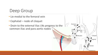
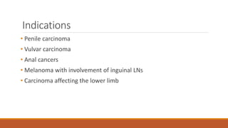
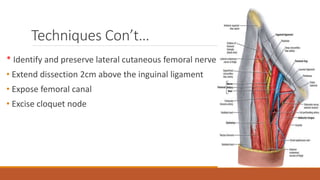
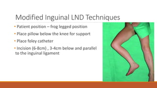
5. Deep Group
• Lie medial to the femoral vein
• Cephalad – node of cloquet
• Drain to the external iliac LNs progress to the
common iliac and para aortic nodes
6. Indications
• Penile carcinoma
• Vulvar carcinoma
• Anal cancers
• Melanoma with involvement of inguinal LNs
• Carcinoma affecting the lower limb
7. Controversies
• Low-volume nodal involvement in a sentinel lymph node
-----------------------------------------------------------------------------------
The Multicenter Selective Lymphadenectomy Trial-2 (MSLT-2 trial)
14. • Pelvic lymph node dissection
- Bulky disease in the superficial groin
- Four or more superficial inguinal nodes are positive
- Positive Cloquet node
- Evidence by PET/CT of disease
15. Modified Inguinal LND (Catalona)
Key aspects
• Shorter skin incision
• Limited dissection
• Preservation of saphenous vein
• Eliminating the need to transpose the Sartorius muscle
16. Modified Inguinal LND Techniques
• Patient position – frog legged position
• Place pillow below the knee for support
• Place foley catheter
• Incision (6-8cm) , 3-4cm below and parallel
to the inguinal ligament
17. Techniques Con’t…
• Incise down to the scarpa’s fascia
• Preserve subcutaneous tissue over the camper’s fascia
• Identify saphenous vein
• Dissect the superficial areolar and nodal tissue off the saphenous
vein to the fascia lata
• Ligate and divide venous branches contributing to saphenous vein
* Saphenous vein is preserved
18. Techniques Con’t…
• Dissection carried superiorly upto 2cm cephalad to the inguinal ligament
• Inferior dissection to ~4cm below the incision
• Specimen sent for frozen section evaluation
• wound irrigated and closed
• A closed suction drainage (5-7 days)
19. Enlarged or Ulcerated nodes
• Indurate skin
• Leave a 2cm margin
• Femoral vessel involvement – anterior
wall resection with reconstruction or
venous ligation
• Large skin defects – tensor fascia lata or
gracilis myocutanous flap
25. Sentinel Lymph Node Biopsy
• Standard of care for patients at high risk for nodal metastases
• In clinical stage I/II melanoma with tumor thickness from 1-4 mm
and clinically negative node basins
• 0.76-1.00 mm with specific features
• >4.00 mm tumors and clinically negative nodes
26. SLNB con’t…
• Preoperative lymphoscintigraphy
• Direct intraoperative visualization of
draining lymphatic patterns using a blue dye
• Identifies 15% to 20% of micrometastatic ds.
• Miss up to 12% of true-positive nodes
- Composed of 4 to 25 LNs that are situated in the deep membranous layer of the superficial fascia of the thigh (camper’s fascia)
Superomedial nodes - around the superficial external pudendal and superficial epigastric veins
Superolateral nodes - around the superficial circumflex vein
Inferolateral nodes - around the lateral femoral cutaneous and superficial circumflex veins
Inferomedial – around the greater saphenous vein
Central nodes - around the saphinofemoral junction
Fewer and lie primarily medial to the femoral vein in the femoral canal
The node of cloquet is the most cephalad of this deep groups and is situated b/n the femoral vein and the inguinal ligament
Common iliac LNs receive drainage from deep inguinal, obturator and hypogastric groups
Dissection of the superficial inguinal lymph nodes is currently a recommended treatment for patients who have evidence of nodal metastatic disease from a variety of malignancies
Generally, it is focused on patients who are presumed through staging to have stage III disease with tumor confined to the lymph node basin
The goals of surgery are locoregional disease control with the possibility of long-term cure.
The most common clinical scenarios for consideration of superficial inguinal lymph node dissection currently arise in patients with melanoma who have a positive sentinel lymph node biopsy (SLNB)
some sarcomas like synovial cell sarcoma
There are currently data from several retrospective studies and one prospective German study to suggest that for patients with low-volume nodal involvement in a sentinel lymph node, it may be unnecessary to perform a completion lymph node dissection
The rationale is that the sentinel lymph node often is the only lymph node to contain tumor when a completion lymph node dissection is performed; and if the patient has additional lymph nodes involved, then he or she frequently will also harbor distant metastatic disease
That randomized melanoma patients with a positive sentinel lymph node to completion lymphadenectomy surgery
At the current time, most surgeons will still recommend a completion lymphadenectomy for patients with a positive SLNB in melanoma
The boundaries of the femoral triangle
Inguinal ligament superiorly, lateral border of the adductor muscle medially and medial border of the Sartorius muscle laterally
In the operation of open inguinal lymphadenectomy, the fatty and lymphatic contents of the femoral triangle are cleared
Next step is palpating the femoral artery and mark the femoral vessels
To avoid wound breakdown and and skin edge necrosis we can resect the overlying skin with our dissection and make sure that the remaining skin can be closed primerly
Infiltrate the incision site with adrenaline containing solution
While a horizontal skin incision has the least morbidity associated with it, a vertical incision is also sometimes used if one is trying to incorporate a vertically placed SLNB incision
With either incision, an attempt should be made to encompass the incision of any previous biopsy that was performed and to try and stay out of the biopsy cavity that may be present from a previous lymph node biopsy
Skin incision is then made and carried out to the Scarpa’s fascia
Flaps are raised from the skin edges deep to Scarpa fascia to the adductor longus medially and Sartorius muscles laterally
The saphenous vein is identified at the junction of the sartorius and adductor muscles inferiorly and ligated
Dissection is continued through the muscle aponeurosis
The superficial nodal tissue is systematically mobilized, beginning in the superomedial quadrant, felling the tissue towards the junction of the saphenous vein with the femoral vein at the fossa ovalis
Carry on from distal to proximal with removal of all the tissue along the vessel
Meticulously ligate and divide any large lymphatics
Visualize the neurovascular structures
Identify the saphenous arch and ligate, preferably with 5-0 proline, and divide it
Flaps are then raised superiorly to the inguinal ligament, and medial to the pubic tubercle
The lateral cutaneous femoral nerve is identified at this point and preserved. It usually runs under the fascia of the sartorius muscle and becomes more superficial about 10 cm below the inguinal ligament where it divides into anterior and lateral branches
Division of the saphenous vein from the femoral vein exposes the femoral sheath and scarpa’s triangle
Femoral sheath is opened from the inguinal ligament to the apex of the femoral triangle, within the sheath are: the femoral vein, femoral artery lateral to it and fatty areolar tissue containing the deep inguinal nodes medially
Dissection over the femoral triangle is accomplished in a lateral to medial direction clearing the lymph nodes from the femoral nerve and femoral artery
All the fibrofatty tissues extending from the external oblique aponeurosis 2 cm above the inguinal ligament to the medial border of the adductor longus muscle medially and sartorius muscle laterally are removed
The femoral canal is then explored to excise Cloquet node
Cloquet node is the first node of the deep system, and to get to it one must tease apart the lacunar ligament that runs medially between the inguinal ligament and the pectineal (Cooper) ligament
One needs to be careful to avoid injuring the femoral circumflex vessels as the node is being dissected out
Once the node is removed, the defect in the lacunar ligament is repaired by using two simple 2-0 prolene stitches to pull the inguinal ligament down to the pectineal ligament.
Care is taken not to close the space in a way that compresses the femoral vein
If performing a sartorius flap, the sartorius muscle is divided in its superior aspect and transposed medially over the vessels and secured to the fascia of the external oblique.
Care must be taken if doing this as the blood supply to the sartorius is segmental; and if one is not careful, devascularization of the muscle will occur.
A closed suction drain is then placed within the femoral triangle.
Iliac and hypogastric node dissections are generally performed for clinically or radiographically evident disease.
Some individuals choose to perform a pelvic lymph node dissection if there is bulky disease in the superficial groin or four or more superficial inguinal nodes are positive due to the high incidence of pelvic nodal disease.
preference is to only perform the deep nodal dissection if there is evidence by PET/CT of disease or Cloquet node is positive.
There are several ways to approach the pelvic lymph nodes. One can extend the superficial skin incision by making a lazy-S type of incision
Transecting the inguinal ligament is generally avoided because it increases morbidity and increases wound healing issues
preferred approach to provide access to the retroperitoneal space is to make a separate incision in the lower abdominal musculature, leaving the inguinal ligament intact
Dissect the external iliac and obturator nodes
Limitation of dissection by excluding the area lateral to the femoral artery and caudal to the fossa ovalis
- Retract scrotum and penis out of the field and draped off
(gentle sponge traction is used to separate the skin edges)
Subcutaneous tissue over the campers layer is carefully preserved and meticulous handling of this tissues and the skin edge must be observed throughout
The saphenous vein and its tributes are identified and the superficial areolar and node bearing tissue is gently dissected off the vein downward to the fascia lata
Large lymphatics and small venous branches must be ligated
Dissection carried superiorly to approximately 2cm cephalad to the inguinal ligament where it is carried down to the fascia of the external oblique muscle
In the catalona operation the lower limit of dissection is the lower border of the fossa ovalis
Some surgeons carry out there dissections slightly lower, although there is not much superficial nodal tissue in this area
FS – if nodes are negative the wound is thoroughly irrigated with sterile water and closed in layers. Taking care to eliminate any potential spaces
A closed suction drainage system is placed and remains 5 to 7 days during which time the patient is maintained at complete bed rest
Induration of skin is suggestive of such local invasion
In this setting the tumor is excised leaving a 2cm margin of normal skin around the indurated or ulcerated area
Involvement of the femoral vessels, particularly the vein. Resection of the anterior wall of the vein with reconstruction using a saphenous vein graft or Gore-tex patch maybe required
In severe involvement the vein might be ligated and excised
Recently a new technical way of preforming an inguinal lymphadenectomy has been described called a videoscopic inguinal lymphadenectomy
This procedure uses laparoscopic equipment to dissect out the lymph node tissue and uses very small skin incisions that are much further from the inguinal crease than the open incision
The initial studies suggest that there are significantly lower wound complications in these patients, but it comes at the expense of a longer operative procedure and longer lymphatic drainage postoperative
We use three incisions at the apex of the femoral triangle – trocar entery
positioning patients supine on a split-leg table
The surgeon is positioned between the patient’s legs, and the assistant stands to the outside of the operative limb. Monitors are placed cephalad above each shoulder, with the laparoscopic tower strategically positioned on the side of the operative limb.
Trocar placement for a three-incision
Inguinal lymph node dissection is associated with moderate morbidity.
Several large centers have reported morbidity of 50% or higher related mainly to wound healing issues in the inguinal area
Skin edge necrosis can occur if skin flaps are made too thin
Can be minimized with selecting the appropriate incision and by carful tissue handling
Transposing the head of the Sartorius muscle to cover the defect left over the femoral vessels
Use of intravenous fluorescein dye and a woods’ lamp intraoperatively to assess viability of wound edge
Many techniques have been reported to reduce postoperative lymphedema, such as preserving the muscle fascia, pedicled omentoplasty, and saphenous vein sparing inguinal lymphadenectomy
systematic reviews and meta-analysis appear to suggest that preserving the saphenous vein has the strongest evidence to help minimize leg edema and that a sartorius transposition does not prevent complications.
Can be reduced by careful attention to intraoperative ligation of lymphatics
By immobilization of the limb in the post operative period
By suction drainage of the lymphedema
Elastic support should be used in the immediate postoperative period and maybe required long term in many patients
WI can be minimized intensive preoperative antibiotic therapy to reduce infection and inflammation from the primary and by the use of prophylactic antibiotics
TE maybe avoided through the use of subcutaneous heparin in perioperative period, particularly when classical inguinal and pelvic dissection is combined with prolonged bed rest postoperatively
The sentinel lymph node is the first lymph node in the drainage basin to receive afferent lymphatic communication from the primary tumor site, prior to spread to the other nodes in this region
It was suggestedthat selective sampling of this important "marker" could serve as an accurate predictor of involvement of the rest of the nodal basin.
Supported by numerous prospective randomized clinical trials, the feasibility and accuracy of sentinel lymph node biopsy has been definitively established
0.76-1.00 mm with features such as ulceration, lymphovascular invasion, age <40 years, significant vertical growth phase, and increased mitotic rate.
The process of sentinel lymph node biopsy involves mapping the sentinel lymph node by two complementary techniques: preoperative lymphoscintigraphy and direct intraoperative visualization of draining lymphatic patterns using a blue dye.
Typically on the morning of surgery, patients receive an injection of r-emitting radioactive colloid (commonly technetium-.99m) around the primary tumor site, followed by serial images of the r-emission pattern
As the colloid enters the lymphatic channels surrounding the lesion, it travels to the first lymph node where it collects, forming a "hot spot" on the emission imaging
FIGURE 14.7. Lymphoscintigraphy demonstrates localization of radioactive colloid to the sentinel lymph node.
This identifies the anatomic location of the sentinel node, but gives no information as to whether it contains metastatic melanoma.
This area is marked and the patient is sent to the operating suite with the images to guide the surgeon in identification of the sentinel node
Following induction of general anesthesia, a second lymphatic mapping technique is used for intraoperative identification of the sentinel node
Blue dye is injected intradermally around the primary melanoma and massaged for approximately 5 minutes to augment dye flow in the lymphatic channels
Based on the post-emission imaging and marking, a limited incision is made over the site of the proposed sentinel node, and dissection into the lymphatic basin is guided by the use of a handheld y-probe and the presence of blue dye in lymphatic channels or nodes
The ideal sentinel node identification would be a single blue lymph node with significant r-emission
The sentinel node is then excised, taking care not to disturb the channels of the surrounding lymph node basin