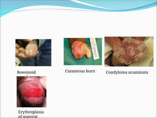
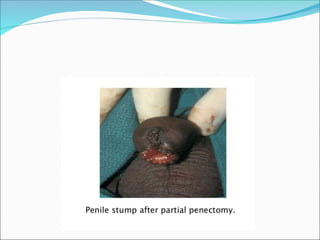
ANATOMY OF PENIS
PENILE CARCINOMA
 It accounts for 0.4-0.6% of all malignant
neoplasms among men in us and europe,it
represents upto 10% of malignant neoplasm in
asian,south american countries
 It is disease of olden men, incidences increases in
sixth decade of life
 More than 95% of lesions are squamous cell
carcinoma
PREMALIGNANT LESIONS
 Cutaneous Horn-rare solid skin over growth ,wide local excision done
 Bowenoid papulosis-multiple papules appears on penile skin or flat
glanular lesion
 Pseusoepitheliomatous micaceous and kerototic balanitits-unusual
hyperkeratotic growths on glans requires excision
 Condyloma acuminata- genital warts,soft multiple lesions on glans
prepuce and shaft
 Leukoplakia-solitary or multiple whitis glanular plaques involve
metus ,excision done
 Balanitis xerotica obliterans- also known as lichen sclerosus et
atrophicus seen over glans and prepuce
 Buschke lowenstein tumor-verucous ca ,aggressive locally advanced
tumor of the glans wide excision done
 Bowens disease-red cutaneous patch on the shaft of penis ,treatment
topical 5 FU,co2 laser ablation or surgical excision
 Erythroplasia of queyrat – red velvet circumscribed painless
lesion ,requires wide local excision
Bowenoid
papules
Cutaneous horn Condyloma acuminata
Erythroplasia
of queyrat
Buschke lowenstein tumor
 Invades locally
 Compresses the adjacent tissues causing urethral
erosion and fistulisation
 Never metastasis
 No sign of malignant change
 C/f bleeding discarge and foul odour
 Treatment is excision
 Laser is effective,topical 5-FU,
PENILE CARCINOMA
 Mc type is SCC {95%}
 Mesenchymal tumors{3%} like kaposi,angiosarcoma etc
 Basal cell ca,malignant melanoma,metastasis
 Carcinoma in situ: Tis of penis is called as erythroplasia
of queyrat it involves glans penis and prepuce
 It consists of red velvety well marginated lesions of glans
penis or may ulcerate with discharge
 Bowens disease it involves penile shaft or
perineum,characterized by sharply defined plaques of
scaly erythema on penile shaft
 Treatment
 Preputial lesion- circumcision or excision with
5mm margins
 Glanular lesion-topical 5 flurouracil cream or
ablation therapy
 Radiotherapy for resistant cases
Penile carcinoma
 Etiology
1.Lack of neonatal circumcision
2.Poor hygeine
3.Phimosis/BXO
4.Hpv infections
5.Exposure to tobacco products
6.Penile trauma mutilating circumcision,penile
tear
 Penile ca arises anywhere on penis but most
commonly on:
1. Glans 48%
2. Prepuce 21%
 Other tumors involve gans and prepuce 9% ,
coronal sulcus 6% and shaft <2%.
Presentation
 Begins as small lesion papillary and
exophytic/flat/ulcerative growth
 Lymphatic spread
 Flat&ulcerative lesions morethan 5 cm and
extending more than 75% of shaft have higher
incidence of metastasis and poor survival
 Bucks fascia protects corporeal invasion
 Distant metastasis uncommon
 Bladder and urethra involvement rare
CLINICAL FEATURES
 Patients present with swelling or ulcer on penis
 Pain is usually not a presenting complaints
 Weakness ,weight loss ,fatigue and significant blood loss from penile lesion or
nodal lesion
 Many present late by the time presentation it ranges from subtle induration to
small papule to warty or exophytic growth or ulcerative growth
 Phimosis may obscure lesion allow tumor progress Erosion through the
prepuce,foul smelling discharge with or without bleeding wit little or no pain
 Mass,ulceration,suppuration or hemorrage in the inguinal area due to
metastasis
 Urinary retention or urethral Fistula due to local corporeal involvement is
rare presentation
 Distant metastasis sites lung bone liver
EXAMINATION
 At presentation most of lesions are confined to penis
 penile lesion assesed with regard to size,location
fixation and involvement of corporeal bodies.
 Inspection of base of penis and scrotum too r/o
extension
 Rectal and bimanual examination provides info
regarding perineal body involvement and presence
of pelvic mass
 Bilateral palpation for inguinal area for adenopathy
Investigations
 Anemia,leucocytosis,hypercalcemia,hypoalbuminemia
 BIOPSY:confirmation of diagnosis
Asessment of depth of invasion,vascular invasion and
histological grade of the lesion mandatory before
initiating any therapy
 Dorsal slit is required for adequate exposure of lesion
 Histologically mostly are scc demonstrating
keratinization,epitelial pearly and degree of mitotic
activity
 Histological types includes 15% papillary,10% warty
and basaloid,3% verrucous and sarcomatoid
 Among these sarcomatoid and basloid (hpv
associaion)are aggresive
BRODERS CLASSIFICATION
 Used in scc to define level of differentiation on
basis of keratinization nuclear pleomorphism
and number of mitosis
 Low grade lesions(grade 1 nd 2) constitue 70-80%
these are well differentiated demonstrate keratin
keratin pearls
 High grade(grade 3 and 4) poorly differentiated
almosts orginates from shaft,10% located in the
prepuce
RADIOLOGICAL DIAGNOSIS
 CXR: for lung metastasis
 USG:cannot delineate invasion into subepithelial connective tissue of
glans penis from corpus spongiosum involvement
 CT SCAN: sensitivity and specificity of ct are 36% and 100%
Mainly for assessment of inguinal & pelvic lymph noe and secondaries
Ct guides biopsy of enlarged pelvic nodes
 MRI : sensitivity and specificity of 100% and 91%
1. Assesses local staging of tumor
2. Assessment of inguinal and pelvic LNs
3. Better with artificial erection
 Newer modalities
1. PET CT of inguinal region detects minimal inguinal metastasis when
lymph nodes are normal on ct/mri
TREATMENT
 Organ preservation
 Penile amputations
Organ preservation:
 Goal : to preserve glans sensation and to maximize
penile shaft length
Indication:
 primary tumor exhibiting favorable histologic
features
 Stages Tis ,Ta,T1 grade 1 and 2 tumors
Circumcision and limited excision
strategies:
1. 2 cm surgical margin required for all patients
undergoing partial penectomy
2. Maximum proximal histologic extent of 5mm for
grade1 and grade 2 tumors and 10mm for grade 3
tumors recommended
 Limitations:
1. proximal and distal deeply invasive tumors
2. High grade tumors
3. Skip lesions
4. Pts with poor compliance who would not be
candidate for salvage procedures
Recurrence rate is 4-6%
DASELER REGION
Inguinal region is divided into four sections by a horizontal and a vertical
line drawn through the fossa ovalis. Five anatomical subgroups with the
central zone being located at the confluence of the greater saphenous vein
and the femoral vein.
CARCINOMA PENIS powerpoint presentations

CARCINOMA PENIS powerpoint presentations

  • 2.
  • 4.
    PENILE CARCINOMA  Itaccounts for 0.4-0.6% of all malignant neoplasms among men in us and europe,it represents upto 10% of malignant neoplasm in asian,south american countries  It is disease of olden men, incidences increases in sixth decade of life  More than 95% of lesions are squamous cell carcinoma
  • 5.
    PREMALIGNANT LESIONS  CutaneousHorn-rare solid skin over growth ,wide local excision done  Bowenoid papulosis-multiple papules appears on penile skin or flat glanular lesion  Pseusoepitheliomatous micaceous and kerototic balanitits-unusual hyperkeratotic growths on glans requires excision  Condyloma acuminata- genital warts,soft multiple lesions on glans prepuce and shaft  Leukoplakia-solitary or multiple whitis glanular plaques involve metus ,excision done  Balanitis xerotica obliterans- also known as lichen sclerosus et atrophicus seen over glans and prepuce  Buschke lowenstein tumor-verucous ca ,aggressive locally advanced tumor of the glans wide excision done  Bowens disease-red cutaneous patch on the shaft of penis ,treatment topical 5 FU,co2 laser ablation or surgical excision  Erythroplasia of queyrat – red velvet circumscribed painless lesion ,requires wide local excision
  • 6.
    Bowenoid papules Cutaneous horn Condylomaacuminata Erythroplasia of queyrat
  • 7.
    Buschke lowenstein tumor Invades locally  Compresses the adjacent tissues causing urethral erosion and fistulisation  Never metastasis  No sign of malignant change  C/f bleeding discarge and foul odour  Treatment is excision  Laser is effective,topical 5-FU,
  • 8.
    PENILE CARCINOMA  Mctype is SCC {95%}  Mesenchymal tumors{3%} like kaposi,angiosarcoma etc  Basal cell ca,malignant melanoma,metastasis  Carcinoma in situ: Tis of penis is called as erythroplasia of queyrat it involves glans penis and prepuce  It consists of red velvety well marginated lesions of glans penis or may ulcerate with discharge  Bowens disease it involves penile shaft or perineum,characterized by sharply defined plaques of scaly erythema on penile shaft
  • 9.
     Treatment  Preputiallesion- circumcision or excision with 5mm margins  Glanular lesion-topical 5 flurouracil cream or ablation therapy  Radiotherapy for resistant cases
  • 10.
    Penile carcinoma  Etiology 1.Lackof neonatal circumcision 2.Poor hygeine 3.Phimosis/BXO 4.Hpv infections 5.Exposure to tobacco products 6.Penile trauma mutilating circumcision,penile tear
  • 11.
     Penile caarises anywhere on penis but most commonly on: 1. Glans 48% 2. Prepuce 21%  Other tumors involve gans and prepuce 9% , coronal sulcus 6% and shaft <2%.
  • 12.
    Presentation  Begins assmall lesion papillary and exophytic/flat/ulcerative growth  Lymphatic spread  Flat&ulcerative lesions morethan 5 cm and extending more than 75% of shaft have higher incidence of metastasis and poor survival  Bucks fascia protects corporeal invasion  Distant metastasis uncommon  Bladder and urethra involvement rare
  • 13.
    CLINICAL FEATURES  Patientspresent with swelling or ulcer on penis  Pain is usually not a presenting complaints  Weakness ,weight loss ,fatigue and significant blood loss from penile lesion or nodal lesion  Many present late by the time presentation it ranges from subtle induration to small papule to warty or exophytic growth or ulcerative growth  Phimosis may obscure lesion allow tumor progress Erosion through the prepuce,foul smelling discharge with or without bleeding wit little or no pain  Mass,ulceration,suppuration or hemorrage in the inguinal area due to metastasis  Urinary retention or urethral Fistula due to local corporeal involvement is rare presentation  Distant metastasis sites lung bone liver
  • 14.
    EXAMINATION  At presentationmost of lesions are confined to penis  penile lesion assesed with regard to size,location fixation and involvement of corporeal bodies.  Inspection of base of penis and scrotum too r/o extension  Rectal and bimanual examination provides info regarding perineal body involvement and presence of pelvic mass  Bilateral palpation for inguinal area for adenopathy
  • 15.
    Investigations  Anemia,leucocytosis,hypercalcemia,hypoalbuminemia  BIOPSY:confirmationof diagnosis Asessment of depth of invasion,vascular invasion and histological grade of the lesion mandatory before initiating any therapy  Dorsal slit is required for adequate exposure of lesion  Histologically mostly are scc demonstrating keratinization,epitelial pearly and degree of mitotic activity  Histological types includes 15% papillary,10% warty and basaloid,3% verrucous and sarcomatoid  Among these sarcomatoid and basloid (hpv associaion)are aggresive
  • 16.
    BRODERS CLASSIFICATION  Usedin scc to define level of differentiation on basis of keratinization nuclear pleomorphism and number of mitosis  Low grade lesions(grade 1 nd 2) constitue 70-80% these are well differentiated demonstrate keratin keratin pearls  High grade(grade 3 and 4) poorly differentiated almosts orginates from shaft,10% located in the prepuce
  • 18.
    RADIOLOGICAL DIAGNOSIS  CXR:for lung metastasis  USG:cannot delineate invasion into subepithelial connective tissue of glans penis from corpus spongiosum involvement  CT SCAN: sensitivity and specificity of ct are 36% and 100% Mainly for assessment of inguinal & pelvic lymph noe and secondaries Ct guides biopsy of enlarged pelvic nodes  MRI : sensitivity and specificity of 100% and 91% 1. Assesses local staging of tumor 2. Assessment of inguinal and pelvic LNs 3. Better with artificial erection  Newer modalities 1. PET CT of inguinal region detects minimal inguinal metastasis when lymph nodes are normal on ct/mri
  • 22.
    TREATMENT  Organ preservation Penile amputations Organ preservation:  Goal : to preserve glans sensation and to maximize penile shaft length Indication:  primary tumor exhibiting favorable histologic features  Stages Tis ,Ta,T1 grade 1 and 2 tumors
  • 24.
    Circumcision and limitedexcision strategies: 1. 2 cm surgical margin required for all patients undergoing partial penectomy 2. Maximum proximal histologic extent of 5mm for grade1 and grade 2 tumors and 10mm for grade 3 tumors recommended  Limitations: 1. proximal and distal deeply invasive tumors 2. High grade tumors 3. Skip lesions 4. Pts with poor compliance who would not be candidate for salvage procedures Recurrence rate is 4-6%
  • 38.
    DASELER REGION Inguinal regionis divided into four sections by a horizontal and a vertical line drawn through the fossa ovalis. Five anatomical subgroups with the central zone being located at the confluence of the greater saphenous vein and the femoral vein.