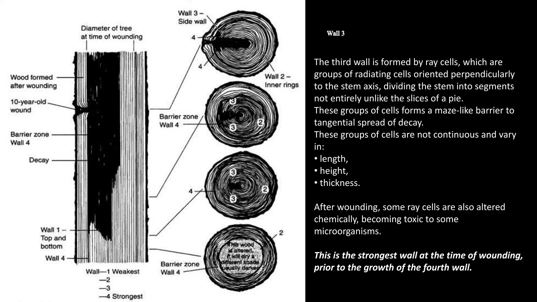
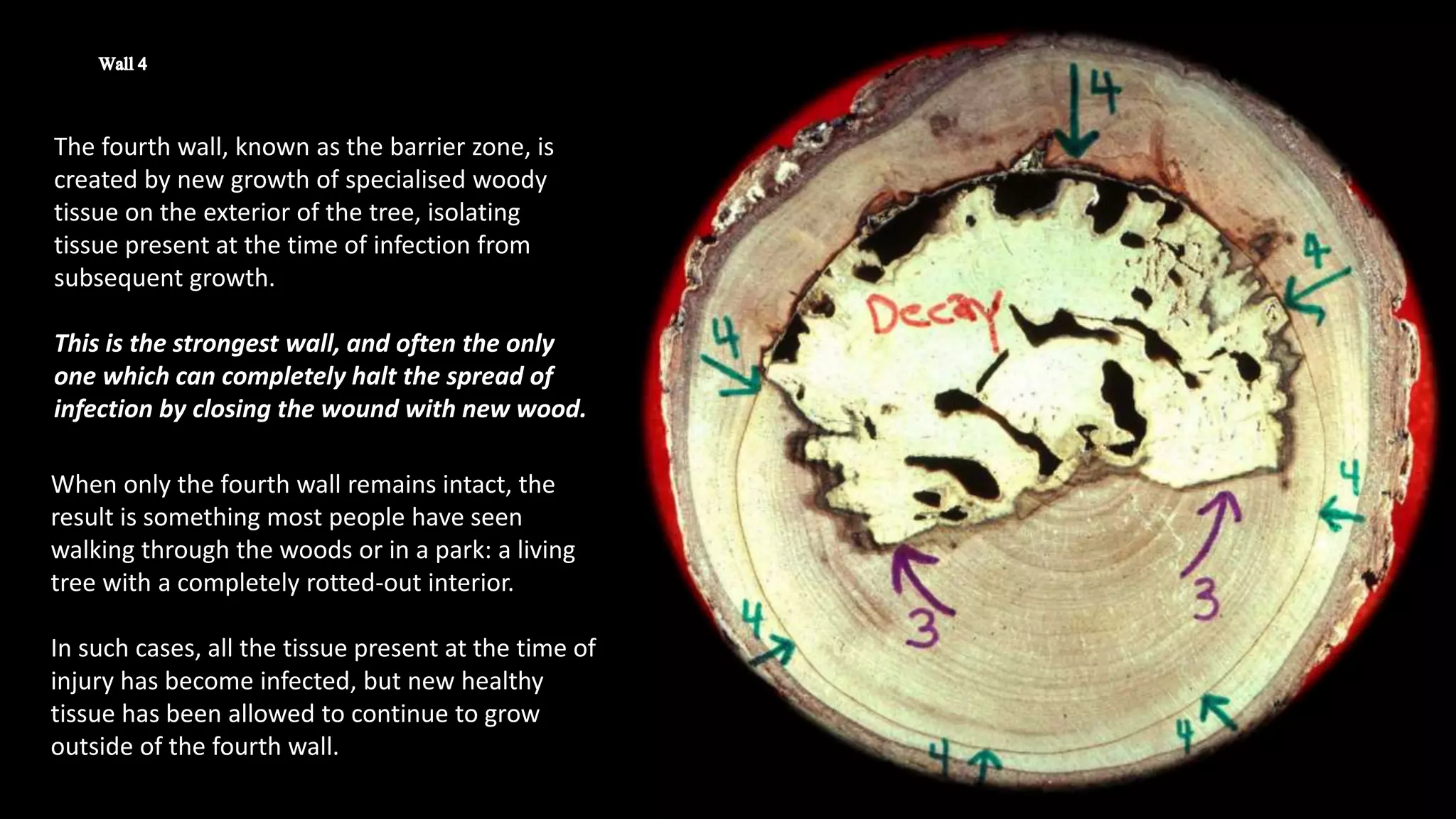
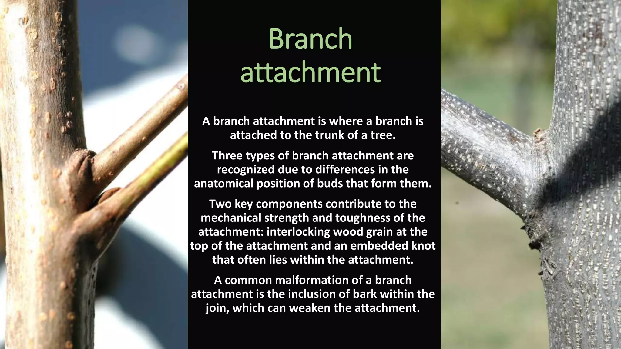
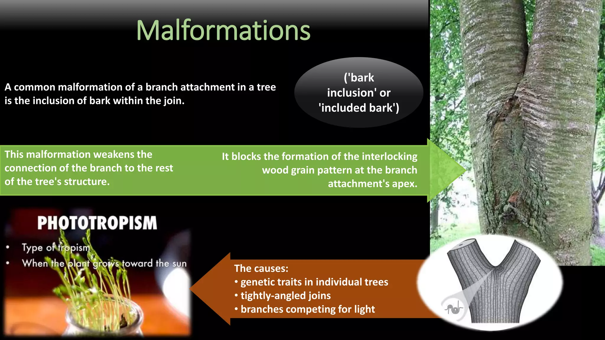
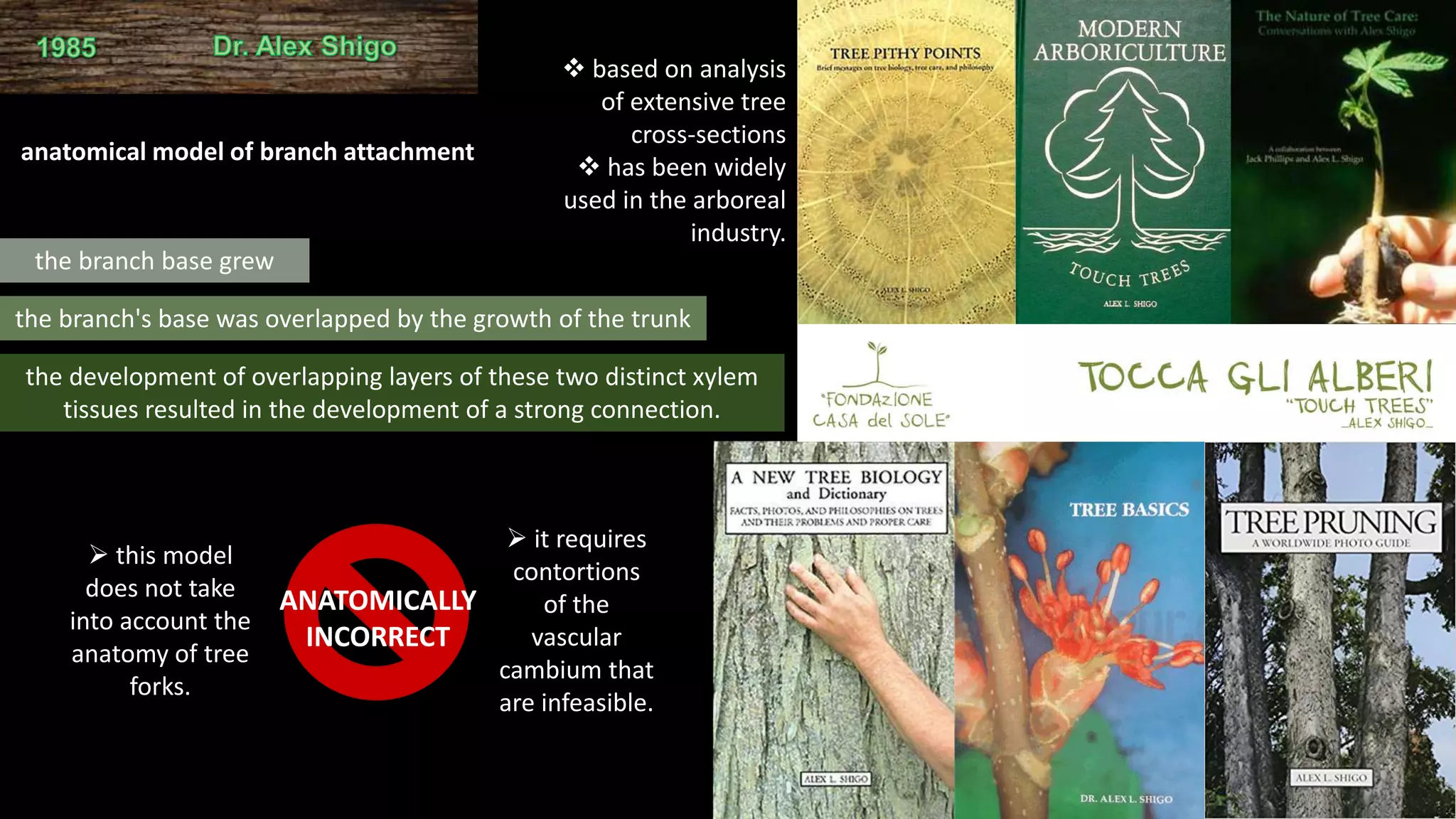
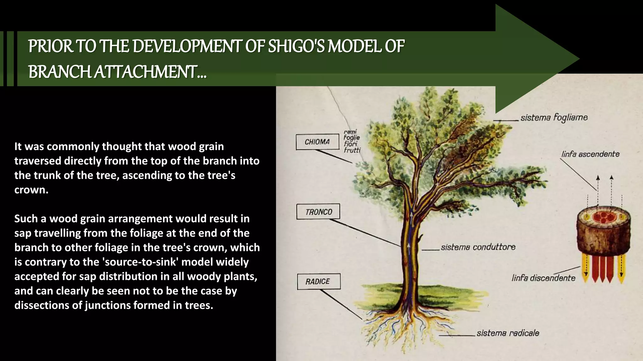
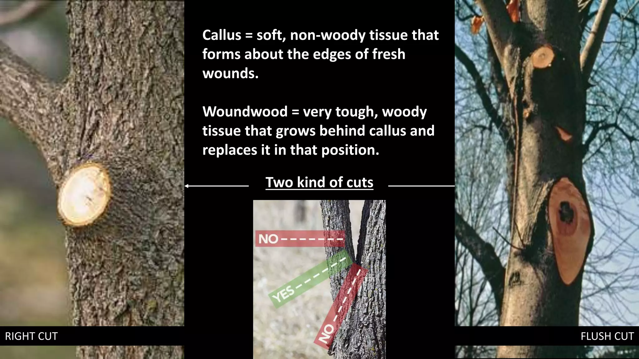
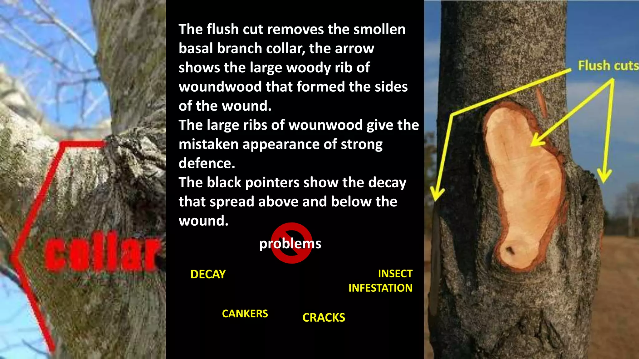
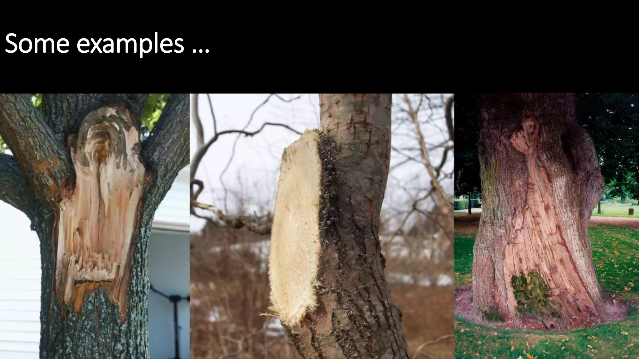
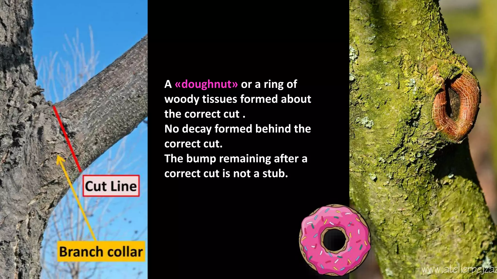
Example of Compartmentalization,
with radial and circular walling, in an
Acer platanoides of 5 years
when a tree is wounded cells undergo changes to
form "walls" around the wound, slowing or
preventing the spread of disease and decay to the rest
of the tree.
o
m
p
a
r
t
m
e
n
t
a
l
i
z
a
t
i
o
n
f e
c
a
y
n r
e
e
s
The first wall is formed by plugging up normally
conductive vascular tissue above and below the wound.
This tissue runs up and down the length of the stem, so
plugging it slows the vertical spread of decay.
Tissues are plugged in various ways, such as with:
• tylosis,
• polyphenolic deposits,
• anti-fungal substances
• in conifers by the closure of the bordered pits linking
vessel cells.
This wall is the weakest.
The second wall is formed by the thick-walled, lignin-
rich cells of the latewood growth ring interior and
exterior to the wound, thus slowing the radial spread of
decay.
This wall is the second weakest, and is continuous
except where intersected by ray cells.
The third wall is formed by ray cells, which are
groups of radiating cells oriented perpendicularly
to the stem axis, dividing the stem into segments
not entirely unlike the slices of a pie.
These groups of cells forms a maze-like barrier to
tangential spread of decay.
These groups of cells are not continuous and vary
in:
• length,
• height,
• thickness.
After wounding, some ray cells are also altered
chemically, becoming toxic to some
microorganisms.
This is the strongest wall at the time of wounding,
prior to the growth of the fourth wall.
The fourth wall, known as the barrier zone, is
created by new growth of specialised woody
tissue on the exterior of the tree, isolating
tissue present at the time of infection from
subsequent growth.
This is the strongest wall, and often the only
one which can completely halt the spread of
infection by closing the wound with new wood.
When only the fourth wall remains intact, the
result is something most people have seen
walking through the woods or in a park: a living
tree with a completely rotted-out interior.
In such cases, all the tissue present at the time of
injury has become infected, but new healthy
tissue has been allowed to continue to grow
outside of the fourth wall.
For example
arborists are frequently called upon to analyze the danger posed
to people or property by a damaged or decaying tree.
In the production of maple syrup holes are drilled
into a tree's vascular tissues, which necessarily
damages the tree.
For example
Pruning Techniques
How Much Should Be Pruned?
Branch
attachment
A branch attachment is where a branch is
attached to the trunk of a tree.
Three types of branch attachment are
recognized due to differences in the
anatomical position of buds that form them.
Two key components contribute to the
mechanical strength and toughness of the
attachment: interlocking wood grain at the
top of the attachment and an embedded knot
that often lies within the attachment.
A common malformation of a branch
attachment is the inclusion of bark within the
join, which can weaken the attachment.
Trunk collars
Branch collars
Cambial zone
Phloem
Bark cambium
Bark branch
ridge
form as branch and trunk tissues meet
above the branch.
the often visible swelling in a woody plant that
forms at the base of a branch where it is attached to
its parent branch or to the tree's trunk.
a cell generator that is between
the wood or xylem and inner bark.
the living tissue that transports the soluble organic
compounds made during photosynthesis, in particular
the sugar sucrose, to parts of the plant where needed.
the outermost layers of stems and
roots of woody plants.
the stem and main wooden axis of a tree.
The xylem tissues in this location are
denser than in surrounding tissues of the
tree's stem or branch, the wood grain
pattern formed is tortuous and in these
tissues there is typically a reduction in
vessel length, diameter and frequency of
occurrence.
This specialized xylem tissue, formed
under the branch bark ridge, provides
unique mechanical properties to the
union of the branch to the trunk,
requiring that wood fibres are stretched
along their length (the axial strength of
the wood) in order to rupture the
attachment apart.
Initially branches are mechanically
attached to the trunks of trees by
forming interlocking wood grain
patterns at the top of the joint.
The base of the smaller branch becomes occluded in the larger trunk of
the tree which is producing a larger increment of growth, and this
occluded part of the branch forms a knot that provides substantial
additional mechanical support to the attachment as it develops.
The combination of the interlocking wood grain at
the apex of the branch and the occluded knot
embedded into the tree's trunk make mature
branch attachments in trees very strong
components of a tree's crown.
Different types of
branch attachments
Botanists commonly
differentiate between
branches
anatomical difference can be
found on dissection between
these branch attachments
an initial knot that originates near
to the stem's pith
a bud trace that originates near the
stem's pith, and adventitious
epicormic branches will exhibit
neither of these internal features.
originate from the initial extension
growth
have developed from the growth of latent
buds or adventitious buds that developed
later on the tree's trunk surface.
Malformations
A common malformation of a branch attachment in a tree
is the inclusion of bark within the join.
('bark
inclusion' or
'included bark')
This malformation weakens the
connection of the branch to the rest
of the tree's structure.
It blocks the formation of the interlocking
wood grain pattern at the branch
attachment's apex.
The causes:
• genetic traits in individual trees
• tightly-angled joins
• branches competing for light
anatomical model of branch attachment
the development of overlapping layers of these two distinct xylem
tissues resulted in the development of a strong connection.
 this model
does not take
into account the
anatomy of tree
forks.
ANATOMICALLY
INCORRECT
 it requires
contortions
of the
vascular
cambium that
are infeasible.
the branch base grew
the branch's base was overlapped by the growth of the trunk
 based on analysis
of extensive tree
cross-sections
 has been widely
used in the arboreal
industry.
It was commonly thought that wood grain
traversed directly from the top of the branch into
the trunk of the tree, ascending to the tree's
crown.
Such a wood grain arrangement would result in
sap travelling from the foliage at the end of the
branch to other foliage in the tree's crown, which
is contrary to the 'source-to-sink' model widely
accepted for sap distribution in all woody plants,
and can clearly be seen not to be the case by
dissections of junctions formed in trees.
PRIOR TO THE DEVELOPMENT OF SHIGO'SMODEL OF
BRANCH ATTACHMENT…
FLUSHCUTS
Callus = soft, non-woody tissue that
forms about the edges of fresh
wounds.
Woundwood = very tough, woody
tissue that grows behind callus and
replaces it in that position.
Two kind of cuts
RIGHT CUT FLUSH CUT
The flush cut removes the smollen
basal branch collar, the arrow
shows the large woody rib of
woundwood that formed the sides
of the wound.
The large ribs of wounwood give the
mistaken appearance of strong
defence.
The black pointers show the decay
that spread above and below the
wound.
problems
DECAY
CANKERS CRACKS
INSECT
INFESTATION
Some examples …
A «doughnut» or a ring of
woody tissues formed about
the correct cut .
No decay formed behind the
correct cut.
The bump remaining after a
correct cut is not a stub.
Some examples …
Inglese alternanza

Inglese alternanza

  • 1.
    Example of Compartmentalization, withradial and circular walling, in an Acer platanoides of 5 years
  • 3.
    when a treeis wounded cells undergo changes to form "walls" around the wound, slowing or preventing the spread of disease and decay to the rest of the tree. o m p a r t m e n t a l i z a t i o n f e c a y n r e e s
  • 4.
    The first wallis formed by plugging up normally conductive vascular tissue above and below the wound. This tissue runs up and down the length of the stem, so plugging it slows the vertical spread of decay. Tissues are plugged in various ways, such as with: • tylosis, • polyphenolic deposits, • anti-fungal substances • in conifers by the closure of the bordered pits linking vessel cells. This wall is the weakest.
  • 5.
    The second wallis formed by the thick-walled, lignin- rich cells of the latewood growth ring interior and exterior to the wound, thus slowing the radial spread of decay. This wall is the second weakest, and is continuous except where intersected by ray cells.
  • 6.
    The third wallis formed by ray cells, which are groups of radiating cells oriented perpendicularly to the stem axis, dividing the stem into segments not entirely unlike the slices of a pie. These groups of cells forms a maze-like barrier to tangential spread of decay. These groups of cells are not continuous and vary in: • length, • height, • thickness. After wounding, some ray cells are also altered chemically, becoming toxic to some microorganisms. This is the strongest wall at the time of wounding, prior to the growth of the fourth wall.
  • 7.
    The fourth wall,known as the barrier zone, is created by new growth of specialised woody tissue on the exterior of the tree, isolating tissue present at the time of infection from subsequent growth. This is the strongest wall, and often the only one which can completely halt the spread of infection by closing the wound with new wood. When only the fourth wall remains intact, the result is something most people have seen walking through the woods or in a park: a living tree with a completely rotted-out interior. In such cases, all the tissue present at the time of injury has become infected, but new healthy tissue has been allowed to continue to grow outside of the fourth wall.
  • 8.
    For example arborists arefrequently called upon to analyze the danger posed to people or property by a damaged or decaying tree. In the production of maple syrup holes are drilled into a tree's vascular tissues, which necessarily damages the tree. For example
  • 13.
  • 14.
    How Much ShouldBe Pruned?
  • 15.
    Branch attachment A branch attachmentis where a branch is attached to the trunk of a tree. Three types of branch attachment are recognized due to differences in the anatomical position of buds that form them. Two key components contribute to the mechanical strength and toughness of the attachment: interlocking wood grain at the top of the attachment and an embedded knot that often lies within the attachment. A common malformation of a branch attachment is the inclusion of bark within the join, which can weaken the attachment.
  • 16.
    Trunk collars Branch collars Cambialzone Phloem Bark cambium Bark branch ridge form as branch and trunk tissues meet above the branch. the often visible swelling in a woody plant that forms at the base of a branch where it is attached to its parent branch or to the tree's trunk. a cell generator that is between the wood or xylem and inner bark. the living tissue that transports the soluble organic compounds made during photosynthesis, in particular the sugar sucrose, to parts of the plant where needed. the outermost layers of stems and roots of woody plants. the stem and main wooden axis of a tree.
  • 17.
    The xylem tissuesin this location are denser than in surrounding tissues of the tree's stem or branch, the wood grain pattern formed is tortuous and in these tissues there is typically a reduction in vessel length, diameter and frequency of occurrence. This specialized xylem tissue, formed under the branch bark ridge, provides unique mechanical properties to the union of the branch to the trunk, requiring that wood fibres are stretched along their length (the axial strength of the wood) in order to rupture the attachment apart.
  • 18.
    Initially branches aremechanically attached to the trunks of trees by forming interlocking wood grain patterns at the top of the joint. The base of the smaller branch becomes occluded in the larger trunk of the tree which is producing a larger increment of growth, and this occluded part of the branch forms a knot that provides substantial additional mechanical support to the attachment as it develops. The combination of the interlocking wood grain at the apex of the branch and the occluded knot embedded into the tree's trunk make mature branch attachments in trees very strong components of a tree's crown.
  • 19.
    Different types of branchattachments Botanists commonly differentiate between branches anatomical difference can be found on dissection between these branch attachments an initial knot that originates near to the stem's pith a bud trace that originates near the stem's pith, and adventitious epicormic branches will exhibit neither of these internal features. originate from the initial extension growth have developed from the growth of latent buds or adventitious buds that developed later on the tree's trunk surface.
  • 20.
    Malformations A common malformationof a branch attachment in a tree is the inclusion of bark within the join. ('bark inclusion' or 'included bark') This malformation weakens the connection of the branch to the rest of the tree's structure. It blocks the formation of the interlocking wood grain pattern at the branch attachment's apex. The causes: • genetic traits in individual trees • tightly-angled joins • branches competing for light
  • 21.
    anatomical model ofbranch attachment the development of overlapping layers of these two distinct xylem tissues resulted in the development of a strong connection.  this model does not take into account the anatomy of tree forks. ANATOMICALLY INCORRECT  it requires contortions of the vascular cambium that are infeasible. the branch base grew the branch's base was overlapped by the growth of the trunk  based on analysis of extensive tree cross-sections  has been widely used in the arboreal industry.
  • 22.
    It was commonlythought that wood grain traversed directly from the top of the branch into the trunk of the tree, ascending to the tree's crown. Such a wood grain arrangement would result in sap travelling from the foliage at the end of the branch to other foliage in the tree's crown, which is contrary to the 'source-to-sink' model widely accepted for sap distribution in all woody plants, and can clearly be seen not to be the case by dissections of junctions formed in trees. PRIOR TO THE DEVELOPMENT OF SHIGO'SMODEL OF BRANCH ATTACHMENT…
  • 23.
  • 24.
    Callus = soft,non-woody tissue that forms about the edges of fresh wounds. Woundwood = very tough, woody tissue that grows behind callus and replaces it in that position. Two kind of cuts RIGHT CUT FLUSH CUT
  • 25.
    The flush cutremoves the smollen basal branch collar, the arrow shows the large woody rib of woundwood that formed the sides of the wound. The large ribs of wounwood give the mistaken appearance of strong defence. The black pointers show the decay that spread above and below the wound. problems DECAY CANKERS CRACKS INSECT INFESTATION
  • 26.
  • 27.
    A «doughnut» ora ring of woody tissues formed about the correct cut . No decay formed behind the correct cut. The bump remaining after a correct cut is not a stub.
  • 28.