SJM College of Pharmacy,
Chitradurga
Prepare By,
AdarshaBP
Ass Professor(Pharmacognosy)
SJM College of Pharmacy
1
PHARMACEUTICAL
BIOTECHNOLOGY
IMMUNOBLOTTING
• Viral antigens are detected with a polyclonal or a MAb
onto nitrocellulose paper.
• After incubation, the protein bands (immune
complexes) are visualized with peroxidase-conjugated
protein and a colour reagent.
• A colour develops in the bands where antibody binds to
the antigen.
TYPES OF BLOTTING TECHNIQUES
1. Southern Blot
2. Northern Blot
3. Western blot
Western blotting
• The Western blot is an analytical technique used to
detect specific proteins in a given sample of tissue
homogenate or extract.
• Immunoblotting: because an antibody is used to
specifically detect its antigen
• Western blot for Proteins was Developed by George
Stark using antibodies to locate Proteins
• Western blotting is based on building antibody-protein
complex through specific binding of antibodies to
proteins immobilized on a membrane.
• Then detecting the bound antibody through
several detection methods
The Western blotting workflow:
• Sample preparation
• Gel electrophoresis
• Transfer
• Blocking &Antibody probing
• Detection
• Analysis
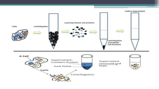
1. Sample preparation
• Samples may be taken from whole tissue, from cell culture, bacteria,
virus or environmental samples.
• In most cases, samples are solid tissues.
• First broken down mechanically using a blender (for larger sample
volumes), using a homogenizer (smaller volumes), or by sonication.
• Cells may also be broken open by one of the above mechanical methods.
• A combination of biochemical and mechanical techniques, including
various types of filtration and centrifugation.
• To encourage lysis of cells and to solubilize
proteins, may be employed : detergents, salts, and
buffers
• To prevent the digestion of the sample by its own
enzymes -Anti Protease and phosphatase
• To avoid protein denaturing-Tissue preparation is
often done at cold temperatures
2. GEL ELECTROPHORESIS:
• PAGE(Polyacrylamide gel electrophoresis) is mainly used for the separation of
protein particles.
• SDS (sodium dodecyl sulfate) is added to the gel and running buffer to denature the protein
molecules which will ensure that proteins are separated solely on the basis of size and not
on three-dimensional structure.
• Molecular weight markers are used to define the size of proteins run in a gel. Markers are
composed of different proteins of known size.
• As proteins are not directly visible in the gel, the gel must be stained. Proteins are usually
stained with dyes such as Blue, Silver stain, or Deep Purple.
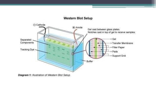
3. Protein Transfer:
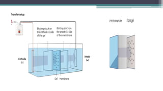
• Protein are transferred from the gel to a solid support membrane such
as nitrocellulose or polyvinylidene difluoride (PVDF).
• The proteins transferred from the gels are immobilized which makes it
possible to detect the proteins on the membrane using specific antibodies.
• Electro-blotting is mainly used which uses electric field to pull
proteins from the gel onto the PVDF or nitrocellulose membrane.
• The proteins move from within the gel onto the membrane while
maintaining the organization they had within the gel
4. Blocking &Antibody Probing
• Western blotting involves the immobilization of
biomolecules on a membrane and blocking the spaces not
already occupied by proteins.
• As non-specific binding of antibodies to the membrane
reduces the specificity and sensitivity of the assay.
• Two main classes of blocking agents (proteins and non-
ionic detergents) are commonly used for western blotting.
5. Antibody Probing
• Western blotting protocols usually uses a non-labeled
primary antibody directed against the target protein and
labeled secondary antibody directed against the constant
region of the primary antibody.
• The secondary antibody serves not only as a carrier of the label
but is also a mechanism to amplify the emitted signals because
secondary antibodies are most often polyclonal and can bind
primary antibody at different epitopes simultaneously.
• Alkaline phosphatase (AP) and horseradish peroxidase
(HRP) are the two most commonly used enzymes for
protein detection using chromogenic substrates.
• Other antibody probes used for protein detection are:
1. Fluorophores
2. Biotinylated secondary antibodies
3. Radioisotopes.
6. Detection
• Different detection system are used in western blotting based on
fluorescence, chromogenic or radio-isotopic detection.
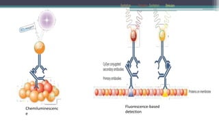
• Enzymatic detection methods require the addition of a substrate
that emits light when it reacts with an enzyme conjugated to a
secondary antibody.
• Fluorescence-based detection requires the excitation of
fluorophore using a light source of a specific wavelength causing
light emission.
Chemiluminescenc
e
Fluorescence-based
detection
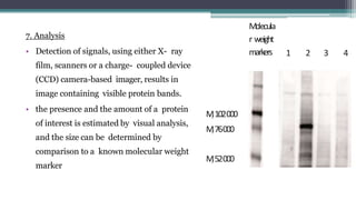
7. Analysis
• Detection of signals, using either X- ray
film, scanners or a charge- coupled device
(CCD) camera-based imager, results in
image containing visible protein bands.
• the presence and the amount of a protein
of interest is estimated by visual analysis,
and the size can be determined by
comparison to a known molecular weight
marker
Molecula
r weight
markers 1 2 3 4
rM102000
rM76000
rM52000
Applications of Western Blotting
• Diagnosis of HIV by ELISA involves the western blotting technique.
• Western blotting technique is also used to detect some forms of Lyme
Diseases.
• Western blotting technique is used in definitive test for BSE.
• Confirmatory test for Hepatitis-B involves western blotting technique.
• Western blotting test is used in the analysis of Biomarkers such as
hormones, growth factors and cytokines.
• This technique is also employed in The Gene expression studies.
Southern Blot
• Southern Blot is the analytical technique used in molecular
biology, immunogenetics and other molecular methods to detect
or identify DNA of interest from a mixture of DNA sample or a
specific base sequence within a strand of DNA.
• The technique was developed by a molecular biologist E.M.
Southern in 1975 for analysing the related genes in a DNA
restriction fragment and thus named as Southern blotting in his
honour.
Principle of Southern Blot:
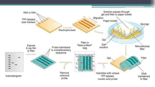
• The process involves the transfer of electrophoresis-separated DNA fragments
to a carrier membrane which is usually nitrocellulose and the subsequent
detection of the target DNA fragment by probe hybridization.
• Hybridization refers to the process of forming a double-stranded DNA molecule
between a single-stranded DNA probe and a single-stranded target DNA.
• Since the probe and target DNA are complementary to each other, the reaction
is specific which aids in the detection of the specific DNA fragment.
Steps Involved in Southern Blot
1. Extraction and purification of DNA from cells:
DNA is first separated from target cells following standard methods
of genomic DNA extraction and then purified.
2. Restriction Digestion or DNA Fragmentation:
Restriction endonucleases are used to cut high-molecular-weight
DNA strands into smaller fragments. One or more restriction
enzymes can be used to achieve such fragments.
3. Separation by Electrophoresis
The separation may be done by agarose gel electrophoresis in
which the negatively charged DNA fragments move towards
the positively charged anode, the distance moved depending
upon its size.
4. Depurination
Partial depurination is done by the use of dilute HCl which
promotes higher efficiency transfer of DNA fragments by it
breaking down into smaller pieces.
5.Denaturation
DNA is then denatured with a mild alkali such as an
alkaline solution of NaOH. This causes the double
stranded DNA to become single-stranded, making
them suitable for hybridization. DNA is then
neutralized with NaCl to prevent re-hybridization
before addition of the probe.
6.Blotting
• The denatured fragments are then transferred onto a nylon
or nitrocellulose filter membrane which is done by placing
the gel on top of a buffer saturated filter paper, then laying
nitrocellulose filter membrane on the top of gel. Finally
some dry filter papers are placed on top of the membrane.
Fragments are pulled towards the nitrocellulose filter
membrane by capillary action and result in the contact
print of the gel.
7. Baking
The nitrocellulose membrane is removed from the
blotting stack, and the membrane with single
stranded DNA bands attached on to it is baked in a
vacuum or regular oven at 80 °C for 2-3 hours or
exposed to ultraviolet radiation to permanently
attach the transferred DNA onto the membrane.
8. Hybridization
The membrane is then exposed to a hybridization probe
which is a single DNA fragment with a specific
sequence whose presence in the target DNA is to be
determined.
The probe DNA is labeled so that it can be detected,
usually by incorporating radioactivity or tagging the
molecule with a fluorescent or chromogenic dye.
9. Washing of unbound probes
After hybridization, the membrane is thoroughly washed with a buffer to remove
the probe that is bound nonspecifically or any unbound probes present.
10. Autoradiograph
The hybridized regions are detected autoradiographically by placing the
nitrocellulose membrane in contact with a photographic film which shows the
hybridized DNA molecules. The pattern of hybridization is visualized on X-ray
film by autoradiography in case of a radioactive or fluorescent probe is used or
by the development of color on the membrane if a chromogenic detection
method is used.
Applications of Southern Blot
• Identifying specific DNA in a DNA sample.
• Preparation of RFLP (Restriction Fragment Length Polymorphism) maps
• Detection of mutations, deletions or gene rearrangements in DNA
• For criminal identification and DNA fingerprinting (VNTR)
• Detection and identification of trans gene in transgenic individual
• Mapping of restriction sites
• For diagnosis of infectious diseases
• Prognosis of cancer and prenatal diagnosis of genetic diseases
• Determination of the molecular weight of a restriction fragment and to measure relative
amounts in different samples.