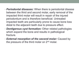
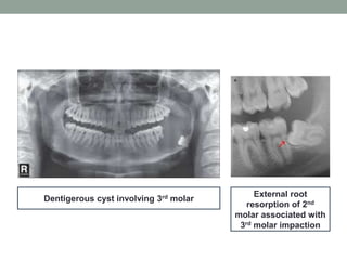
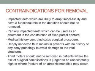
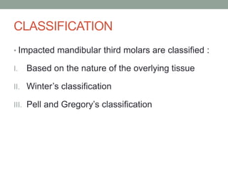
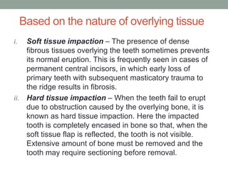
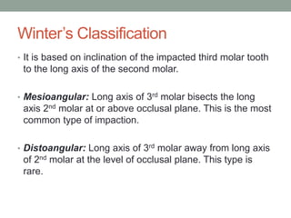
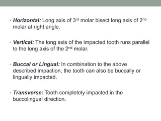
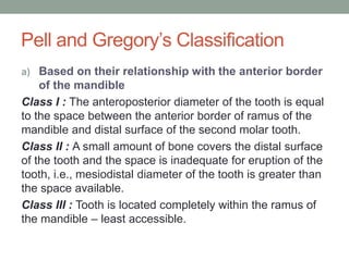
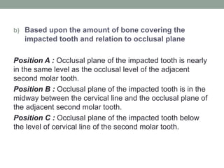
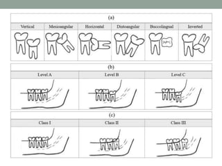
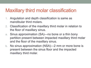
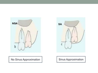
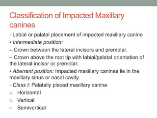
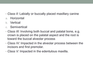
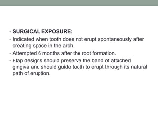
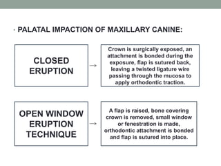
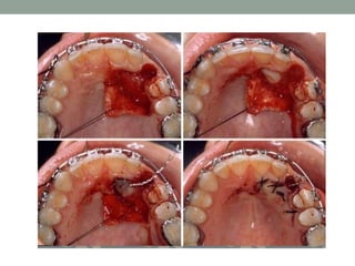
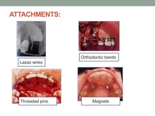
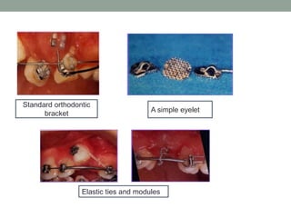
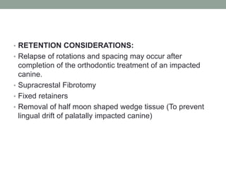
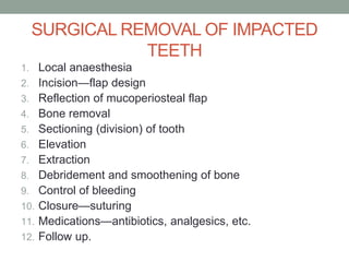
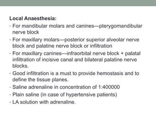
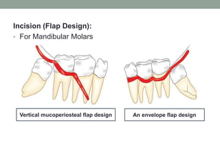
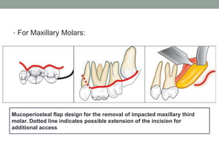
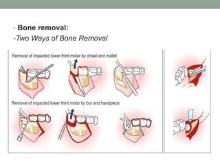
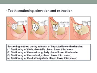
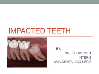
2. CONTENTS
• Definition
• Frequency of impacted teeth
• Theories of mandibular impaction
• Etiology of Impaction
• Indications for removal of impacted teeth
• Contraindications for removal of impacted teeth
• Classification
• Radiological Examination
• Management of Impacted canine
• Surgical removal of impacted teeth
• Complications
• References
3. DEFINITION
• Impacted tooth is defined as a tooth which is
completely or partially unerupted, is positioned
against another tooth, bone or soft tissue so that
its further eruption is unlikely and described
according to its anatomic position.
4. FREQUENCY OF IMPACTED TEETH
• Frequency of impacted teeth occurs in the
following order:
1. Mandibular third molars
2. Maxillary third molars
3. Maxillary cuspids
4. Mandibular bicuspids
5. Mandibular cuspids
6. Maxillary bicuspids
7. Maxillary central incisors
8. Maxillary lateral incisors
5. THEORIES OF MANDIBULAR
IMPACTION
• One of the most popular theories is insufficient
development of the retromolar space. Few other theories
are described below:
1. Orthodontic theory(small jaw-decreased space):
• Jaws develop in downward and forward direction, while as
the teeth move in a forward direction; the presence of
dense bone decreases the movement of teeth in a
forward direction. Growth of the jaw and movement of
teeth occurs in forward direction, anything that interferes
with such moment will cause an impaction (small jaw-
decreased space). A dense bone decreases the
movement of the teeth in forward direction.
6. • Causes for the increased density of bone are
1) Acute infection
2) Local inflammation of PDL
3) Malocclusion
4) Trauma
5) Early loss of primary teeth and arrested growth of the
jaw, leading to insufficient space available for normal
eruption.
• This condition is without doubt the cause of impaction.
7. 2. Nodine’s Phylogenic theory (Nodine 1943) [More-
functional masticatory force – better the
development of the jaw]:
• Nature tries to eliminate the disused organs. This causes
elimination of the unused teeth which causes congenital
absence of third molars. It is also known that in about
10% of the Caucasian race, third molars are lacking, as
the mandible and maxilla decreased in size leaving
insufficient room for third molars i.e., used makes the
organ develop better, disuse causes slow regression of
the organ. Due to changing nutritional habits, our
civilisation have practically eliminated needs for large
powerful jaws, thus, over centuries the mandible and
maxilla decreased in size leaving insufficient room for
third molars.
8. 3. Mendelian theory:
• Heredity is the most common cause. An individual may
inherit small jaws from one parent and a complement of
large teeth from the other, i.e., hereditary transmission of
small jaws and large teeth from parents to children. This
may be an important etiological factor in the occurrence of
impaction, i.e., small mandible with impaction of second
and third molars.
4. Pathological theory:
• Osteosclerosis in the third molar area, caused by the
early disease of adjacent molars, cause chronic infections
affecting an individual and may bring the condensation of
osseous tissue further preventing the growth and
development of the jaws.
9. 5. Endocrinal theory:
• Increase or decrease in the growth hormone secretion
may affect the size of the jaws. An imbalance of endocrine
activity leads to the lack of growth of the jaws, this lack
provides a cause for impaction.
6. Nature and nurture theory:
• Described by A. J. MacGregor explains that impaction can
occur due to a mismatch in size and shape of teeth and
jaws. This can be due to
a) Primate evolution owing to the increase in brain size
with the sacrifice of the jaw size without reduction in the
teeth size.
b) Change of diet of the modern civilisation due to under
used teeth.
10. c) Loss of use of weapons by the civilisation as cooking
decreases tough and abrasive food.
• With all these changes it is important to note that the
dentition has not reduced in size as it should have in
attrition. Attrition causes increased muscle activity that
stimulates jaw growth but instead there has been an
increase in impaction. This is due to the absence of
constant chewing due to high-calorie cooked food.
11. ETIOLOGY OF IMPACTION
• Berger lists the following local and systemic causes of
impaction.
LOCAL CAUSES:
• Irregularity in the position and pressure of the adjacent
tooth.
• Density of the overlying or surrounding bone.
• Localised chronic inflammation with resultant increase in
density of the overlying mucous membrane.
• Lack of space due to underdeveloped jaws. Arch length
and tooth size discrepancy.
12. • Obstructions: It can be a soft or hard tissue obstruction
caused by retained deciduous teeth, thick fibrous alveolar
mucosa or chronically inflamed mucosa, dense bone with
inflammatory changes due to odontome, cyst or
odontogenic tumour that prevents eruption at the
chronological age.
• Dilaceration: Abnormal path of eruption of the tooth due to
traumatic forces during the eruption period.
• Over retained deciduous teeth.
• Ectopic position of tooth bud.
13. A calcifying odontogenic cyst associated with the right maxillary impacted
third molar with dense opaque foci throughout the cystic cavity.
14. A panoramic view shows a unicystic ameloblastoma presenting as a
unilocular radiolucent lesion associated with the right mandibular
impacted third molar.
15. A multilocular extensive ameloblastoma is found on the right aspect and
ramus of the mandible in relation to an impacted molar tooth.
16. This panoramic view shows an adenomatoid odontogenic tumor
occurring as a unilocular radiolucent lesion surrounding the crown of the
right maxillary impacted permanent canine.
17. An extensive keratocystic odontogenic tumor as a multilocular radiolucent
lesion in the left mandibular ramus in close approximation to the impacted
third molar.
18. A calcifying epithelial odontogenic tumor is seen in the left posterior
mandible as a mixed lesion with dense opaque foci associated with an
impacted tooth.
19. This panoramic view shows a large complex odontoma associated with the
impacted left mandibular third molar.
20. SYSTEMIC CAUSES:
General causes
A. Prenatal causes: Heredity
B. Postnatal causes: All those conditions that may
interfere with the development of the child, such as:
1. Rickets
2. Anaemia
3. Congenital syphilis
4. Tuberculosis
5. Endocrine dysfunctions
6. Malnutrition
22. INDICATIONS FOR REMOVAL
• Infections: Removal of any symptomatic impacted tooth
should be considered with food impaction causing
halitosis, especially where there have been one or more
episodes of infection such as pericoronitis(75-80% of
patients with impacted third molar develop pericoronitis),
cellulitis, abscess formation; or untreatable
pulpal/periapical pathology.
• Unrestorable caries: Removal should be considered
when there is caries in the impacted tooth or when there
is caries in the adjacent second molar tooth which cannot
be satisfactorily treated without removal of the third molar.
24. • Periodontal diseases: When there is periodontal disease
between the third and second molar, early removal of the
impacted third molar will result in repair of the injured
periodontium and is therefore beneficial. Untreated
impacted teeth are particularly prone to cause bone loss
distal to the adjacent teeth due to pressure effect.
• Dentigerous cyst formation: Other related pathologies
which expand the bone and results in pathological
fracture.
• External resorption of the second molar: Caused by
the pressure of the third molar on 2nd molar.
25. Dentigerous cyst involving 3rd molar
External root
resorption of 2nd
molar associated with
3rd molar impaction
26. • Buccoverted impacted molars: May cause cheek bite,
frictional keratosis or traumatic fibroma mandating
extraction.
• Third molar removal may occasionally be indicated for
orthodontic reasons.
• Removal of the third molar may be indicated prior to
orthognathic surgery, e.g. when a sagittal split
osteotomy is planned, removal of the third molar
diminishes the risk of surgical complications with regard to
that of osteotomy.
• Prophylactic removal in presence of specific medical
and surgical conditions.
• Fracture of the mandible in the third molar region or
when a tooth is involved in tumour resection.
27. • Atypical pain from an unerupted third molar is the most
unusual situation and it is essential to avoid any confusion
with temporomandibular joint (TMJ) or muscle dysfunction
before considering removal.
• Impacted teeth in edentulous ridge that causes
ulcerations in the mucosa under a denture.
• Third molar removal may be considered for autogenous
transplantation to a first molar socket.
28. CONTRAINDICATIONS FOR REMOVAL
• Impacted teeth which are likely to erupt successfully and
have a functional role in the dentition should not be
removed.
• Partially impacted teeth which can be used as an
abutment in the construction of fixed partial denture.
• Medical history contraindicates surgical procedure.
• Deeply impacted third molars in patients with no history of
any bony pathology to avoid damage to the vital
structures.
• Third molars should not be removed in patients where the
risk of surgical complications is judged to be unacceptably
high or where fracture of an atrophic mandible may occur.
29. CLASSIFICATION
• Impacted mandibular third molars are classified :
I. Based on the nature of the overlying tissue
II. Winter’s classification
III. Pell and Gregory’s classification
30. Based on the nature of overlying tissue
i. Soft tissue impaction – The presence of dense
fibrous tissues overlying the teeth sometimes prevents
its normal eruption. This is frequently seen in cases of
permanent central incisors, in which early loss of
primary teeth with subsequent masticatory trauma to
the ridge results in fibrosis.
ii. Hard tissue impaction – When the teeth fail to erupt
due to obstruction caused by the overlying bone, it is
known as hard tissue impaction. Here the impacted
tooth is completely encased in bone so that, when the
soft tissue flap is reflected, the tooth is not visible.
Extensive amount of bone must be removed and the
tooth may require sectioning before removal.
31. Winter’s Classification
• It is based on inclination of the impacted third molar tooth
to the long axis of the second molar.
• Mesioangular: Long axis of 3rd molar bisects the long
axis 2nd molar at or above occlusal plane. This is the most
common type of impaction.
• Distoangular: Long axis of 3rd molar away from long axis
of 2nd molar at the level of occlusal plane. This type is
rare.
32. • Horizontal: Long axis of 3rd molar bisect long axis of 2nd
molar at right angle.
• Vertical: The long axis of the impacted tooth runs parallel
to the long axis of the 2nd molar.
• Buccal or Lingual: In combination to the above
described impaction, the tooth can also be buccally or
lingually impacted.
• Transverse: Tooth completely impacted in the
buccolingual direction.
33. Pell and Gregory’s Classification
a) Based on their relationship with the anterior border
of the mandible
Class I : The anteroposterior diameter of the tooth is equal
to the space between the anterior border of ramus of the
mandible and distal surface of the second molar tooth.
Class II : A small amount of bone covers the distal surface
of the tooth and the space is inadequate for eruption of the
tooth, i.e., mesiodistal diameter of the tooth is greater than
the space available.
Class III : Tooth is located completely within the ramus of
the mandible – least accessible.
34. b) Based upon the amount of bone covering the
impacted tooth and relation to occlusal plane
Position A : Occlusal plane of the impacted tooth is nearly
in the same level as the occlusal level of the adjacent
second molar tooth.
Position B : Occlusal plane of the impacted tooth is in the
midway between the cervical line and the occlusal plane of
the adjacent second molar tooth.
Position C : Occlusal plane of the impacted tooth below
the level of cervical line of the second molar tooth.
35.
36. Maxillary third molar classification
1. Angulation and depth classification is same as
mandibular third molars.
2. Classification of the maxillary third molar in relation to
the floor of maxillary sinus.
a. Sinus approximation (SA)—no bone or a thin bony
partition present between impacted maxillary third molar
and the floor of the maxillary sinus.
b. No sinus approximation (NSA)—2 mm or more bone is
present between the sinus floor and the impacted
maxillary third molar.
38. Classification of Impacted Maxillary
canines
• Labial or palatal placement of impacted maxillary canine
• Intermediate position:
– Crown between the lateral incisors and premolar.
– Crown above the root tip with labial/palatal orientation of
the lateral incisor or premolar.
• Aberrant position: Impacted maxillary canines lie in the
maxillary sinus or nasal cavity.
• Class I: Palatally placed maxillary canine
a. Horizontal
b. Vertical
c. Semivertical
39. • Class II: Labially or buccally placed maxillary canine
a. Horizontal
b. Vertical
c. Semivertical
• Class III: Involving both buccal and palatal bone, e.g.
crown is placed on the palatal aspect and the root is
toward the buccal alveolar process
• Class IV: Impacted in the alveolar process between the
incisors and first premolar.
• Class V: Impacted in the edentulous maxilla.
40. RADIOLOGICAL EXAMINATION
• INTRAORAL X-RAY
- The position and depth of the tooth can be assessed by
taking intraoral X-ray or even lateral extraoral X-ray and
tracing can be done, which was originally advocated by
George Winter.
- Three imaginary lines are drawn which are known as
Winter’s lines.
• White line corresponds to the occlusal plane. The line is
drawn touching the occlusal surfaces of first and second
molar and is extended posteriorly over the third molar
region. It indicates the difference in occlusal level of
second and third molars.
41. • Amber line represents the bone level. The line is drawn
from the crest of the interdental septum between the
molars and extended posteriorly distal to third molar or to
the ascending ramus. This line denotes the alveolar bone
covering the impacted tooth and the portion of tooth not
covered by the bone.
• Red line is drawn perpendicular from the amber line to an
imaginary point of application of the elevator. It indicates
the amount of bone that will have to be removed before
elevation, i.e. the depth of the tooth in bone and the
difficulty encountered in removing the tooth.
If the length of the red line is more than 5 mm then the
extraction is difficult. Every additional millimeter renders
the removal of the impacted tooth three times more
difficult (more than 9 mm—below the level of the apices of
the second molar).
42.
43. • EXTRAORAL X-RAYS
• For mandibular teeth
• OPG
• Lateral oblique view mandible
• For maxillary teeth
• OPG
• PA view Water’s position
44. MANAGEMENT OF IMPACTED CANINE
1. Interceptive treatment
2. Treatment of labial impaction
3. Treatment of palatal impaction
4. Methods of applying traction
5. Retention consideration
45. • INTERCEPTIVE TREATMENT:
• When the clinician detects early signs of ectopic eruption
of canines, an attempt should be made to prevent their
impaction and its potential sequelae.
• Selective extraction of the deciduous canines as early as
8 or 9 years of age.
• Normalise the eruption of ectopically erupting permanent
canine.
46. • LABIAL IMPACTION OF IMPACTED CANINE:
• Due to ectopic migration of canine crown over the root of
lateral incisor or insufficient space in the arch caused by
midline shift of dental origin.
• Arch length – tooth material discrepancy is the most
common cause.
• Extraction of deciduous canine at early age of 8 or 9
years will enhance eruption and self correction of labial
impaction.
47.
48. • SURGICAL EXPOSURE:
• Indicated when tooth does not erupt spontaneously after
creating space in the arch.
• Attempted 6 months after the root formation.
• Flap designs should preserve the band of attached
gingiva and should guide tooth to erupt through its natural
path of eruption.
49. • PALATAL IMPACTION OF MAXILLARY CANINE:
CLOSED
ERUPTION
Crown is surgically exposed, an
attachment is bonded during the
exposure, flap is sutured back,
leaving a twisted ligature wire
passing through the mucosa to
apply orthodontic traction.
OPEN WINDOW
ERUPTION
TECHNIQUE
A flap is raised, bone covering
crown is removed, small window
or fenestration is made,
orthodontic attachment is bonded
and flap is sutured into place.
53. • RETENTION CONSIDERATIONS:
• Relapse of rotations and spacing may occur after
completion of the orthodontic treatment of an impacted
canine.
• Supracrestal Fibrotomy
• Fixed retainers
• Removal of half moon shaped wedge tissue (To prevent
lingual drift of palatally impacted canine)
54. SURGICAL REMOVAL OF IMPACTED
TEETH
1. Local anaesthesia
2. Incision—flap design
3. Reflection of mucoperiosteal flap
4. Bone removal
5. Sectioning (division) of tooth
6. Elevation
7. Extraction
8. Debridement and smoothening of bone
9. Control of bleeding
10. Closure—suturing
11. Medications—antibiotics, analgesics, etc.
12. Follow up.
55. Local Anaesthesia:
• For mandibular molars and canines—pterygomandibular
nerve block
• For maxillary molars—posterior superior alveolar nerve
block and palatine nerve block or infiltration
• For maxillary canines—infraorbital nerve block + palatal
infiltration of incisive canal and bilateral palatine nerve
blocks.
• Good infiltration is a must to provide hemostasis and to
define the tissue planes.
• Saline adrenaline in concentration of 1:400000
• Plain saline (in case of hypertensive patients)
• LA solution with adrenaline.
56. Incision (Flap Design):
• For Mandibular Molars
Vertical mucoperiosteal flap design An envelope flap design
57. • For Maxillary Molars:
Mucoperiosteal flap design for the removal of impacted maxillary third
molar. Dotted line indicates possible extension of the incision for
additional access
59. • Tooth sectioning, elevation and extraction
Sectioning method during removal of impacted lower third molar:
(1) Sectioning of the horizontally placed lower third molar,
(2) Sectioning of the mesioangularly placed lower third molar,
(3) Sectioning of the vertically placed lower third molar,
(4) Sectioning of the distoangularly placed lower third molar
60. • Elevation
• Coupland elevator—placed at the base of the crown.
• Winter Cryer’s—may be used in wedging action/buccal
elevation. Buccal elevation may be done in molar and
canines by drilling a purchase point in the roots just below
CEJ.
• Wedging action is useful, when molar crown is split
vertically down to bifurcation of roots.
- Important precautions: Support the inferior border and
lingual cortex of the bone in the mandibular impaction.
Support to the palatal bone in the maxillary third molar or
canine impactions during elevation should be given.
Support the neighbouring tooth to prevent luxation of the
same.
61. • Debridement and smoothening of Bone Margins
• Irrigation of the socket
• Curetting to remove any remaining dental follicle and
epithelium
• Look for pieces of coronal portion (especially in carious
teeth/sectioned teeth), check for remnants of
bone/granulation tissue, bleeding points
• Check for caries (root/crown)/erosion/damage to the
adjacent teeth
• Round off the margins of the socket with large vulcanite
round bur or bone file
•Irrigate the socket again
• Control bleeding before suturing.
62. • Closure
• 3-0 black silk is used. Interrupted sutures given and
maintained for 7 days. In case of molars, suture distal to
second molar should be placed first and should be water
tight to prevent pocket formation.
• In case of palatally impacted canines, incisive papilla
should be sutured carefully to reduce postoperative
bleeding.
63. COMPLICATIONS
• Intraoperative Complications:
• During Incision
- For molars, facial vessel or buccal vessel may be cut. For
lower canines–mental vessels and for upper canines—
incisive canal or greater palatine vessels may be
damaged.
• During Bone Removal
- Damage to the second molar, damage to the roots of
overlying teeth, slipping of the bur into the soft tissues,
fracture of the mandible when using chisel and mallet.
64. • During Elevation
• Luxation of neighbouring/overlying tooth
• Fracture of the adjoining bone
• Fracture of the tuberosity
• Slipping of the tooth into pterygomandibular/temporal
spaces , sublingual pouch and/maxillary sinus.
• Damage to nasal wall/overlying teeth/lingual, inferior
alveolar or mental nerve.
• During Debridement
• Damage to inferior alveolar nerve/lingual nerve.
• Damage to maxillary sinus.
65. • Postoperative complications
- Pain, swelling, trismus, hypoesthesia, sensitivity, loss of
vitality of neighbouring teeth.
• Pocket formation.
• Sinus tract formation, oroantral fistula, oronasal fistula.
66. REFERENCES
• Textbook of Oral and Maxillofacial Surgery – Third Edition
– Neelima Anil Malik
• Textbook of Oral and Maxillofacial Surgery – Third Edition
– S M Balaji
• Shafer’s Textbook of Oral Pathology
• Jaw lesions associated with impacted tooth: A
radiographic diagnostic guide retrieved from
https://www.ncbi.nlm.nih.gov/pmc/articles/PMC5035719/