SlideShare a Scribd company logo
1 of 9
Download to read offline
ANTIBODY VALIDATION REPORT
Report Number 96933-a
Application Immunofluorescence
Model Number STJ96933
Antibody Name Anti-PCNA antibody
Host Mouse
Clonality Monoclonal
Clone ID 12D10
Species HUMAN Tissue LUNG CANCER
Image
Description
Immunofluorescence analysis of Human
lung cancer tissue. 1: PCNA Monoclonal
Antibody(12D10)(red) was diluted at
1:200 (4 degree Celsius,overnight). 2:
Cy3 labled Secondary antibody was
diluted at 1:300 (room temperature,
50min).3: Picture B: DAPI(blue) 10min.
Picture A:Target. Picture B: DAPI.
Picture C: merge of A+B.
Primary Antibody Incubation
After blocking solution was removed a 1:200 primary antibody/PBS
solution was added on the slide, and incubated overnight at 4°C (a
small amount of distilled water was added into the incubation box to
prevent evaporation of antibody).
Secondary Antibody Incubation
slides were washed with PBS on a shaker for 5min, and repeated 3
times. Shortly after the slides were dried and corresponding
secondary antibody solution was added (HRP labelled), covering the
tissues, and incubated at room temperature for 50min.
DAPI Counter-Staining
slides were washed with PBS on a shaker for 5min, repeated 3 times
and then dried. DAPI staining solution was added inside the PAP
circles and incubated for 10 min at room temperature without light
exposure.
Mounting
Slides were washed with PBS on a shaker for 5min, and repeated 3
times. Shortly after slides were dried, anti-quench mountings were
used to mount slides.
Visualization
The slides were observed and placed under a NIKON inverted
fluorescence microscope (Ultra violet excitation 330-380nm,
emission 420nm; FITC green excitation 465-495nm, emission 515-
555 nm; CY3 red excitation 510-560nm, emission 590nm)
Immunofluorescence Protocol
Tissue Processing
Slides were incubated sequentially into: Xylene - 15min, Anhydrous
ethanol – 15 min, Anhydrous ethanol – 5 min, 85% alcohol – 5 min,
75% alcohol – 5 min & washed with distilled water – 5 min.
Antigen Retrieval
Tissue slides were incubated with citric acid (PH6.0) antigen
retrieval buffer, and microwaved for antigen retrieval (heated until
boiled and then stop heating) for 8min. Slides were then heated with
medium power for 7min. During this process slides are kept from
drying out. After cooling down at room temperature, slides were
washed with PBS on a shaker for 5min, and repeated 3 times.
Anti-Quench
shortly after slides were dried, a PAP pen was used to draw circles
around the tissues (to prevent draining of the antibody). Inside the
circles, anti-quench mountings were added and incubated for 5 min,
and then flushed with water for 10min.
BSA Blocking
Inside the circles, BSA was used to cover the tissue evenly, blocking
for 30min.
St John's Laboratory Ltd.
www.stjohnslabs.com
ANTIBODY VALIDATION REPORT
Report Number 96933-b
Application Immunofluorescence
Model Number STJ96933
Antibody Name Anti-PCNA antibody
Host Mouse
Clonality Monoclonal
Clone ID 12D10
Species HUMAN Tissue LUNG CANCER
Image
Description
Immunofluorescence analysis of Human
lung cancer tissue. 1: PCNA Monoclonal
Antibody(12D10)(red) was diluted at
1:200 (4 degree Celsius,overnight). 2:
Cy3 labled Secondary antibody was
diluted at 1:300 (room temperature,
50min).3: Picture B: DAPI(blue) 10min.
Picture A:Target. Picture B: DAPI.
Picture C: merge of A+B.
Primary Antibody Incubation
After blocking solution was removed a 1:200 primary antibody/PBS
solution was added on the slide, and incubated overnight at 4°C (a
small amount of distilled water was added into the incubation box to
prevent evaporation of antibody).
Secondary Antibody Incubation
slides were washed with PBS on a shaker for 5min, and repeated 3
times. Shortly after the slides were dried and corresponding
secondary antibody solution was added (HRP labelled), covering the
tissues, and incubated at room temperature for 50min.
DAPI Counter-Staining
slides were washed with PBS on a shaker for 5min, repeated 3 times
and then dried. DAPI staining solution was added inside the PAP
circles and incubated for 10 min at room temperature without light
exposure.
Mounting
Slides were washed with PBS on a shaker for 5min, and repeated 3
times. Shortly after slides were dried, anti-quench mountings were
used to mount slides.
Visualization
The slides were observed and placed under a NIKON inverted
fluorescence microscope (Ultra violet excitation 330-380nm,
emission 420nm; FITC green excitation 465-495nm, emission 515-
555 nm; CY3 red excitation 510-560nm, emission 590nm)
Immunofluorescence Protocol
Tissue Processing
Slides were incubated sequentially into: Xylene - 15min, Anhydrous
ethanol – 15 min, Anhydrous ethanol – 5 min, 85% alcohol – 5 min,
75% alcohol – 5 min & washed with distilled water – 5 min.
Antigen Retrieval
Tissue slides were incubated with citric acid (PH6.0) antigen
retrieval buffer, and microwaved for antigen retrieval (heated until
boiled and then stop heating) for 8min. Slides were then heated with
medium power for 7min. During this process slides are kept from
drying out. After cooling down at room temperature, slides were
washed with PBS on a shaker for 5min, and repeated 3 times.
Anti-Quench
shortly after slides were dried, a PAP pen was used to draw circles
around the tissues (to prevent draining of the antibody). Inside the
circles, anti-quench mountings were added and incubated for 5 min,
and then flushed with water for 10min.
BSA Blocking
Inside the circles, BSA was used to cover the tissue evenly, blocking
for 30min.
St John's Laboratory Ltd.
www.stjohnslabs.com
ANTIBODY VALIDATION REPORT
Report Number 96933-c
Application Immunofluorescence
Model Number STJ96933
Antibody Name Anti-PCNA antibody
Host Mouse
Clonality Monoclonal
Clone ID 12D10
Species HUMAN Tissue LUNG CANCER
Image
Description
Immunofluorescence analysis of Human
lung cancer tissue. 1: PCNA Monoclonal
Antibody(12D10)(red) was diluted at
1:200 (4 degree Celsius,overnight). 2:
Cy3 labled Secondary antibody was
diluted at 1:300 (room temperature,
50min).3: Picture B: DAPI(blue) 10min.
Picture A:Target. Picture B: DAPI.
Picture C: merge of A+B.
Primary Antibody Incubation
After blocking solution was removed a 1:200 primary antibody/PBS
solution was added on the slide, and incubated overnight at 4°C (a
small amount of distilled water was added into the incubation box to
prevent evaporation of antibody).
Secondary Antibody Incubation
slides were washed with PBS on a shaker for 5min, and repeated 3
times. Shortly after the slides were dried and corresponding
secondary antibody solution was added (HRP labelled), covering the
tissues, and incubated at room temperature for 50min.
DAPI Counter-Staining
slides were washed with PBS on a shaker for 5min, repeated 3 times
and then dried. DAPI staining solution was added inside the PAP
circles and incubated for 10 min at room temperature without light
exposure.
Mounting
Slides were washed with PBS on a shaker for 5min, and repeated 3
times. Shortly after slides were dried, anti-quench mountings were
used to mount slides.
Visualization
The slides were observed and placed under a NIKON inverted
fluorescence microscope (Ultra violet excitation 330-380nm,
emission 420nm; FITC green excitation 465-495nm, emission 515-
555 nm; CY3 red excitation 510-560nm, emission 590nm)
Immunofluorescence Protocol
Tissue Processing
Slides were incubated sequentially into: Xylene - 15min, Anhydrous
ethanol – 15 min, Anhydrous ethanol – 5 min, 85% alcohol – 5 min,
75% alcohol – 5 min & washed with distilled water – 5 min.
Antigen Retrieval
Tissue slides were incubated with citric acid (PH6.0) antigen
retrieval buffer, and microwaved for antigen retrieval (heated until
boiled and then stop heating) for 8min. Slides were then heated with
medium power for 7min. During this process slides are kept from
drying out. After cooling down at room temperature, slides were
washed with PBS on a shaker for 5min, and repeated 3 times.
Anti-Quench
shortly after slides were dried, a PAP pen was used to draw circles
around the tissues (to prevent draining of the antibody). Inside the
circles, anti-quench mountings were added and incubated for 5 min,
and then flushed with water for 10min.
BSA Blocking
Inside the circles, BSA was used to cover the tissue evenly, blocking
for 30min.
St John's Laboratory Ltd.
www.stjohnslabs.com
ANTIBODY VALIDATION REPORT
Report Number 96933-d
Application Immunofluorescence
Model Number STJ96933
Antibody Name Anti-PCNA antibody
Host Mouse
Clonality Monoclonal
Clone ID 12D10
Species MOUSE Tissue LUNG
Image
Description
Immunofluorescence analysis of Mouse
lung tissue. 1: PCNA Monoclonal
Antibody(12D10)(red) was diluted at
1:200 (4 degree Celsius,overnight). 2:
Cy3 labled Secondary antibody was
diluted at 1:300 (room temperature,
50min).3: Picture B: DAPI(blue) 10min.
Picture A:Target. Picture B: DAPI.
Picture C: merge of A+B.
Primary Antibody Incubation
After blocking solution was removed a 1:200 primary antibody/PBS
solution was added on the slide, and incubated overnight at 4°C (a
small amount of distilled water was added into the incubation box to
prevent evaporation of antibody).
Secondary Antibody Incubation
slides were washed with PBS on a shaker for 5min, and repeated 3
times. Shortly after the slides were dried and corresponding
secondary antibody solution was added (HRP labelled), covering the
tissues, and incubated at room temperature for 50min.
DAPI Counter-Staining
slides were washed with PBS on a shaker for 5min, repeated 3 times
and then dried. DAPI staining solution was added inside the PAP
circles and incubated for 10 min at room temperature without light
exposure.
Mounting
Slides were washed with PBS on a shaker for 5min, and repeated 3
times. Shortly after slides were dried, anti-quench mountings were
used to mount slides.
Visualization
The slides were observed and placed under a NIKON inverted
fluorescence microscope (Ultra violet excitation 330-380nm,
emission 420nm; FITC green excitation 465-495nm, emission 515-
555 nm; CY3 red excitation 510-560nm, emission 590nm)
Immunofluorescence Protocol
Tissue Processing
Slides were incubated sequentially into: Xylene - 15min, Anhydrous
ethanol – 15 min, Anhydrous ethanol – 5 min, 85% alcohol – 5 min,
75% alcohol – 5 min & washed with distilled water – 5 min.
Antigen Retrieval
Tissue slides were incubated with citric acid (PH6.0) antigen
retrieval buffer, and microwaved for antigen retrieval (heated until
boiled and then stop heating) for 8min. Slides were then heated with
medium power for 7min. During this process slides are kept from
drying out. After cooling down at room temperature, slides were
washed with PBS on a shaker for 5min, and repeated 3 times.
Anti-Quench
shortly after slides were dried, a PAP pen was used to draw circles
around the tissues (to prevent draining of the antibody). Inside the
circles, anti-quench mountings were added and incubated for 5 min,
and then flushed with water for 10min.
BSA Blocking
Inside the circles, BSA was used to cover the tissue evenly, blocking
for 30min.
St John's Laboratory Ltd.
www.stjohnslabs.com
ANTIBODY VALIDATION REPORT
Report Number 96933-e
Application Immunofluorescence
Model Number STJ96933
Antibody Name Anti-PCNA antibody
Host Mouse
Clonality Monoclonal
Clone ID 12D10
Species MOUSE Tissue LUNG
Image
Description
Immunofluorescence analysis of Mouse
lung tissue. 1: PCNA Monoclonal
Antibody(12D10)(red) was diluted at
1:200 (4 degree Celsius,overnight). 2:
Cy3 labled Secondary antibody was
diluted at 1:300 (room temperature,
50min).3: Picture B: DAPI(blue) 10min.
Picture A:Target. Picture B: DAPI.
Picture C: merge of A+B.
Primary Antibody Incubation
After blocking solution was removed a 1:200 primary antibody/PBS
solution was added on the slide, and incubated overnight at 4°C (a
small amount of distilled water was added into the incubation box to
prevent evaporation of antibody).
Secondary Antibody Incubation
slides were washed with PBS on a shaker for 5min, and repeated 3
times. Shortly after the slides were dried and corresponding
secondary antibody solution was added (HRP labelled), covering the
tissues, and incubated at room temperature for 50min.
DAPI Counter-Staining
slides were washed with PBS on a shaker for 5min, repeated 3 times
and then dried. DAPI staining solution was added inside the PAP
circles and incubated for 10 min at room temperature without light
exposure.
Mounting
Slides were washed with PBS on a shaker for 5min, and repeated 3
times. Shortly after slides were dried, anti-quench mountings were
used to mount slides.
Visualization
The slides were observed and placed under a NIKON inverted
fluorescence microscope (Ultra violet excitation 330-380nm,
emission 420nm; FITC green excitation 465-495nm, emission 515-
555 nm; CY3 red excitation 510-560nm, emission 590nm)
Immunofluorescence Protocol
Tissue Processing
Slides were incubated sequentially into: Xylene - 15min, Anhydrous
ethanol – 15 min, Anhydrous ethanol – 5 min, 85% alcohol – 5 min,
75% alcohol – 5 min & washed with distilled water – 5 min.
Antigen Retrieval
Tissue slides were incubated with citric acid (PH6.0) antigen
retrieval buffer, and microwaved for antigen retrieval (heated until
boiled and then stop heating) for 8min. Slides were then heated with
medium power for 7min. During this process slides are kept from
drying out. After cooling down at room temperature, slides were
washed with PBS on a shaker for 5min, and repeated 3 times.
Anti-Quench
shortly after slides were dried, a PAP pen was used to draw circles
around the tissues (to prevent draining of the antibody). Inside the
circles, anti-quench mountings were added and incubated for 5 min,
and then flushed with water for 10min.
BSA Blocking
Inside the circles, BSA was used to cover the tissue evenly, blocking
for 30min.
St John's Laboratory Ltd.
www.stjohnslabs.com
ANTIBODY VALIDATION REPORT
Report Number 96933-f
Application Immunofluorescence
Model Number STJ96933
Antibody Name Anti-PCNA antibody
Host Mouse
Clonality Monoclonal
Clone ID 12D10
Species MOUSE Tissue LUNG
Image
Description
Immunofluorescence analysis of Mouse
lung tissue. 1: PCNA Monoclonal
Antibody(12D10)(red) was diluted at
1:200 (4 degree Celsius,overnight). 2:
Cy3 labled Secondary antibody was
diluted at 1:300 (room temperature,
50min).3: Picture B: DAPI(blue) 10min.
Picture A:Target. Picture B: DAPI.
Picture C: merge of A+B.
Primary Antibody Incubation
After blocking solution was removed a 1:200 primary antibody/PBS
solution was added on the slide, and incubated overnight at 4°C (a
small amount of distilled water was added into the incubation box to
prevent evaporation of antibody).
Secondary Antibody Incubation
slides were washed with PBS on a shaker for 5min, and repeated 3
times. Shortly after the slides were dried and corresponding
secondary antibody solution was added (HRP labelled), covering the
tissues, and incubated at room temperature for 50min.
DAPI Counter-Staining
slides were washed with PBS on a shaker for 5min, repeated 3 times
and then dried. DAPI staining solution was added inside the PAP
circles and incubated for 10 min at room temperature without light
exposure.
Mounting
Slides were washed with PBS on a shaker for 5min, and repeated 3
times. Shortly after slides were dried, anti-quench mountings were
used to mount slides.
Visualization
The slides were observed and placed under a NIKON inverted
fluorescence microscope (Ultra violet excitation 330-380nm,
emission 420nm; FITC green excitation 465-495nm, emission 515-
555 nm; CY3 red excitation 510-560nm, emission 590nm)
Immunofluorescence Protocol
Tissue Processing
Slides were incubated sequentially into: Xylene - 15min, Anhydrous
ethanol – 15 min, Anhydrous ethanol – 5 min, 85% alcohol – 5 min,
75% alcohol – 5 min & washed with distilled water – 5 min.
Antigen Retrieval
Tissue slides were incubated with citric acid (PH6.0) antigen
retrieval buffer, and microwaved for antigen retrieval (heated until
boiled and then stop heating) for 8min. Slides were then heated with
medium power for 7min. During this process slides are kept from
drying out. After cooling down at room temperature, slides were
washed with PBS on a shaker for 5min, and repeated 3 times.
Anti-Quench
shortly after slides were dried, a PAP pen was used to draw circles
around the tissues (to prevent draining of the antibody). Inside the
circles, anti-quench mountings were added and incubated for 5 min,
and then flushed with water for 10min.
BSA Blocking
Inside the circles, BSA was used to cover the tissue evenly, blocking
for 30min.
St John's Laboratory Ltd.
www.stjohnslabs.com
ANTIBODY VALIDATION REPORT
Report Number 96933-g
Application Immunofluorescence
Model Number STJ96933
Antibody Name Anti-PCNA antibody
Host Mouse
Clonality Monoclonal
Clone ID 12D10
Species RAT Tissue TESTIS
Image
Description
Immunofluorescence analysis of Rat
testis tissue. 1: PCNA Monoclonal
Antibody(12D10)(red) was diluted at
1:200 (4 degree Celsius,overnight). 2:
Cy3 labled Secondary antibody was
diluted at 1:300 (room temperature,
50min).3: Picture B: DAPI(blue) 10min.
Picture A:Target. Picture B: DAPI.
Picture C: merge of A+B.
Primary Antibody Incubation
After blocking solution was removed a 1:200 primary antibody/PBS
solution was added on the slide, and incubated overnight at 4°C (a
small amount of distilled water was added into the incubation box to
prevent evaporation of antibody).
Secondary Antibody Incubation
slides were washed with PBS on a shaker for 5min, and repeated 3
times. Shortly after the slides were dried and corresponding
secondary antibody solution was added (HRP labelled), covering the
tissues, and incubated at room temperature for 50min.
DAPI Counter-Staining
slides were washed with PBS on a shaker for 5min, repeated 3 times
and then dried. DAPI staining solution was added inside the PAP
circles and incubated for 10 min at room temperature without light
exposure.
Mounting
Slides were washed with PBS on a shaker for 5min, and repeated 3
times. Shortly after slides were dried, anti-quench mountings were
used to mount slides.
Visualization
The slides were observed and placed under a NIKON inverted
fluorescence microscope (Ultra violet excitation 330-380nm,
emission 420nm; FITC green excitation 465-495nm, emission 515-
555 nm; CY3 red excitation 510-560nm, emission 590nm)
Immunofluorescence Protocol
Tissue Processing
Slides were incubated sequentially into: Xylene - 15min, Anhydrous
ethanol – 15 min, Anhydrous ethanol – 5 min, 85% alcohol – 5 min,
75% alcohol – 5 min & washed with distilled water – 5 min.
Antigen Retrieval
Tissue slides were incubated with citric acid (PH6.0) antigen
retrieval buffer, and microwaved for antigen retrieval (heated until
boiled and then stop heating) for 8min. Slides were then heated with
medium power for 7min. During this process slides are kept from
drying out. After cooling down at room temperature, slides were
washed with PBS on a shaker for 5min, and repeated 3 times.
Anti-Quench
shortly after slides were dried, a PAP pen was used to draw circles
around the tissues (to prevent draining of the antibody). Inside the
circles, anti-quench mountings were added and incubated for 5 min,
and then flushed with water for 10min.
BSA Blocking
Inside the circles, BSA was used to cover the tissue evenly, blocking
for 30min.
St John's Laboratory Ltd.
www.stjohnslabs.com
ANTIBODY VALIDATION REPORT
Report Number 96933-h
Application Immunofluorescence
Model Number STJ96933
Antibody Name Anti-PCNA antibody
Host Mouse
Clonality Monoclonal
Clone ID 12D10
Species RAT Tissue TESTIS
Image
Description
Immunofluorescence analysis of Rat
testis tissue. 1: PCNA Monoclonal
Antibody(12D10)(red) was diluted at
1:200 (4 degree Celsius,overnight). 2:
Cy3 labled Secondary antibody was
diluted at 1:300 (room temperature,
50min).3: Picture B: DAPI(blue) 10min.
Picture A:Target. Picture B: DAPI.
Picture C: merge of A+B.
Primary Antibody Incubation
After blocking solution was removed a 1:200 primary antibody/PBS
solution was added on the slide, and incubated overnight at 4°C (a
small amount of distilled water was added into the incubation box to
prevent evaporation of antibody).
Secondary Antibody Incubation
slides were washed with PBS on a shaker for 5min, and repeated 3
times. Shortly after the slides were dried and corresponding
secondary antibody solution was added (HRP labelled), covering the
tissues, and incubated at room temperature for 50min.
DAPI Counter-Staining
slides were washed with PBS on a shaker for 5min, repeated 3 times
and then dried. DAPI staining solution was added inside the PAP
circles and incubated for 10 min at room temperature without light
exposure.
Mounting
Slides were washed with PBS on a shaker for 5min, and repeated 3
times. Shortly after slides were dried, anti-quench mountings were
used to mount slides.
Visualization
The slides were observed and placed under a NIKON inverted
fluorescence microscope (Ultra violet excitation 330-380nm,
emission 420nm; FITC green excitation 465-495nm, emission 515-
555 nm; CY3 red excitation 510-560nm, emission 590nm)
Immunofluorescence Protocol
Tissue Processing
Slides were incubated sequentially into: Xylene - 15min, Anhydrous
ethanol – 15 min, Anhydrous ethanol – 5 min, 85% alcohol – 5 min,
75% alcohol – 5 min & washed with distilled water – 5 min.
Antigen Retrieval
Tissue slides were incubated with citric acid (PH6.0) antigen
retrieval buffer, and microwaved for antigen retrieval (heated until
boiled and then stop heating) for 8min. Slides were then heated with
medium power for 7min. During this process slides are kept from
drying out. After cooling down at room temperature, slides were
washed with PBS on a shaker for 5min, and repeated 3 times.
Anti-Quench
shortly after slides were dried, a PAP pen was used to draw circles
around the tissues (to prevent draining of the antibody). Inside the
circles, anti-quench mountings were added and incubated for 5 min,
and then flushed with water for 10min.
BSA Blocking
Inside the circles, BSA was used to cover the tissue evenly, blocking
for 30min.
St John's Laboratory Ltd.
www.stjohnslabs.com
ANTIBODY VALIDATION REPORT
Report Number 96933-i
Application Immunofluorescence
Model Number STJ96933
Antibody Name Anti-PCNA antibody
Host Mouse
Clonality Monoclonal
Clone ID 12D10
Species RAT Tissue TESTIS
Image
Description
Immunofluorescence analysis of Rat
testis tissue. 1: PCNA Monoclonal
Antibody(12D10)(red) was diluted at
1:200 (4 degree Celsius,overnight). 2:
Cy3 labled Secondary antibody was
diluted at 1:300 (room temperature,
50min).3: Picture B: DAPI(blue) 10min.
Picture A:Target. Picture B: DAPI.
Picture C: merge of A+B.
Primary Antibody Incubation
After blocking solution was removed a 1:200 primary antibody/PBS
solution was added on the slide, and incubated overnight at 4°C (a
small amount of distilled water was added into the incubation box to
prevent evaporation of antibody).
Secondary Antibody Incubation
slides were washed with PBS on a shaker for 5min, and repeated 3
times. Shortly after the slides were dried and corresponding
secondary antibody solution was added (HRP labelled), covering the
tissues, and incubated at room temperature for 50min.
DAPI Counter-Staining
slides were washed with PBS on a shaker for 5min, repeated 3 times
and then dried. DAPI staining solution was added inside the PAP
circles and incubated for 10 min at room temperature without light
exposure.
Mounting
Slides were washed with PBS on a shaker for 5min, and repeated 3
times. Shortly after slides were dried, anti-quench mountings were
used to mount slides.
Visualization
The slides were observed and placed under a NIKON inverted
fluorescence microscope (Ultra violet excitation 330-380nm,
emission 420nm; FITC green excitation 465-495nm, emission 515-
555 nm; CY3 red excitation 510-560nm, emission 590nm)
Immunofluorescence Protocol
Tissue Processing
Slides were incubated sequentially into: Xylene - 15min, Anhydrous
ethanol – 15 min, Anhydrous ethanol – 5 min, 85% alcohol – 5 min,
75% alcohol – 5 min & washed with distilled water – 5 min.
Antigen Retrieval
Tissue slides were incubated with citric acid (PH6.0) antigen
retrieval buffer, and microwaved for antigen retrieval (heated until
boiled and then stop heating) for 8min. Slides were then heated with
medium power for 7min. During this process slides are kept from
drying out. After cooling down at room temperature, slides were
washed with PBS on a shaker for 5min, and repeated 3 times.
Anti-Quench
shortly after slides were dried, a PAP pen was used to draw circles
around the tissues (to prevent draining of the antibody). Inside the
circles, anti-quench mountings were added and incubated for 5 min,
and then flushed with water for 10min.
BSA Blocking
Inside the circles, BSA was used to cover the tissue evenly, blocking
for 30min.
St John's Laboratory Ltd.
www.stjohnslabs.com

More Related Content

What's hot

Immunofluorescence Antibody Validation Report for Anti-CK17 Antibody (STJ96983)
Immunofluorescence Antibody Validation Report for Anti-CK17 Antibody (STJ96983)Immunofluorescence Antibody Validation Report for Anti-CK17 Antibody (STJ96983)
Immunofluorescence Antibody Validation Report for Anti-CK17 Antibody (STJ96983)St John's Laboratory Ltd
 
Immunofluorescence Antibody Validation Report for Anti-Ub Antibody (STJ97236)
Immunofluorescence Antibody Validation Report for Anti-Ub Antibody (STJ97236)Immunofluorescence Antibody Validation Report for Anti-Ub Antibody (STJ97236)
Immunofluorescence Antibody Validation Report for Anti-Ub Antibody (STJ97236)St John's Laboratory Ltd
 
Immunofluorescence Antibody Validation Report for Anti-Smad3 Antibody (STJ95699)
Immunofluorescence Antibody Validation Report for Anti-Smad3 Antibody (STJ95699)Immunofluorescence Antibody Validation Report for Anti-Smad3 Antibody (STJ95699)
Immunofluorescence Antibody Validation Report for Anti-Smad3 Antibody (STJ95699)St John's Laboratory Ltd
 
Immunofluorescence Antibody Validation Report for Anti-CD45 Antibody (STJ96948)
Immunofluorescence Antibody Validation Report for Anti-CD45 Antibody (STJ96948)Immunofluorescence Antibody Validation Report for Anti-CD45 Antibody (STJ96948)
Immunofluorescence Antibody Validation Report for Anti-CD45 Antibody (STJ96948)St John's Laboratory Ltd
 
Immunofluorescence Antibody Validation Report for Anti-CYCS Antibody (STJ97419)
Immunofluorescence Antibody Validation Report for Anti-CYCS Antibody (STJ97419)Immunofluorescence Antibody Validation Report for Anti-CYCS Antibody (STJ97419)
Immunofluorescence Antibody Validation Report for Anti-CYCS Antibody (STJ97419)St John's Laboratory Ltd
 
Immunofluorescence Antibody Validation Report for Anti-N-cadherin Antibody (S...
Immunofluorescence Antibody Validation Report for Anti-N-cadherin Antibody (S...Immunofluorescence Antibody Validation Report for Anti-N-cadherin Antibody (S...
Immunofluorescence Antibody Validation Report for Anti-N-cadherin Antibody (S...St John's Laboratory Ltd
 
Immunofluorescence Antibody Validation Report for Anti-Smad2 Antibody (STJ95694)
Immunofluorescence Antibody Validation Report for Anti-Smad2 Antibody (STJ95694)Immunofluorescence Antibody Validation Report for Anti-Smad2 Antibody (STJ95694)
Immunofluorescence Antibody Validation Report for Anti-Smad2 Antibody (STJ95694)St John's Laboratory Ltd
 
Immunofluorescence Antibody Validation Report for Anti-Caspase-8 Antibody (ST...
Immunofluorescence Antibody Validation Report for Anti-Caspase-8 Antibody (ST...Immunofluorescence Antibody Validation Report for Anti-Caspase-8 Antibody (ST...
Immunofluorescence Antibody Validation Report for Anti-Caspase-8 Antibody (ST...St John's Laboratory Ltd
 
Immunofluorescence Antibody Validation Report for Anti-PDGF-B Antibody (STJ94...
Immunofluorescence Antibody Validation Report for Anti-PDGF-B Antibody (STJ94...Immunofluorescence Antibody Validation Report for Anti-PDGF-B Antibody (STJ94...
Immunofluorescence Antibody Validation Report for Anti-PDGF-B Antibody (STJ94...St John's Laboratory Ltd
 
Immunofluorescence Antibody Validation Report for Anti-CD10 Antibody (STJ96974)
Immunofluorescence Antibody Validation Report for Anti-CD10 Antibody (STJ96974)Immunofluorescence Antibody Validation Report for Anti-CD10 Antibody (STJ96974)
Immunofluorescence Antibody Validation Report for Anti-CD10 Antibody (STJ96974)St John's Laboratory Ltd
 
Immunofluorescence Antibody Validation Report for Anti-CD63 Antibody (STJ96890)
Immunofluorescence Antibody Validation Report for Anti-CD63 Antibody (STJ96890)Immunofluorescence Antibody Validation Report for Anti-CD63 Antibody (STJ96890)
Immunofluorescence Antibody Validation Report for Anti-CD63 Antibody (STJ96890)St John's Laboratory Ltd
 
Immunofluorescence Antibody Validation Report for Anti-NSE Antibody (STJ96968)
Immunofluorescence Antibody Validation Report for Anti-NSE Antibody (STJ96968)Immunofluorescence Antibody Validation Report for Anti-NSE Antibody (STJ96968)
Immunofluorescence Antibody Validation Report for Anti-NSE Antibody (STJ96968)St John's Laboratory Ltd
 
Immunofluorescence Antibody Validation Report for Anti-Transferrin Antibody (...
Immunofluorescence Antibody Validation Report for Anti-Transferrin Antibody (...Immunofluorescence Antibody Validation Report for Anti-Transferrin Antibody (...
Immunofluorescence Antibody Validation Report for Anti-Transferrin Antibody (...St John's Laboratory Ltd
 
Immunofluorescence Antibody Validation Report for Anti-ABCG2 Antibody (STJ97253)
Immunofluorescence Antibody Validation Report for Anti-ABCG2 Antibody (STJ97253)Immunofluorescence Antibody Validation Report for Anti-ABCG2 Antibody (STJ97253)
Immunofluorescence Antibody Validation Report for Anti-ABCG2 Antibody (STJ97253)St John's Laboratory Ltd
 
Immunofluorescence Antibody Validation Report for Anti-HSP90β Antibody (STJ97...
Immunofluorescence Antibody Validation Report for Anti-HSP90β Antibody (STJ97...Immunofluorescence Antibody Validation Report for Anti-HSP90β Antibody (STJ97...
Immunofluorescence Antibody Validation Report for Anti-HSP90β Antibody (STJ97...St John's Laboratory Ltd
 
Immunofluorescence Antibody Validation Report for Anti-BMP-2 Antibody (STJ97346)
Immunofluorescence Antibody Validation Report for Anti-BMP-2 Antibody (STJ97346)Immunofluorescence Antibody Validation Report for Anti-BMP-2 Antibody (STJ97346)
Immunofluorescence Antibody Validation Report for Anti-BMP-2 Antibody (STJ97346)St John's Laboratory Ltd
 
Immunofluorescence Antibody Validation Report for Anti-PPAR-γ Antibody (STJ95...
Immunofluorescence Antibody Validation Report for Anti-PPAR-γ Antibody (STJ95...Immunofluorescence Antibody Validation Report for Anti-PPAR-γ Antibody (STJ95...
Immunofluorescence Antibody Validation Report for Anti-PPAR-γ Antibody (STJ95...St John's Laboratory Ltd
 
Immunofluorescence Antibody Validation Report for Anti-Rho A Antibody (STJ95442)
Immunofluorescence Antibody Validation Report for Anti-Rho A Antibody (STJ95442)Immunofluorescence Antibody Validation Report for Anti-Rho A Antibody (STJ95442)
Immunofluorescence Antibody Validation Report for Anti-Rho A Antibody (STJ95442)St John's Laboratory Ltd
 
Immunofluorescence Antibody Validation Report for Anti-Dnmt3b Antibody (STJ92...
Immunofluorescence Antibody Validation Report for Anti-Dnmt3b Antibody (STJ92...Immunofluorescence Antibody Validation Report for Anti-Dnmt3b Antibody (STJ92...
Immunofluorescence Antibody Validation Report for Anti-Dnmt3b Antibody (STJ92...St John's Laboratory Ltd
 
Immunofluorescence Antibody Validation Report for Anti-FAS Antibody (STJ93041)
Immunofluorescence Antibody Validation Report for Anti-FAS Antibody (STJ93041)Immunofluorescence Antibody Validation Report for Anti-FAS Antibody (STJ93041)
Immunofluorescence Antibody Validation Report for Anti-FAS Antibody (STJ93041)St John's Laboratory Ltd
 

What's hot (20)

Immunofluorescence Antibody Validation Report for Anti-CK17 Antibody (STJ96983)
Immunofluorescence Antibody Validation Report for Anti-CK17 Antibody (STJ96983)Immunofluorescence Antibody Validation Report for Anti-CK17 Antibody (STJ96983)
Immunofluorescence Antibody Validation Report for Anti-CK17 Antibody (STJ96983)
 
Immunofluorescence Antibody Validation Report for Anti-Ub Antibody (STJ97236)
Immunofluorescence Antibody Validation Report for Anti-Ub Antibody (STJ97236)Immunofluorescence Antibody Validation Report for Anti-Ub Antibody (STJ97236)
Immunofluorescence Antibody Validation Report for Anti-Ub Antibody (STJ97236)
 
Immunofluorescence Antibody Validation Report for Anti-Smad3 Antibody (STJ95699)
Immunofluorescence Antibody Validation Report for Anti-Smad3 Antibody (STJ95699)Immunofluorescence Antibody Validation Report for Anti-Smad3 Antibody (STJ95699)
Immunofluorescence Antibody Validation Report for Anti-Smad3 Antibody (STJ95699)
 
Immunofluorescence Antibody Validation Report for Anti-CD45 Antibody (STJ96948)
Immunofluorescence Antibody Validation Report for Anti-CD45 Antibody (STJ96948)Immunofluorescence Antibody Validation Report for Anti-CD45 Antibody (STJ96948)
Immunofluorescence Antibody Validation Report for Anti-CD45 Antibody (STJ96948)
 
Immunofluorescence Antibody Validation Report for Anti-CYCS Antibody (STJ97419)
Immunofluorescence Antibody Validation Report for Anti-CYCS Antibody (STJ97419)Immunofluorescence Antibody Validation Report for Anti-CYCS Antibody (STJ97419)
Immunofluorescence Antibody Validation Report for Anti-CYCS Antibody (STJ97419)
 
Immunofluorescence Antibody Validation Report for Anti-N-cadherin Antibody (S...
Immunofluorescence Antibody Validation Report for Anti-N-cadherin Antibody (S...Immunofluorescence Antibody Validation Report for Anti-N-cadherin Antibody (S...
Immunofluorescence Antibody Validation Report for Anti-N-cadherin Antibody (S...
 
Immunofluorescence Antibody Validation Report for Anti-Smad2 Antibody (STJ95694)
Immunofluorescence Antibody Validation Report for Anti-Smad2 Antibody (STJ95694)Immunofluorescence Antibody Validation Report for Anti-Smad2 Antibody (STJ95694)
Immunofluorescence Antibody Validation Report for Anti-Smad2 Antibody (STJ95694)
 
Immunofluorescence Antibody Validation Report for Anti-Caspase-8 Antibody (ST...
Immunofluorescence Antibody Validation Report for Anti-Caspase-8 Antibody (ST...Immunofluorescence Antibody Validation Report for Anti-Caspase-8 Antibody (ST...
Immunofluorescence Antibody Validation Report for Anti-Caspase-8 Antibody (ST...
 
Immunofluorescence Antibody Validation Report for Anti-PDGF-B Antibody (STJ94...
Immunofluorescence Antibody Validation Report for Anti-PDGF-B Antibody (STJ94...Immunofluorescence Antibody Validation Report for Anti-PDGF-B Antibody (STJ94...
Immunofluorescence Antibody Validation Report for Anti-PDGF-B Antibody (STJ94...
 
Immunofluorescence Antibody Validation Report for Anti-CD10 Antibody (STJ96974)
Immunofluorescence Antibody Validation Report for Anti-CD10 Antibody (STJ96974)Immunofluorescence Antibody Validation Report for Anti-CD10 Antibody (STJ96974)
Immunofluorescence Antibody Validation Report for Anti-CD10 Antibody (STJ96974)
 
Immunofluorescence Antibody Validation Report for Anti-CD63 Antibody (STJ96890)
Immunofluorescence Antibody Validation Report for Anti-CD63 Antibody (STJ96890)Immunofluorescence Antibody Validation Report for Anti-CD63 Antibody (STJ96890)
Immunofluorescence Antibody Validation Report for Anti-CD63 Antibody (STJ96890)
 
Immunofluorescence Antibody Validation Report for Anti-NSE Antibody (STJ96968)
Immunofluorescence Antibody Validation Report for Anti-NSE Antibody (STJ96968)Immunofluorescence Antibody Validation Report for Anti-NSE Antibody (STJ96968)
Immunofluorescence Antibody Validation Report for Anti-NSE Antibody (STJ96968)
 
Immunofluorescence Antibody Validation Report for Anti-Transferrin Antibody (...
Immunofluorescence Antibody Validation Report for Anti-Transferrin Antibody (...Immunofluorescence Antibody Validation Report for Anti-Transferrin Antibody (...
Immunofluorescence Antibody Validation Report for Anti-Transferrin Antibody (...
 
Immunofluorescence Antibody Validation Report for Anti-ABCG2 Antibody (STJ97253)
Immunofluorescence Antibody Validation Report for Anti-ABCG2 Antibody (STJ97253)Immunofluorescence Antibody Validation Report for Anti-ABCG2 Antibody (STJ97253)
Immunofluorescence Antibody Validation Report for Anti-ABCG2 Antibody (STJ97253)
 
Immunofluorescence Antibody Validation Report for Anti-HSP90β Antibody (STJ97...
Immunofluorescence Antibody Validation Report for Anti-HSP90β Antibody (STJ97...Immunofluorescence Antibody Validation Report for Anti-HSP90β Antibody (STJ97...
Immunofluorescence Antibody Validation Report for Anti-HSP90β Antibody (STJ97...
 
Immunofluorescence Antibody Validation Report for Anti-BMP-2 Antibody (STJ97346)
Immunofluorescence Antibody Validation Report for Anti-BMP-2 Antibody (STJ97346)Immunofluorescence Antibody Validation Report for Anti-BMP-2 Antibody (STJ97346)
Immunofluorescence Antibody Validation Report for Anti-BMP-2 Antibody (STJ97346)
 
Immunofluorescence Antibody Validation Report for Anti-PPAR-γ Antibody (STJ95...
Immunofluorescence Antibody Validation Report for Anti-PPAR-γ Antibody (STJ95...Immunofluorescence Antibody Validation Report for Anti-PPAR-γ Antibody (STJ95...
Immunofluorescence Antibody Validation Report for Anti-PPAR-γ Antibody (STJ95...
 
Immunofluorescence Antibody Validation Report for Anti-Rho A Antibody (STJ95442)
Immunofluorescence Antibody Validation Report for Anti-Rho A Antibody (STJ95442)Immunofluorescence Antibody Validation Report for Anti-Rho A Antibody (STJ95442)
Immunofluorescence Antibody Validation Report for Anti-Rho A Antibody (STJ95442)
 
Immunofluorescence Antibody Validation Report for Anti-Dnmt3b Antibody (STJ92...
Immunofluorescence Antibody Validation Report for Anti-Dnmt3b Antibody (STJ92...Immunofluorescence Antibody Validation Report for Anti-Dnmt3b Antibody (STJ92...
Immunofluorescence Antibody Validation Report for Anti-Dnmt3b Antibody (STJ92...
 
Immunofluorescence Antibody Validation Report for Anti-FAS Antibody (STJ93041)
Immunofluorescence Antibody Validation Report for Anti-FAS Antibody (STJ93041)Immunofluorescence Antibody Validation Report for Anti-FAS Antibody (STJ93041)
Immunofluorescence Antibody Validation Report for Anti-FAS Antibody (STJ93041)
 

Viewers also liked

Immunofluorescence Antibody Validation Report for Anti-β-Tubulin Antibody (ST...
Immunofluorescence Antibody Validation Report for Anti-β-Tubulin Antibody (ST...Immunofluorescence Antibody Validation Report for Anti-β-Tubulin Antibody (ST...
Immunofluorescence Antibody Validation Report for Anti-β-Tubulin Antibody (ST...St John's Laboratory Ltd
 
Immunofluorescence Antibody Validation Report for Anti-Lamin B1 Antibody (STJ...
Immunofluorescence Antibody Validation Report for Anti-Lamin B1 Antibody (STJ...Immunofluorescence Antibody Validation Report for Anti-Lamin B1 Antibody (STJ...
Immunofluorescence Antibody Validation Report for Anti-Lamin B1 Antibody (STJ...St John's Laboratory Ltd
 
Immunofluorescence Antibody Validation Report for Anti-β I tubulin Antibody (...
Immunofluorescence Antibody Validation Report for Anti-β I tubulin Antibody (...Immunofluorescence Antibody Validation Report for Anti-β I tubulin Antibody (...
Immunofluorescence Antibody Validation Report for Anti-β I tubulin Antibody (...St John's Laboratory Ltd
 
Immunofluorescence Antibody Validation Report for Anti-Histone H3 Antibody (S...
Immunofluorescence Antibody Validation Report for Anti-Histone H3 Antibody (S...Immunofluorescence Antibody Validation Report for Anti-Histone H3 Antibody (S...
Immunofluorescence Antibody Validation Report for Anti-Histone H3 Antibody (S...St John's Laboratory Ltd
 
Immunofluorescence Antibody Validation Report for Anti-Jagged1 Antibody (STJ9...
Immunofluorescence Antibody Validation Report for Anti-Jagged1 Antibody (STJ9...Immunofluorescence Antibody Validation Report for Anti-Jagged1 Antibody (STJ9...
Immunofluorescence Antibody Validation Report for Anti-Jagged1 Antibody (STJ9...St John's Laboratory Ltd
 
Immunofluorescence Antibody Validation Report for Anti-GFAP Antibody (STJ96961)
Immunofluorescence Antibody Validation Report for Anti-GFAP Antibody (STJ96961)Immunofluorescence Antibody Validation Report for Anti-GFAP Antibody (STJ96961)
Immunofluorescence Antibody Validation Report for Anti-GFAP Antibody (STJ96961)St John's Laboratory Ltd
 
Immunofluorescence Antibody Validation Report for Anti-GAPDH Antibody (STJ96931)
Immunofluorescence Antibody Validation Report for Anti-GAPDH Antibody (STJ96931)Immunofluorescence Antibody Validation Report for Anti-GAPDH Antibody (STJ96931)
Immunofluorescence Antibody Validation Report for Anti-GAPDH Antibody (STJ96931)St John's Laboratory Ltd
 
Immunocytochemistry Antibody Customer Review for Anti β-actin Antibody (STJ96...
Immunocytochemistry Antibody Customer Review for Anti β-actin Antibody (STJ96...Immunocytochemistry Antibody Customer Review for Anti β-actin Antibody (STJ96...
Immunocytochemistry Antibody Customer Review for Anti β-actin Antibody (STJ96...St John's Laboratory Ltd
 
Immunofluorescence Antibody Validation Report for Anti-β-actin Antibody (STJ9...
Immunofluorescence Antibody Validation Report for Anti-β-actin Antibody (STJ9...Immunofluorescence Antibody Validation Report for Anti-β-actin Antibody (STJ9...
Immunofluorescence Antibody Validation Report for Anti-β-actin Antibody (STJ9...St John's Laboratory Ltd
 
Immunohistochemistry Antibody Validation Report for Anti-PCNA Antibody (STJ96...
Immunohistochemistry Antibody Validation Report for Anti-PCNA Antibody (STJ96...Immunohistochemistry Antibody Validation Report for Anti-PCNA Antibody (STJ96...
Immunohistochemistry Antibody Validation Report for Anti-PCNA Antibody (STJ96...St John's Laboratory Ltd
 
Immunohistochemistry Antibody Validation Report for Anti-Lamin B1 Antibody (S...
Immunohistochemistry Antibody Validation Report for Anti-Lamin B1 Antibody (S...Immunohistochemistry Antibody Validation Report for Anti-Lamin B1 Antibody (S...
Immunohistochemistry Antibody Validation Report for Anti-Lamin B1 Antibody (S...St John's Laboratory Ltd
 
Immunohistochemistry Antibody Validation Report for Anti-Histone H3 Antibody ...
Immunohistochemistry Antibody Validation Report for Anti-Histone H3 Antibody ...Immunohistochemistry Antibody Validation Report for Anti-Histone H3 Antibody ...
Immunohistochemistry Antibody Validation Report for Anti-Histone H3 Antibody ...St John's Laboratory Ltd
 
Immunohistochemistry Antibody Validation Report for Anti-GFAP Antibody (STJ96...
Immunohistochemistry Antibody Validation Report for Anti-GFAP Antibody (STJ96...Immunohistochemistry Antibody Validation Report for Anti-GFAP Antibody (STJ96...
Immunohistochemistry Antibody Validation Report for Anti-GFAP Antibody (STJ96...St John's Laboratory Ltd
 
Immunohistochemistry Antibody Validation Report for Anti-Jagged1 Antibody (ST...
Immunohistochemistry Antibody Validation Report for Anti-Jagged1 Antibody (ST...Immunohistochemistry Antibody Validation Report for Anti-Jagged1 Antibody (ST...
Immunohistochemistry Antibody Validation Report for Anti-Jagged1 Antibody (ST...St John's Laboratory Ltd
 
Immunohistochemistry Antibody Validation Report for Anti-β-actin Antibody (ST...
Immunohistochemistry Antibody Validation Report for Anti-β-actin Antibody (ST...Immunohistochemistry Antibody Validation Report for Anti-β-actin Antibody (ST...
Immunohistochemistry Antibody Validation Report for Anti-β-actin Antibody (ST...St John's Laboratory Ltd
 
Immunohistochemistry Antibody Validation Report for Anti-β-Tubulin Antibody (...
Immunohistochemistry Antibody Validation Report for Anti-β-Tubulin Antibody (...Immunohistochemistry Antibody Validation Report for Anti-β-Tubulin Antibody (...
Immunohistochemistry Antibody Validation Report for Anti-β-Tubulin Antibody (...St John's Laboratory Ltd
 
Immunohistochemistry Antibody Validation Report for Anti-GAPDH Antibody (STJ9...
Immunohistochemistry Antibody Validation Report for Anti-GAPDH Antibody (STJ9...Immunohistochemistry Antibody Validation Report for Anti-GAPDH Antibody (STJ9...
Immunohistochemistry Antibody Validation Report for Anti-GAPDH Antibody (STJ9...St John's Laboratory Ltd
 
Immunohistochemistry Antibody Validation Report for Anti-Phospho-ERK 1/2 (T20...
Immunohistochemistry Antibody Validation Report for Anti-Phospho-ERK 1/2 (T20...Immunohistochemistry Antibody Validation Report for Anti-Phospho-ERK 1/2 (T20...
Immunohistochemistry Antibody Validation Report for Anti-Phospho-ERK 1/2 (T20...St John's Laboratory Ltd
 
Immunofluorescence Antibody Validation Report for Anti-Phospho-Stat3 (Y705) A...
Immunofluorescence Antibody Validation Report for Anti-Phospho-Stat3 (Y705) A...Immunofluorescence Antibody Validation Report for Anti-Phospho-Stat3 (Y705) A...
Immunofluorescence Antibody Validation Report for Anti-Phospho-Stat3 (Y705) A...St John's Laboratory Ltd
 
Immunofluorescence Antibody Validation Report for Anti-Phospho-ERK 1/2 (Y222/...
Immunofluorescence Antibody Validation Report for Anti-Phospho-ERK 1/2 (Y222/...Immunofluorescence Antibody Validation Report for Anti-Phospho-ERK 1/2 (Y222/...
Immunofluorescence Antibody Validation Report for Anti-Phospho-ERK 1/2 (Y222/...St John's Laboratory Ltd
 

Viewers also liked (20)

Immunofluorescence Antibody Validation Report for Anti-β-Tubulin Antibody (ST...
Immunofluorescence Antibody Validation Report for Anti-β-Tubulin Antibody (ST...Immunofluorescence Antibody Validation Report for Anti-β-Tubulin Antibody (ST...
Immunofluorescence Antibody Validation Report for Anti-β-Tubulin Antibody (ST...
 
Immunofluorescence Antibody Validation Report for Anti-Lamin B1 Antibody (STJ...
Immunofluorescence Antibody Validation Report for Anti-Lamin B1 Antibody (STJ...Immunofluorescence Antibody Validation Report for Anti-Lamin B1 Antibody (STJ...
Immunofluorescence Antibody Validation Report for Anti-Lamin B1 Antibody (STJ...
 
Immunofluorescence Antibody Validation Report for Anti-β I tubulin Antibody (...
Immunofluorescence Antibody Validation Report for Anti-β I tubulin Antibody (...Immunofluorescence Antibody Validation Report for Anti-β I tubulin Antibody (...
Immunofluorescence Antibody Validation Report for Anti-β I tubulin Antibody (...
 
Immunofluorescence Antibody Validation Report for Anti-Histone H3 Antibody (S...
Immunofluorescence Antibody Validation Report for Anti-Histone H3 Antibody (S...Immunofluorescence Antibody Validation Report for Anti-Histone H3 Antibody (S...
Immunofluorescence Antibody Validation Report for Anti-Histone H3 Antibody (S...
 
Immunofluorescence Antibody Validation Report for Anti-Jagged1 Antibody (STJ9...
Immunofluorescence Antibody Validation Report for Anti-Jagged1 Antibody (STJ9...Immunofluorescence Antibody Validation Report for Anti-Jagged1 Antibody (STJ9...
Immunofluorescence Antibody Validation Report for Anti-Jagged1 Antibody (STJ9...
 
Immunofluorescence Antibody Validation Report for Anti-GFAP Antibody (STJ96961)
Immunofluorescence Antibody Validation Report for Anti-GFAP Antibody (STJ96961)Immunofluorescence Antibody Validation Report for Anti-GFAP Antibody (STJ96961)
Immunofluorescence Antibody Validation Report for Anti-GFAP Antibody (STJ96961)
 
Immunofluorescence Antibody Validation Report for Anti-GAPDH Antibody (STJ96931)
Immunofluorescence Antibody Validation Report for Anti-GAPDH Antibody (STJ96931)Immunofluorescence Antibody Validation Report for Anti-GAPDH Antibody (STJ96931)
Immunofluorescence Antibody Validation Report for Anti-GAPDH Antibody (STJ96931)
 
Immunocytochemistry Antibody Customer Review for Anti β-actin Antibody (STJ96...
Immunocytochemistry Antibody Customer Review for Anti β-actin Antibody (STJ96...Immunocytochemistry Antibody Customer Review for Anti β-actin Antibody (STJ96...
Immunocytochemistry Antibody Customer Review for Anti β-actin Antibody (STJ96...
 
Immunofluorescence Antibody Validation Report for Anti-β-actin Antibody (STJ9...
Immunofluorescence Antibody Validation Report for Anti-β-actin Antibody (STJ9...Immunofluorescence Antibody Validation Report for Anti-β-actin Antibody (STJ9...
Immunofluorescence Antibody Validation Report for Anti-β-actin Antibody (STJ9...
 
Immunohistochemistry Antibody Validation Report for Anti-PCNA Antibody (STJ96...
Immunohistochemistry Antibody Validation Report for Anti-PCNA Antibody (STJ96...Immunohistochemistry Antibody Validation Report for Anti-PCNA Antibody (STJ96...
Immunohistochemistry Antibody Validation Report for Anti-PCNA Antibody (STJ96...
 
Immunohistochemistry Antibody Validation Report for Anti-Lamin B1 Antibody (S...
Immunohistochemistry Antibody Validation Report for Anti-Lamin B1 Antibody (S...Immunohistochemistry Antibody Validation Report for Anti-Lamin B1 Antibody (S...
Immunohistochemistry Antibody Validation Report for Anti-Lamin B1 Antibody (S...
 
Immunohistochemistry Antibody Validation Report for Anti-Histone H3 Antibody ...
Immunohistochemistry Antibody Validation Report for Anti-Histone H3 Antibody ...Immunohistochemistry Antibody Validation Report for Anti-Histone H3 Antibody ...
Immunohistochemistry Antibody Validation Report for Anti-Histone H3 Antibody ...
 
Immunohistochemistry Antibody Validation Report for Anti-GFAP Antibody (STJ96...
Immunohistochemistry Antibody Validation Report for Anti-GFAP Antibody (STJ96...Immunohistochemistry Antibody Validation Report for Anti-GFAP Antibody (STJ96...
Immunohistochemistry Antibody Validation Report for Anti-GFAP Antibody (STJ96...
 
Immunohistochemistry Antibody Validation Report for Anti-Jagged1 Antibody (ST...
Immunohistochemistry Antibody Validation Report for Anti-Jagged1 Antibody (ST...Immunohistochemistry Antibody Validation Report for Anti-Jagged1 Antibody (ST...
Immunohistochemistry Antibody Validation Report for Anti-Jagged1 Antibody (ST...
 
Immunohistochemistry Antibody Validation Report for Anti-β-actin Antibody (ST...
Immunohistochemistry Antibody Validation Report for Anti-β-actin Antibody (ST...Immunohistochemistry Antibody Validation Report for Anti-β-actin Antibody (ST...
Immunohistochemistry Antibody Validation Report for Anti-β-actin Antibody (ST...
 
Immunohistochemistry Antibody Validation Report for Anti-β-Tubulin Antibody (...
Immunohistochemistry Antibody Validation Report for Anti-β-Tubulin Antibody (...Immunohistochemistry Antibody Validation Report for Anti-β-Tubulin Antibody (...
Immunohistochemistry Antibody Validation Report for Anti-β-Tubulin Antibody (...
 
Immunohistochemistry Antibody Validation Report for Anti-GAPDH Antibody (STJ9...
Immunohistochemistry Antibody Validation Report for Anti-GAPDH Antibody (STJ9...Immunohistochemistry Antibody Validation Report for Anti-GAPDH Antibody (STJ9...
Immunohistochemistry Antibody Validation Report for Anti-GAPDH Antibody (STJ9...
 
Immunohistochemistry Antibody Validation Report for Anti-Phospho-ERK 1/2 (T20...
Immunohistochemistry Antibody Validation Report for Anti-Phospho-ERK 1/2 (T20...Immunohistochemistry Antibody Validation Report for Anti-Phospho-ERK 1/2 (T20...
Immunohistochemistry Antibody Validation Report for Anti-Phospho-ERK 1/2 (T20...
 
Immunofluorescence Antibody Validation Report for Anti-Phospho-Stat3 (Y705) A...
Immunofluorescence Antibody Validation Report for Anti-Phospho-Stat3 (Y705) A...Immunofluorescence Antibody Validation Report for Anti-Phospho-Stat3 (Y705) A...
Immunofluorescence Antibody Validation Report for Anti-Phospho-Stat3 (Y705) A...
 
Immunofluorescence Antibody Validation Report for Anti-Phospho-ERK 1/2 (Y222/...
Immunofluorescence Antibody Validation Report for Anti-Phospho-ERK 1/2 (Y222/...Immunofluorescence Antibody Validation Report for Anti-Phospho-ERK 1/2 (Y222/...
Immunofluorescence Antibody Validation Report for Anti-Phospho-ERK 1/2 (Y222/...
 

Similar to Immunofluorescence Antibody Validation Report for Anti-PCNA Antibody (STJ96933)

Immunofluorescence Antibody Validation Report for Anti-CA IX Antibody (STJ96978)
Immunofluorescence Antibody Validation Report for Anti-CA IX Antibody (STJ96978)Immunofluorescence Antibody Validation Report for Anti-CA IX Antibody (STJ96978)
Immunofluorescence Antibody Validation Report for Anti-CA IX Antibody (STJ96978)St John's Laboratory Ltd
 
Immunofluorescence Antibody Validation Report for Anti-LC3A Antibody (STJ97755)
Immunofluorescence Antibody Validation Report for Anti-LC3A Antibody (STJ97755)Immunofluorescence Antibody Validation Report for Anti-LC3A Antibody (STJ97755)
Immunofluorescence Antibody Validation Report for Anti-LC3A Antibody (STJ97755)St John's Laboratory Ltd
 
Immunofluorescence Antibody Validation Report for Anti-VE-Cadherin Antibody (...
Immunofluorescence Antibody Validation Report for Anti-VE-Cadherin Antibody (...Immunofluorescence Antibody Validation Report for Anti-VE-Cadherin Antibody (...
Immunofluorescence Antibody Validation Report for Anti-VE-Cadherin Antibody (...St John's Laboratory Ltd
 
Immunofluorescence Antibody Validation Report for Anti-CK7 Antibody (STJ96956)
Immunofluorescence Antibody Validation Report for Anti-CK7 Antibody (STJ96956)Immunofluorescence Antibody Validation Report for Anti-CK7 Antibody (STJ96956)
Immunofluorescence Antibody Validation Report for Anti-CK7 Antibody (STJ96956)St John's Laboratory Ltd
 
Immunofluorescence Antibody Validation Report for Anti-Caspase 9 Antibody (ST...
Immunofluorescence Antibody Validation Report for Anti-Caspase 9 Antibody (ST...Immunofluorescence Antibody Validation Report for Anti-Caspase 9 Antibody (ST...
Immunofluorescence Antibody Validation Report for Anti-Caspase 9 Antibody (ST...St John's Laboratory Ltd
 
Immunofluorescence Antibody Validation Report for Anti-ABCB5 Antibody (STJ96976)
Immunofluorescence Antibody Validation Report for Anti-ABCB5 Antibody (STJ96976)Immunofluorescence Antibody Validation Report for Anti-ABCB5 Antibody (STJ96976)
Immunofluorescence Antibody Validation Report for Anti-ABCB5 Antibody (STJ96976)St John's Laboratory Ltd
 
Immunofluorescence Antibody Validation Report for Anti-XRCC4 Antibody (STJ96988)
Immunofluorescence Antibody Validation Report for Anti-XRCC4 Antibody (STJ96988)Immunofluorescence Antibody Validation Report for Anti-XRCC4 Antibody (STJ96988)
Immunofluorescence Antibody Validation Report for Anti-XRCC4 Antibody (STJ96988)St John's Laboratory Ltd
 
Immunofluorescence Antibody Validation Report for Anti-HSP70 Antibody (STJ96944)
Immunofluorescence Antibody Validation Report for Anti-HSP70 Antibody (STJ96944)Immunofluorescence Antibody Validation Report for Anti-HSP70 Antibody (STJ96944)
Immunofluorescence Antibody Validation Report for Anti-HSP70 Antibody (STJ96944)St John's Laboratory Ltd
 
Immunofluorescence Antibody Validation Report for Anti-Cleaved-Caspase-3 p17 ...
Immunofluorescence Antibody Validation Report for Anti-Cleaved-Caspase-3 p17 ...Immunofluorescence Antibody Validation Report for Anti-Cleaved-Caspase-3 p17 ...
Immunofluorescence Antibody Validation Report for Anti-Cleaved-Caspase-3 p17 ...St John's Laboratory Ltd
 
Immunofluorescence Antibody Validation Report for Anti-CYCS Antibody (STJ98953)
Immunofluorescence Antibody Validation Report for Anti-CYCS Antibody (STJ98953)Immunofluorescence Antibody Validation Report for Anti-CYCS Antibody (STJ98953)
Immunofluorescence Antibody Validation Report for Anti-CYCS Antibody (STJ98953)St John's Laboratory Ltd
 
Immunofluorescence Antibody Validation Report for Anti-TTR Antibody (STJ98876)
Immunofluorescence Antibody Validation Report for Anti-TTR Antibody (STJ98876)Immunofluorescence Antibody Validation Report for Anti-TTR Antibody (STJ98876)
Immunofluorescence Antibody Validation Report for Anti-TTR Antibody (STJ98876)St John's Laboratory Ltd
 
Immunofluorescence Antibody Validation Report for Anti-Luciferase Antibody (S...
Immunofluorescence Antibody Validation Report for Anti-Luciferase Antibody (S...Immunofluorescence Antibody Validation Report for Anti-Luciferase Antibody (S...
Immunofluorescence Antibody Validation Report for Anti-Luciferase Antibody (S...St John's Laboratory Ltd
 
Immunofluorescence Antibody Validation Report for Anti-α-tubulin Antibody (ST...
Immunofluorescence Antibody Validation Report for Anti-α-tubulin Antibody (ST...Immunofluorescence Antibody Validation Report for Anti-α-tubulin Antibody (ST...
Immunofluorescence Antibody Validation Report for Anti-α-tubulin Antibody (ST...St John's Laboratory Ltd
 
Immunofluorescence Antibody Validation Report for Anti-CK16 Antibody (STJ96958)
Immunofluorescence Antibody Validation Report for Anti-CK16 Antibody (STJ96958)Immunofluorescence Antibody Validation Report for Anti-CK16 Antibody (STJ96958)
Immunofluorescence Antibody Validation Report for Anti-CK16 Antibody (STJ96958)St John's Laboratory Ltd
 
Immunofluorescence Antibody Validation Report for Anti-Cleaved-Caspase-1 (D21...
Immunofluorescence Antibody Validation Report for Anti-Cleaved-Caspase-1 (D21...Immunofluorescence Antibody Validation Report for Anti-Cleaved-Caspase-1 (D21...
Immunofluorescence Antibody Validation Report for Anti-Cleaved-Caspase-1 (D21...St John's Laboratory Ltd
 
Immunofluorescence Antibody Validation Report for Anti-c-Myc Antibody (STJ92356)
Immunofluorescence Antibody Validation Report for Anti-c-Myc Antibody (STJ92356)Immunofluorescence Antibody Validation Report for Anti-c-Myc Antibody (STJ92356)
Immunofluorescence Antibody Validation Report for Anti-c-Myc Antibody (STJ92356)St John's Laboratory Ltd
 
Immunofluorescence Antibody Validation Report for Anti-Cleaved-Notch 2 (D1733...
Immunofluorescence Antibody Validation Report for Anti-Cleaved-Notch 2 (D1733...Immunofluorescence Antibody Validation Report for Anti-Cleaved-Notch 2 (D1733...
Immunofluorescence Antibody Validation Report for Anti-Cleaved-Notch 2 (D1733...St John's Laboratory Ltd
 
Immunofluorescence Antibody Validation Report for Anti-Cystatin C Antibody (S...
Immunofluorescence Antibody Validation Report for Anti-Cystatin C Antibody (S...Immunofluorescence Antibody Validation Report for Anti-Cystatin C Antibody (S...
Immunofluorescence Antibody Validation Report for Anti-Cystatin C Antibody (S...St John's Laboratory Ltd
 
Immunofluorescence Antibody Validation Report for Anti-Cleaved-Caspase-3 p12 ...
Immunofluorescence Antibody Validation Report for Anti-Cleaved-Caspase-3 p12 ...Immunofluorescence Antibody Validation Report for Anti-Cleaved-Caspase-3 p12 ...
Immunofluorescence Antibody Validation Report for Anti-Cleaved-Caspase-3 p12 ...St John's Laboratory Ltd
 

Similar to Immunofluorescence Antibody Validation Report for Anti-PCNA Antibody (STJ96933) (19)

Immunofluorescence Antibody Validation Report for Anti-CA IX Antibody (STJ96978)
Immunofluorescence Antibody Validation Report for Anti-CA IX Antibody (STJ96978)Immunofluorescence Antibody Validation Report for Anti-CA IX Antibody (STJ96978)
Immunofluorescence Antibody Validation Report for Anti-CA IX Antibody (STJ96978)
 
Immunofluorescence Antibody Validation Report for Anti-LC3A Antibody (STJ97755)
Immunofluorescence Antibody Validation Report for Anti-LC3A Antibody (STJ97755)Immunofluorescence Antibody Validation Report for Anti-LC3A Antibody (STJ97755)
Immunofluorescence Antibody Validation Report for Anti-LC3A Antibody (STJ97755)
 
Immunofluorescence Antibody Validation Report for Anti-VE-Cadherin Antibody (...
Immunofluorescence Antibody Validation Report for Anti-VE-Cadherin Antibody (...Immunofluorescence Antibody Validation Report for Anti-VE-Cadherin Antibody (...
Immunofluorescence Antibody Validation Report for Anti-VE-Cadherin Antibody (...
 
Immunofluorescence Antibody Validation Report for Anti-CK7 Antibody (STJ96956)
Immunofluorescence Antibody Validation Report for Anti-CK7 Antibody (STJ96956)Immunofluorescence Antibody Validation Report for Anti-CK7 Antibody (STJ96956)
Immunofluorescence Antibody Validation Report for Anti-CK7 Antibody (STJ96956)
 
Immunofluorescence Antibody Validation Report for Anti-Caspase 9 Antibody (ST...
Immunofluorescence Antibody Validation Report for Anti-Caspase 9 Antibody (ST...Immunofluorescence Antibody Validation Report for Anti-Caspase 9 Antibody (ST...
Immunofluorescence Antibody Validation Report for Anti-Caspase 9 Antibody (ST...
 
Immunofluorescence Antibody Validation Report for Anti-ABCB5 Antibody (STJ96976)
Immunofluorescence Antibody Validation Report for Anti-ABCB5 Antibody (STJ96976)Immunofluorescence Antibody Validation Report for Anti-ABCB5 Antibody (STJ96976)
Immunofluorescence Antibody Validation Report for Anti-ABCB5 Antibody (STJ96976)
 
Immunofluorescence Antibody Validation Report for Anti-XRCC4 Antibody (STJ96988)
Immunofluorescence Antibody Validation Report for Anti-XRCC4 Antibody (STJ96988)Immunofluorescence Antibody Validation Report for Anti-XRCC4 Antibody (STJ96988)
Immunofluorescence Antibody Validation Report for Anti-XRCC4 Antibody (STJ96988)
 
Immunofluorescence Antibody Validation Report for Anti-HSP70 Antibody (STJ96944)
Immunofluorescence Antibody Validation Report for Anti-HSP70 Antibody (STJ96944)Immunofluorescence Antibody Validation Report for Anti-HSP70 Antibody (STJ96944)
Immunofluorescence Antibody Validation Report for Anti-HSP70 Antibody (STJ96944)
 
Immunofluorescence Antibody Validation Report for Anti-Cleaved-Caspase-3 p17 ...
Immunofluorescence Antibody Validation Report for Anti-Cleaved-Caspase-3 p17 ...Immunofluorescence Antibody Validation Report for Anti-Cleaved-Caspase-3 p17 ...
Immunofluorescence Antibody Validation Report for Anti-Cleaved-Caspase-3 p17 ...
 
Immunofluorescence Antibody Validation Report for Anti-CYCS Antibody (STJ98953)
Immunofluorescence Antibody Validation Report for Anti-CYCS Antibody (STJ98953)Immunofluorescence Antibody Validation Report for Anti-CYCS Antibody (STJ98953)
Immunofluorescence Antibody Validation Report for Anti-CYCS Antibody (STJ98953)
 
Immunofluorescence Antibody Validation Report for Anti-TTR Antibody (STJ98876)
Immunofluorescence Antibody Validation Report for Anti-TTR Antibody (STJ98876)Immunofluorescence Antibody Validation Report for Anti-TTR Antibody (STJ98876)
Immunofluorescence Antibody Validation Report for Anti-TTR Antibody (STJ98876)
 
Immunofluorescence Antibody Validation Report for Anti-Luciferase Antibody (S...
Immunofluorescence Antibody Validation Report for Anti-Luciferase Antibody (S...Immunofluorescence Antibody Validation Report for Anti-Luciferase Antibody (S...
Immunofluorescence Antibody Validation Report for Anti-Luciferase Antibody (S...
 
Immunofluorescence Antibody Validation Report for Anti-α-tubulin Antibody (ST...
Immunofluorescence Antibody Validation Report for Anti-α-tubulin Antibody (ST...Immunofluorescence Antibody Validation Report for Anti-α-tubulin Antibody (ST...
Immunofluorescence Antibody Validation Report for Anti-α-tubulin Antibody (ST...
 
Immunofluorescence Antibody Validation Report for Anti-CK16 Antibody (STJ96958)
Immunofluorescence Antibody Validation Report for Anti-CK16 Antibody (STJ96958)Immunofluorescence Antibody Validation Report for Anti-CK16 Antibody (STJ96958)
Immunofluorescence Antibody Validation Report for Anti-CK16 Antibody (STJ96958)
 
Immunofluorescence Antibody Validation Report for Anti-Cleaved-Caspase-1 (D21...
Immunofluorescence Antibody Validation Report for Anti-Cleaved-Caspase-1 (D21...Immunofluorescence Antibody Validation Report for Anti-Cleaved-Caspase-1 (D21...
Immunofluorescence Antibody Validation Report for Anti-Cleaved-Caspase-1 (D21...
 
Immunofluorescence Antibody Validation Report for Anti-c-Myc Antibody (STJ92356)
Immunofluorescence Antibody Validation Report for Anti-c-Myc Antibody (STJ92356)Immunofluorescence Antibody Validation Report for Anti-c-Myc Antibody (STJ92356)
Immunofluorescence Antibody Validation Report for Anti-c-Myc Antibody (STJ92356)
 
Immunofluorescence Antibody Validation Report for Anti-Cleaved-Notch 2 (D1733...
Immunofluorescence Antibody Validation Report for Anti-Cleaved-Notch 2 (D1733...Immunofluorescence Antibody Validation Report for Anti-Cleaved-Notch 2 (D1733...
Immunofluorescence Antibody Validation Report for Anti-Cleaved-Notch 2 (D1733...
 
Immunofluorescence Antibody Validation Report for Anti-Cystatin C Antibody (S...
Immunofluorescence Antibody Validation Report for Anti-Cystatin C Antibody (S...Immunofluorescence Antibody Validation Report for Anti-Cystatin C Antibody (S...
Immunofluorescence Antibody Validation Report for Anti-Cystatin C Antibody (S...
 
Immunofluorescence Antibody Validation Report for Anti-Cleaved-Caspase-3 p12 ...
Immunofluorescence Antibody Validation Report for Anti-Cleaved-Caspase-3 p12 ...Immunofluorescence Antibody Validation Report for Anti-Cleaved-Caspase-3 p12 ...
Immunofluorescence Antibody Validation Report for Anti-Cleaved-Caspase-3 p12 ...
 

More from St John's Laboratory Ltd

Immunohistochemistry Customer Review Anti-Smo antibody (STJ95710)
Immunohistochemistry Customer Review Anti-Smo antibody (STJ95710)Immunohistochemistry Customer Review Anti-Smo antibody (STJ95710)
Immunohistochemistry Customer Review Anti-Smo antibody (STJ95710)St John's Laboratory Ltd
 
Western Blot Customer Review Anti Glucocorticoid Receptor antibody (STJ97101)
Western Blot Customer Review Anti Glucocorticoid Receptor antibody (STJ97101)Western Blot Customer Review Anti Glucocorticoid Receptor antibody (STJ97101)
Western Blot Customer Review Anti Glucocorticoid Receptor antibody (STJ97101)St John's Laboratory Ltd
 
Western Blot Customer Review Anti-Phospho-Cofilin (S3) Antibody (STJ90230)
Western Blot Customer Review Anti-Phospho-Cofilin (S3) Antibody (STJ90230)Western Blot Customer Review Anti-Phospho-Cofilin (S3) Antibody (STJ90230)
Western Blot Customer Review Anti-Phospho-Cofilin (S3) Antibody (STJ90230)St John's Laboratory Ltd
 
Western Blot Customer Review Anti-WAVE2 Antibody (STJ96263)
Western Blot Customer Review Anti-WAVE2 Antibody (STJ96263)Western Blot Customer Review Anti-WAVE2 Antibody (STJ96263)
Western Blot Customer Review Anti-WAVE2 Antibody (STJ96263)St John's Laboratory Ltd
 
Western Blot Customer Review Anti-Endophilin I Antibody (STJ92924)
Western Blot Customer Review Anti-Endophilin I Antibody (STJ92924)Western Blot Customer Review Anti-Endophilin I Antibody (STJ92924)
Western Blot Customer Review Anti-Endophilin I Antibody (STJ92924)St John's Laboratory Ltd
 
Western Blot Customer Review Anti-β-tubulin Antibody (STJ97037)
Western Blot Customer Review Anti-β-tubulin Antibody (STJ97037)Western Blot Customer Review Anti-β-tubulin Antibody (STJ97037)
Western Blot Customer Review Anti-β-tubulin Antibody (STJ97037)St John's Laboratory Ltd
 
Immunohistochemistry Antibody Validation Report for Anti-Caspase-8 Antibody (...
Immunohistochemistry Antibody Validation Report for Anti-Caspase-8 Antibody (...Immunohistochemistry Antibody Validation Report for Anti-Caspase-8 Antibody (...
Immunohistochemistry Antibody Validation Report for Anti-Caspase-8 Antibody (...St John's Laboratory Ltd
 
Immunohistochemistry Antibody Validation Report for Anti-Gamma Tubulin Antibo...
Immunohistochemistry Antibody Validation Report for Anti-Gamma Tubulin Antibo...Immunohistochemistry Antibody Validation Report for Anti-Gamma Tubulin Antibo...
Immunohistochemistry Antibody Validation Report for Anti-Gamma Tubulin Antibo...St John's Laboratory Ltd
 
Immunohistochemistry Antibody Validation Report for Anti-Epsilon Tubulin Anti...
Immunohistochemistry Antibody Validation Report for Anti-Epsilon Tubulin Anti...Immunohistochemistry Antibody Validation Report for Anti-Epsilon Tubulin Anti...
Immunohistochemistry Antibody Validation Report for Anti-Epsilon Tubulin Anti...St John's Laboratory Ltd
 
Immunohistochemistry Antibody Validation Report for Anti-Luciferase Antibody ...
Immunohistochemistry Antibody Validation Report for Anti-Luciferase Antibody ...Immunohistochemistry Antibody Validation Report for Anti-Luciferase Antibody ...
Immunohistochemistry Antibody Validation Report for Anti-Luciferase Antibody ...St John's Laboratory Ltd
 
Immunohistochemistry Antibody Validation Report for Anti-LC3A Antibody (STJ97...
Immunohistochemistry Antibody Validation Report for Anti-LC3A Antibody (STJ97...Immunohistochemistry Antibody Validation Report for Anti-LC3A Antibody (STJ97...
Immunohistochemistry Antibody Validation Report for Anti-LC3A Antibody (STJ97...St John's Laboratory Ltd
 
Immunohistochemistry Antibody Validation Report for Anti-CHOP Antibody (STJ97...
Immunohistochemistry Antibody Validation Report for Anti-CHOP Antibody (STJ97...Immunohistochemistry Antibody Validation Report for Anti-CHOP Antibody (STJ97...
Immunohistochemistry Antibody Validation Report for Anti-CHOP Antibody (STJ97...St John's Laboratory Ltd
 
Immunohistochemistry Antibody Validation Report for Anti-phospho-MLKL (S358) ...
Immunohistochemistry Antibody Validation Report for Anti-phospho-MLKL (S358) ...Immunohistochemistry Antibody Validation Report for Anti-phospho-MLKL (S358) ...
Immunohistochemistry Antibody Validation Report for Anti-phospho-MLKL (S358) ...St John's Laboratory Ltd
 
Immunohistochemistry Antibody Validation Report for Anti-ERK1 Antibody (STJ97...
Immunohistochemistry Antibody Validation Report for Anti-ERK1 Antibody (STJ97...Immunohistochemistry Antibody Validation Report for Anti-ERK1 Antibody (STJ97...
Immunohistochemistry Antibody Validation Report for Anti-ERK1 Antibody (STJ97...St John's Laboratory Ltd
 
Immunohistochemistry Antibody Validation Report for Anti-PDGFRα Antibody (STJ...
Immunohistochemistry Antibody Validation Report for Anti-PDGFRα Antibody (STJ...Immunohistochemistry Antibody Validation Report for Anti-PDGFRα Antibody (STJ...
Immunohistochemistry Antibody Validation Report for Anti-PDGFRα Antibody (STJ...St John's Laboratory Ltd
 
Immunohistochemistry Antibody Validation Report for Anti-Caspase-1 Antibody (...
Immunohistochemistry Antibody Validation Report for Anti-Caspase-1 Antibody (...Immunohistochemistry Antibody Validation Report for Anti-Caspase-1 Antibody (...
Immunohistochemistry Antibody Validation Report for Anti-Caspase-1 Antibody (...St John's Laboratory Ltd
 
Immunohistochemistry Antibody Validation Report for Anti-Amyloid-β Antibody (...
Immunohistochemistry Antibody Validation Report for Anti-Amyloid-β Antibody (...Immunohistochemistry Antibody Validation Report for Anti-Amyloid-β Antibody (...
Immunohistochemistry Antibody Validation Report for Anti-Amyloid-β Antibody (...St John's Laboratory Ltd
 
Immunohistochemistry Antibody Validation Report for Anti-Bcl-2 Antibody (STJ9...
Immunohistochemistry Antibody Validation Report for Anti-Bcl-2 Antibody (STJ9...Immunohistochemistry Antibody Validation Report for Anti-Bcl-2 Antibody (STJ9...
Immunohistochemistry Antibody Validation Report for Anti-Bcl-2 Antibody (STJ9...St John's Laboratory Ltd
 
Immunohistochemistry Antibody Validation Report for Anti-α-SMA Antibody (STJ9...
Immunohistochemistry Antibody Validation Report for Anti-α-SMA Antibody (STJ9...Immunohistochemistry Antibody Validation Report for Anti-α-SMA Antibody (STJ9...
Immunohistochemistry Antibody Validation Report for Anti-α-SMA Antibody (STJ9...St John's Laboratory Ltd
 

More from St John's Laboratory Ltd (20)

Immunohistochemistry Customer Review Anti-Smo antibody (STJ95710)
Immunohistochemistry Customer Review Anti-Smo antibody (STJ95710)Immunohistochemistry Customer Review Anti-Smo antibody (STJ95710)
Immunohistochemistry Customer Review Anti-Smo antibody (STJ95710)
 
Western Blot Customer Review Anti Glucocorticoid Receptor antibody (STJ97101)
Western Blot Customer Review Anti Glucocorticoid Receptor antibody (STJ97101)Western Blot Customer Review Anti Glucocorticoid Receptor antibody (STJ97101)
Western Blot Customer Review Anti Glucocorticoid Receptor antibody (STJ97101)
 
Western Blot Customer Review Anti-Phospho-Cofilin (S3) Antibody (STJ90230)
Western Blot Customer Review Anti-Phospho-Cofilin (S3) Antibody (STJ90230)Western Blot Customer Review Anti-Phospho-Cofilin (S3) Antibody (STJ90230)
Western Blot Customer Review Anti-Phospho-Cofilin (S3) Antibody (STJ90230)
 
Scientist of the Month - May Byron Baron
Scientist of the Month - May Byron BaronScientist of the Month - May Byron Baron
Scientist of the Month - May Byron Baron
 
Western Blot Customer Review Anti-WAVE2 Antibody (STJ96263)
Western Blot Customer Review Anti-WAVE2 Antibody (STJ96263)Western Blot Customer Review Anti-WAVE2 Antibody (STJ96263)
Western Blot Customer Review Anti-WAVE2 Antibody (STJ96263)
 
Western Blot Customer Review Anti-Endophilin I Antibody (STJ92924)
Western Blot Customer Review Anti-Endophilin I Antibody (STJ92924)Western Blot Customer Review Anti-Endophilin I Antibody (STJ92924)
Western Blot Customer Review Anti-Endophilin I Antibody (STJ92924)
 
Western Blot Customer Review Anti-β-tubulin Antibody (STJ97037)
Western Blot Customer Review Anti-β-tubulin Antibody (STJ97037)Western Blot Customer Review Anti-β-tubulin Antibody (STJ97037)
Western Blot Customer Review Anti-β-tubulin Antibody (STJ97037)
 
Immunohistochemistry Antibody Validation Report for Anti-Caspase-8 Antibody (...
Immunohistochemistry Antibody Validation Report for Anti-Caspase-8 Antibody (...Immunohistochemistry Antibody Validation Report for Anti-Caspase-8 Antibody (...
Immunohistochemistry Antibody Validation Report for Anti-Caspase-8 Antibody (...
 
Immunohistochemistry Antibody Validation Report for Anti-Gamma Tubulin Antibo...
Immunohistochemistry Antibody Validation Report for Anti-Gamma Tubulin Antibo...Immunohistochemistry Antibody Validation Report for Anti-Gamma Tubulin Antibo...
Immunohistochemistry Antibody Validation Report for Anti-Gamma Tubulin Antibo...
 
Immunohistochemistry Antibody Validation Report for Anti-Epsilon Tubulin Anti...
Immunohistochemistry Antibody Validation Report for Anti-Epsilon Tubulin Anti...Immunohistochemistry Antibody Validation Report for Anti-Epsilon Tubulin Anti...
Immunohistochemistry Antibody Validation Report for Anti-Epsilon Tubulin Anti...
 
Immunohistochemistry Antibody Validation Report for Anti-Luciferase Antibody ...
Immunohistochemistry Antibody Validation Report for Anti-Luciferase Antibody ...Immunohistochemistry Antibody Validation Report for Anti-Luciferase Antibody ...
Immunohistochemistry Antibody Validation Report for Anti-Luciferase Antibody ...
 
Immunohistochemistry Antibody Validation Report for Anti-LC3A Antibody (STJ97...
Immunohistochemistry Antibody Validation Report for Anti-LC3A Antibody (STJ97...Immunohistochemistry Antibody Validation Report for Anti-LC3A Antibody (STJ97...
Immunohistochemistry Antibody Validation Report for Anti-LC3A Antibody (STJ97...
 
Immunohistochemistry Antibody Validation Report for Anti-CHOP Antibody (STJ97...
Immunohistochemistry Antibody Validation Report for Anti-CHOP Antibody (STJ97...Immunohistochemistry Antibody Validation Report for Anti-CHOP Antibody (STJ97...
Immunohistochemistry Antibody Validation Report for Anti-CHOP Antibody (STJ97...
 
Immunohistochemistry Antibody Validation Report for Anti-phospho-MLKL (S358) ...
Immunohistochemistry Antibody Validation Report for Anti-phospho-MLKL (S358) ...Immunohistochemistry Antibody Validation Report for Anti-phospho-MLKL (S358) ...
Immunohistochemistry Antibody Validation Report for Anti-phospho-MLKL (S358) ...
 
Immunohistochemistry Antibody Validation Report for Anti-ERK1 Antibody (STJ97...
Immunohistochemistry Antibody Validation Report for Anti-ERK1 Antibody (STJ97...Immunohistochemistry Antibody Validation Report for Anti-ERK1 Antibody (STJ97...
Immunohistochemistry Antibody Validation Report for Anti-ERK1 Antibody (STJ97...
 
Immunohistochemistry Antibody Validation Report for Anti-PDGFRα Antibody (STJ...
Immunohistochemistry Antibody Validation Report for Anti-PDGFRα Antibody (STJ...Immunohistochemistry Antibody Validation Report for Anti-PDGFRα Antibody (STJ...
Immunohistochemistry Antibody Validation Report for Anti-PDGFRα Antibody (STJ...
 
Immunohistochemistry Antibody Validation Report for Anti-Caspase-1 Antibody (...
Immunohistochemistry Antibody Validation Report for Anti-Caspase-1 Antibody (...Immunohistochemistry Antibody Validation Report for Anti-Caspase-1 Antibody (...
Immunohistochemistry Antibody Validation Report for Anti-Caspase-1 Antibody (...
 
Immunohistochemistry Antibody Validation Report for Anti-Amyloid-β Antibody (...
Immunohistochemistry Antibody Validation Report for Anti-Amyloid-β Antibody (...Immunohistochemistry Antibody Validation Report for Anti-Amyloid-β Antibody (...
Immunohistochemistry Antibody Validation Report for Anti-Amyloid-β Antibody (...
 
Immunohistochemistry Antibody Validation Report for Anti-Bcl-2 Antibody (STJ9...
Immunohistochemistry Antibody Validation Report for Anti-Bcl-2 Antibody (STJ9...Immunohistochemistry Antibody Validation Report for Anti-Bcl-2 Antibody (STJ9...
Immunohistochemistry Antibody Validation Report for Anti-Bcl-2 Antibody (STJ9...
 
Immunohistochemistry Antibody Validation Report for Anti-α-SMA Antibody (STJ9...
Immunohistochemistry Antibody Validation Report for Anti-α-SMA Antibody (STJ9...Immunohistochemistry Antibody Validation Report for Anti-α-SMA Antibody (STJ9...
Immunohistochemistry Antibody Validation Report for Anti-α-SMA Antibody (STJ9...
 

Recently uploaded

Energy is the beat of life irrespective of the domains. ATP- the energy curre...
Energy is the beat of life irrespective of the domains. ATP- the energy curre...Energy is the beat of life irrespective of the domains. ATP- the energy curre...
Energy is the beat of life irrespective of the domains. ATP- the energy curre...Nistarini College, Purulia (W.B) India
 
CYTOGENETIC MAP................ ppt.pptx
CYTOGENETIC MAP................ ppt.pptxCYTOGENETIC MAP................ ppt.pptx
CYTOGENETIC MAP................ ppt.pptxCherry
 
TransientOffsetin14CAftertheCarringtonEventRecordedbyPolarTreeRings
TransientOffsetin14CAftertheCarringtonEventRecordedbyPolarTreeRingsTransientOffsetin14CAftertheCarringtonEventRecordedbyPolarTreeRings
TransientOffsetin14CAftertheCarringtonEventRecordedbyPolarTreeRingsSérgio Sacani
 
Understanding Partial Differential Equations: Types and Solution Methods
Understanding Partial Differential Equations: Types and Solution MethodsUnderstanding Partial Differential Equations: Types and Solution Methods
Understanding Partial Differential Equations: Types and Solution Methodsimroshankoirala
 
Climate Change Impacts on Terrestrial and Aquatic Ecosystems.pptx
Climate Change Impacts on Terrestrial and Aquatic Ecosystems.pptxClimate Change Impacts on Terrestrial and Aquatic Ecosystems.pptx
Climate Change Impacts on Terrestrial and Aquatic Ecosystems.pptxDiariAli
 
POGONATUM : morphology, anatomy, reproduction etc.
POGONATUM : morphology, anatomy, reproduction etc.POGONATUM : morphology, anatomy, reproduction etc.
POGONATUM : morphology, anatomy, reproduction etc.Cherry
 
GBSN - Microbiology (Unit 3)Defense Mechanism of the body
GBSN - Microbiology (Unit 3)Defense Mechanism of the body GBSN - Microbiology (Unit 3)Defense Mechanism of the body
GBSN - Microbiology (Unit 3)Defense Mechanism of the body Areesha Ahmad
 
Cyanide resistant respiration pathway.pptx
Cyanide resistant respiration pathway.pptxCyanide resistant respiration pathway.pptx
Cyanide resistant respiration pathway.pptxCherry
 
Digital Dentistry.Digital Dentistryvv.pptx
Digital Dentistry.Digital Dentistryvv.pptxDigital Dentistry.Digital Dentistryvv.pptx
Digital Dentistry.Digital Dentistryvv.pptxMohamedFarag457087
 
ONLINE VOTING SYSTEM SE Project for vote
ONLINE VOTING SYSTEM SE Project for voteONLINE VOTING SYSTEM SE Project for vote
ONLINE VOTING SYSTEM SE Project for voteRaunakRastogi4
 
CURRENT SCENARIO OF POULTRY PRODUCTION IN INDIA
CURRENT SCENARIO OF POULTRY PRODUCTION IN INDIACURRENT SCENARIO OF POULTRY PRODUCTION IN INDIA
CURRENT SCENARIO OF POULTRY PRODUCTION IN INDIADr. TATHAGAT KHOBRAGADE
 
Module for Grade 9 for Asynchronous/Distance learning
Module for Grade 9 for Asynchronous/Distance learningModule for Grade 9 for Asynchronous/Distance learning
Module for Grade 9 for Asynchronous/Distance learninglevieagacer
 
Efficient spin-up of Earth System Models usingsequence acceleration
Efficient spin-up of Earth System Models usingsequence accelerationEfficient spin-up of Earth System Models usingsequence acceleration
Efficient spin-up of Earth System Models usingsequence accelerationSérgio Sacani
 
GBSN - Biochemistry (Unit 3) Metabolism
GBSN - Biochemistry (Unit 3) MetabolismGBSN - Biochemistry (Unit 3) Metabolism
GBSN - Biochemistry (Unit 3) MetabolismAreesha Ahmad
 
Role of AI in seed science Predictive modelling and Beyond.pptx
Role of AI in seed science  Predictive modelling and  Beyond.pptxRole of AI in seed science  Predictive modelling and  Beyond.pptx
Role of AI in seed science Predictive modelling and Beyond.pptxArvind Kumar
 
GBSN - Microbiology (Unit 5) Concept of isolation
GBSN - Microbiology (Unit 5) Concept of isolationGBSN - Microbiology (Unit 5) Concept of isolation
GBSN - Microbiology (Unit 5) Concept of isolationAreesha Ahmad
 
Use of mutants in understanding seedling development.pptx
Use of mutants in understanding seedling development.pptxUse of mutants in understanding seedling development.pptx
Use of mutants in understanding seedling development.pptxRenuJangid3
 
FAIRSpectra - Enabling the FAIRification of Spectroscopy and Spectrometry
FAIRSpectra - Enabling the FAIRification of Spectroscopy and SpectrometryFAIRSpectra - Enabling the FAIRification of Spectroscopy and Spectrometry
FAIRSpectra - Enabling the FAIRification of Spectroscopy and SpectrometryAlex Henderson
 
Porella : features, morphology, anatomy, reproduction etc.
Porella : features, morphology, anatomy, reproduction etc.Porella : features, morphology, anatomy, reproduction etc.
Porella : features, morphology, anatomy, reproduction etc.Cherry
 
Site specific recombination and transposition.........pdf
Site specific recombination and transposition.........pdfSite specific recombination and transposition.........pdf
Site specific recombination and transposition.........pdfCherry
 

Recently uploaded (20)

Energy is the beat of life irrespective of the domains. ATP- the energy curre...
Energy is the beat of life irrespective of the domains. ATP- the energy curre...Energy is the beat of life irrespective of the domains. ATP- the energy curre...
Energy is the beat of life irrespective of the domains. ATP- the energy curre...
 
CYTOGENETIC MAP................ ppt.pptx
CYTOGENETIC MAP................ ppt.pptxCYTOGENETIC MAP................ ppt.pptx
CYTOGENETIC MAP................ ppt.pptx
 
TransientOffsetin14CAftertheCarringtonEventRecordedbyPolarTreeRings
TransientOffsetin14CAftertheCarringtonEventRecordedbyPolarTreeRingsTransientOffsetin14CAftertheCarringtonEventRecordedbyPolarTreeRings
TransientOffsetin14CAftertheCarringtonEventRecordedbyPolarTreeRings
 
Understanding Partial Differential Equations: Types and Solution Methods
Understanding Partial Differential Equations: Types and Solution MethodsUnderstanding Partial Differential Equations: Types and Solution Methods
Understanding Partial Differential Equations: Types and Solution Methods
 
Climate Change Impacts on Terrestrial and Aquatic Ecosystems.pptx
Climate Change Impacts on Terrestrial and Aquatic Ecosystems.pptxClimate Change Impacts on Terrestrial and Aquatic Ecosystems.pptx
Climate Change Impacts on Terrestrial and Aquatic Ecosystems.pptx
 
POGONATUM : morphology, anatomy, reproduction etc.
POGONATUM : morphology, anatomy, reproduction etc.POGONATUM : morphology, anatomy, reproduction etc.
POGONATUM : morphology, anatomy, reproduction etc.
 
GBSN - Microbiology (Unit 3)Defense Mechanism of the body
GBSN - Microbiology (Unit 3)Defense Mechanism of the body GBSN - Microbiology (Unit 3)Defense Mechanism of the body
GBSN - Microbiology (Unit 3)Defense Mechanism of the body
 
Cyanide resistant respiration pathway.pptx
Cyanide resistant respiration pathway.pptxCyanide resistant respiration pathway.pptx
Cyanide resistant respiration pathway.pptx
 
Digital Dentistry.Digital Dentistryvv.pptx
Digital Dentistry.Digital Dentistryvv.pptxDigital Dentistry.Digital Dentistryvv.pptx
Digital Dentistry.Digital Dentistryvv.pptx
 
ONLINE VOTING SYSTEM SE Project for vote
ONLINE VOTING SYSTEM SE Project for voteONLINE VOTING SYSTEM SE Project for vote
ONLINE VOTING SYSTEM SE Project for vote
 
CURRENT SCENARIO OF POULTRY PRODUCTION IN INDIA
CURRENT SCENARIO OF POULTRY PRODUCTION IN INDIACURRENT SCENARIO OF POULTRY PRODUCTION IN INDIA
CURRENT SCENARIO OF POULTRY PRODUCTION IN INDIA
 
Module for Grade 9 for Asynchronous/Distance learning
Module for Grade 9 for Asynchronous/Distance learningModule for Grade 9 for Asynchronous/Distance learning
Module for Grade 9 for Asynchronous/Distance learning
 
Efficient spin-up of Earth System Models usingsequence acceleration
Efficient spin-up of Earth System Models usingsequence accelerationEfficient spin-up of Earth System Models usingsequence acceleration
Efficient spin-up of Earth System Models usingsequence acceleration
 
GBSN - Biochemistry (Unit 3) Metabolism
GBSN - Biochemistry (Unit 3) MetabolismGBSN - Biochemistry (Unit 3) Metabolism
GBSN - Biochemistry (Unit 3) Metabolism
 
Role of AI in seed science Predictive modelling and Beyond.pptx
Role of AI in seed science  Predictive modelling and  Beyond.pptxRole of AI in seed science  Predictive modelling and  Beyond.pptx
Role of AI in seed science Predictive modelling and Beyond.pptx
 
GBSN - Microbiology (Unit 5) Concept of isolation
GBSN - Microbiology (Unit 5) Concept of isolationGBSN - Microbiology (Unit 5) Concept of isolation
GBSN - Microbiology (Unit 5) Concept of isolation
 
Use of mutants in understanding seedling development.pptx
Use of mutants in understanding seedling development.pptxUse of mutants in understanding seedling development.pptx
Use of mutants in understanding seedling development.pptx
 
FAIRSpectra - Enabling the FAIRification of Spectroscopy and Spectrometry
FAIRSpectra - Enabling the FAIRification of Spectroscopy and SpectrometryFAIRSpectra - Enabling the FAIRification of Spectroscopy and Spectrometry
FAIRSpectra - Enabling the FAIRification of Spectroscopy and Spectrometry
 
Porella : features, morphology, anatomy, reproduction etc.
Porella : features, morphology, anatomy, reproduction etc.Porella : features, morphology, anatomy, reproduction etc.
Porella : features, morphology, anatomy, reproduction etc.
 
Site specific recombination and transposition.........pdf
Site specific recombination and transposition.........pdfSite specific recombination and transposition.........pdf
Site specific recombination and transposition.........pdf
 

Immunofluorescence Antibody Validation Report for Anti-PCNA Antibody (STJ96933)

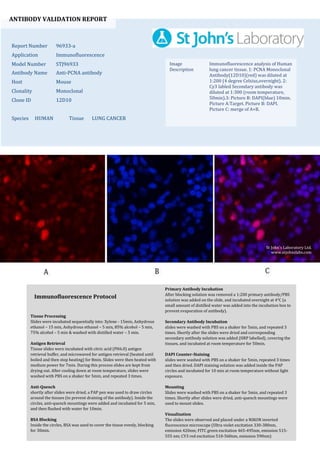
  • 1. ANTIBODY VALIDATION REPORT Report Number 96933-a Application Immunofluorescence Model Number STJ96933 Antibody Name Anti-PCNA antibody Host Mouse Clonality Monoclonal Clone ID 12D10 Species HUMAN Tissue LUNG CANCER Image Description Immunofluorescence analysis of Human lung cancer tissue. 1: PCNA Monoclonal Antibody(12D10)(red) was diluted at 1:200 (4 degree Celsius,overnight). 2: Cy3 labled Secondary antibody was diluted at 1:300 (room temperature, 50min).3: Picture B: DAPI(blue) 10min. Picture A:Target. Picture B: DAPI. Picture C: merge of A+B. Primary Antibody Incubation After blocking solution was removed a 1:200 primary antibody/PBS solution was added on the slide, and incubated overnight at 4°C (a small amount of distilled water was added into the incubation box to prevent evaporation of antibody). Secondary Antibody Incubation slides were washed with PBS on a shaker for 5min, and repeated 3 times. Shortly after the slides were dried and corresponding secondary antibody solution was added (HRP labelled), covering the tissues, and incubated at room temperature for 50min. DAPI Counter-Staining slides were washed with PBS on a shaker for 5min, repeated 3 times and then dried. DAPI staining solution was added inside the PAP circles and incubated for 10 min at room temperature without light exposure. Mounting Slides were washed with PBS on a shaker for 5min, and repeated 3 times. Shortly after slides were dried, anti-quench mountings were used to mount slides. Visualization The slides were observed and placed under a NIKON inverted fluorescence microscope (Ultra violet excitation 330-380nm, emission 420nm; FITC green excitation 465-495nm, emission 515- 555 nm; CY3 red excitation 510-560nm, emission 590nm) Immunofluorescence Protocol Tissue Processing Slides were incubated sequentially into: Xylene - 15min, Anhydrous ethanol – 15 min, Anhydrous ethanol – 5 min, 85% alcohol – 5 min, 75% alcohol – 5 min & washed with distilled water – 5 min. Antigen Retrieval Tissue slides were incubated with citric acid (PH6.0) antigen retrieval buffer, and microwaved for antigen retrieval (heated until boiled and then stop heating) for 8min. Slides were then heated with medium power for 7min. During this process slides are kept from drying out. After cooling down at room temperature, slides were washed with PBS on a shaker for 5min, and repeated 3 times. Anti-Quench shortly after slides were dried, a PAP pen was used to draw circles around the tissues (to prevent draining of the antibody). Inside the circles, anti-quench mountings were added and incubated for 5 min, and then flushed with water for 10min. BSA Blocking Inside the circles, BSA was used to cover the tissue evenly, blocking for 30min. St John's Laboratory Ltd. www.stjohnslabs.com
  • 2. ANTIBODY VALIDATION REPORT Report Number 96933-b Application Immunofluorescence Model Number STJ96933 Antibody Name Anti-PCNA antibody Host Mouse Clonality Monoclonal Clone ID 12D10 Species HUMAN Tissue LUNG CANCER Image Description Immunofluorescence analysis of Human lung cancer tissue. 1: PCNA Monoclonal Antibody(12D10)(red) was diluted at 1:200 (4 degree Celsius,overnight). 2: Cy3 labled Secondary antibody was diluted at 1:300 (room temperature, 50min).3: Picture B: DAPI(blue) 10min. Picture A:Target. Picture B: DAPI. Picture C: merge of A+B. Primary Antibody Incubation After blocking solution was removed a 1:200 primary antibody/PBS solution was added on the slide, and incubated overnight at 4°C (a small amount of distilled water was added into the incubation box to prevent evaporation of antibody). Secondary Antibody Incubation slides were washed with PBS on a shaker for 5min, and repeated 3 times. Shortly after the slides were dried and corresponding secondary antibody solution was added (HRP labelled), covering the tissues, and incubated at room temperature for 50min. DAPI Counter-Staining slides were washed with PBS on a shaker for 5min, repeated 3 times and then dried. DAPI staining solution was added inside the PAP circles and incubated for 10 min at room temperature without light exposure. Mounting Slides were washed with PBS on a shaker for 5min, and repeated 3 times. Shortly after slides were dried, anti-quench mountings were used to mount slides. Visualization The slides were observed and placed under a NIKON inverted fluorescence microscope (Ultra violet excitation 330-380nm, emission 420nm; FITC green excitation 465-495nm, emission 515- 555 nm; CY3 red excitation 510-560nm, emission 590nm) Immunofluorescence Protocol Tissue Processing Slides were incubated sequentially into: Xylene - 15min, Anhydrous ethanol – 15 min, Anhydrous ethanol – 5 min, 85% alcohol – 5 min, 75% alcohol – 5 min & washed with distilled water – 5 min. Antigen Retrieval Tissue slides were incubated with citric acid (PH6.0) antigen retrieval buffer, and microwaved for antigen retrieval (heated until boiled and then stop heating) for 8min. Slides were then heated with medium power for 7min. During this process slides are kept from drying out. After cooling down at room temperature, slides were washed with PBS on a shaker for 5min, and repeated 3 times. Anti-Quench shortly after slides were dried, a PAP pen was used to draw circles around the tissues (to prevent draining of the antibody). Inside the circles, anti-quench mountings were added and incubated for 5 min, and then flushed with water for 10min. BSA Blocking Inside the circles, BSA was used to cover the tissue evenly, blocking for 30min. St John's Laboratory Ltd. www.stjohnslabs.com
  • 3. ANTIBODY VALIDATION REPORT Report Number 96933-c Application Immunofluorescence Model Number STJ96933 Antibody Name Anti-PCNA antibody Host Mouse Clonality Monoclonal Clone ID 12D10 Species HUMAN Tissue LUNG CANCER Image Description Immunofluorescence analysis of Human lung cancer tissue. 1: PCNA Monoclonal Antibody(12D10)(red) was diluted at 1:200 (4 degree Celsius,overnight). 2: Cy3 labled Secondary antibody was diluted at 1:300 (room temperature, 50min).3: Picture B: DAPI(blue) 10min. Picture A:Target. Picture B: DAPI. Picture C: merge of A+B. Primary Antibody Incubation After blocking solution was removed a 1:200 primary antibody/PBS solution was added on the slide, and incubated overnight at 4°C (a small amount of distilled water was added into the incubation box to prevent evaporation of antibody). Secondary Antibody Incubation slides were washed with PBS on a shaker for 5min, and repeated 3 times. Shortly after the slides were dried and corresponding secondary antibody solution was added (HRP labelled), covering the tissues, and incubated at room temperature for 50min. DAPI Counter-Staining slides were washed with PBS on a shaker for 5min, repeated 3 times and then dried. DAPI staining solution was added inside the PAP circles and incubated for 10 min at room temperature without light exposure. Mounting Slides were washed with PBS on a shaker for 5min, and repeated 3 times. Shortly after slides were dried, anti-quench mountings were used to mount slides. Visualization The slides were observed and placed under a NIKON inverted fluorescence microscope (Ultra violet excitation 330-380nm, emission 420nm; FITC green excitation 465-495nm, emission 515- 555 nm; CY3 red excitation 510-560nm, emission 590nm) Immunofluorescence Protocol Tissue Processing Slides were incubated sequentially into: Xylene - 15min, Anhydrous ethanol – 15 min, Anhydrous ethanol – 5 min, 85% alcohol – 5 min, 75% alcohol – 5 min & washed with distilled water – 5 min. Antigen Retrieval Tissue slides were incubated with citric acid (PH6.0) antigen retrieval buffer, and microwaved for antigen retrieval (heated until boiled and then stop heating) for 8min. Slides were then heated with medium power for 7min. During this process slides are kept from drying out. After cooling down at room temperature, slides were washed with PBS on a shaker for 5min, and repeated 3 times. Anti-Quench shortly after slides were dried, a PAP pen was used to draw circles around the tissues (to prevent draining of the antibody). Inside the circles, anti-quench mountings were added and incubated for 5 min, and then flushed with water for 10min. BSA Blocking Inside the circles, BSA was used to cover the tissue evenly, blocking for 30min. St John's Laboratory Ltd. www.stjohnslabs.com
  • 4. ANTIBODY VALIDATION REPORT Report Number 96933-d Application Immunofluorescence Model Number STJ96933 Antibody Name Anti-PCNA antibody Host Mouse Clonality Monoclonal Clone ID 12D10 Species MOUSE Tissue LUNG Image Description Immunofluorescence analysis of Mouse lung tissue. 1: PCNA Monoclonal Antibody(12D10)(red) was diluted at 1:200 (4 degree Celsius,overnight). 2: Cy3 labled Secondary antibody was diluted at 1:300 (room temperature, 50min).3: Picture B: DAPI(blue) 10min. Picture A:Target. Picture B: DAPI. Picture C: merge of A+B. Primary Antibody Incubation After blocking solution was removed a 1:200 primary antibody/PBS solution was added on the slide, and incubated overnight at 4°C (a small amount of distilled water was added into the incubation box to prevent evaporation of antibody). Secondary Antibody Incubation slides were washed with PBS on a shaker for 5min, and repeated 3 times. Shortly after the slides were dried and corresponding secondary antibody solution was added (HRP labelled), covering the tissues, and incubated at room temperature for 50min. DAPI Counter-Staining slides were washed with PBS on a shaker for 5min, repeated 3 times and then dried. DAPI staining solution was added inside the PAP circles and incubated for 10 min at room temperature without light exposure. Mounting Slides were washed with PBS on a shaker for 5min, and repeated 3 times. Shortly after slides were dried, anti-quench mountings were used to mount slides. Visualization The slides were observed and placed under a NIKON inverted fluorescence microscope (Ultra violet excitation 330-380nm, emission 420nm; FITC green excitation 465-495nm, emission 515- 555 nm; CY3 red excitation 510-560nm, emission 590nm) Immunofluorescence Protocol Tissue Processing Slides were incubated sequentially into: Xylene - 15min, Anhydrous ethanol – 15 min, Anhydrous ethanol – 5 min, 85% alcohol – 5 min, 75% alcohol – 5 min & washed with distilled water – 5 min. Antigen Retrieval Tissue slides were incubated with citric acid (PH6.0) antigen retrieval buffer, and microwaved for antigen retrieval (heated until boiled and then stop heating) for 8min. Slides were then heated with medium power for 7min. During this process slides are kept from drying out. After cooling down at room temperature, slides were washed with PBS on a shaker for 5min, and repeated 3 times. Anti-Quench shortly after slides were dried, a PAP pen was used to draw circles around the tissues (to prevent draining of the antibody). Inside the circles, anti-quench mountings were added and incubated for 5 min, and then flushed with water for 10min. BSA Blocking Inside the circles, BSA was used to cover the tissue evenly, blocking for 30min. St John's Laboratory Ltd. www.stjohnslabs.com
  • 5. ANTIBODY VALIDATION REPORT Report Number 96933-e Application Immunofluorescence Model Number STJ96933 Antibody Name Anti-PCNA antibody Host Mouse Clonality Monoclonal Clone ID 12D10 Species MOUSE Tissue LUNG Image Description Immunofluorescence analysis of Mouse lung tissue. 1: PCNA Monoclonal Antibody(12D10)(red) was diluted at 1:200 (4 degree Celsius,overnight). 2: Cy3 labled Secondary antibody was diluted at 1:300 (room temperature, 50min).3: Picture B: DAPI(blue) 10min. Picture A:Target. Picture B: DAPI. Picture C: merge of A+B. Primary Antibody Incubation After blocking solution was removed a 1:200 primary antibody/PBS solution was added on the slide, and incubated overnight at 4°C (a small amount of distilled water was added into the incubation box to prevent evaporation of antibody). Secondary Antibody Incubation slides were washed with PBS on a shaker for 5min, and repeated 3 times. Shortly after the slides were dried and corresponding secondary antibody solution was added (HRP labelled), covering the tissues, and incubated at room temperature for 50min. DAPI Counter-Staining slides were washed with PBS on a shaker for 5min, repeated 3 times and then dried. DAPI staining solution was added inside the PAP circles and incubated for 10 min at room temperature without light exposure. Mounting Slides were washed with PBS on a shaker for 5min, and repeated 3 times. Shortly after slides were dried, anti-quench mountings were used to mount slides. Visualization The slides were observed and placed under a NIKON inverted fluorescence microscope (Ultra violet excitation 330-380nm, emission 420nm; FITC green excitation 465-495nm, emission 515- 555 nm; CY3 red excitation 510-560nm, emission 590nm) Immunofluorescence Protocol Tissue Processing Slides were incubated sequentially into: Xylene - 15min, Anhydrous ethanol – 15 min, Anhydrous ethanol – 5 min, 85% alcohol – 5 min, 75% alcohol – 5 min & washed with distilled water – 5 min. Antigen Retrieval Tissue slides were incubated with citric acid (PH6.0) antigen retrieval buffer, and microwaved for antigen retrieval (heated until boiled and then stop heating) for 8min. Slides were then heated with medium power for 7min. During this process slides are kept from drying out. After cooling down at room temperature, slides were washed with PBS on a shaker for 5min, and repeated 3 times. Anti-Quench shortly after slides were dried, a PAP pen was used to draw circles around the tissues (to prevent draining of the antibody). Inside the circles, anti-quench mountings were added and incubated for 5 min, and then flushed with water for 10min. BSA Blocking Inside the circles, BSA was used to cover the tissue evenly, blocking for 30min. St John's Laboratory Ltd. www.stjohnslabs.com
  • 6. ANTIBODY VALIDATION REPORT Report Number 96933-f Application Immunofluorescence Model Number STJ96933 Antibody Name Anti-PCNA antibody Host Mouse Clonality Monoclonal Clone ID 12D10 Species MOUSE Tissue LUNG Image Description Immunofluorescence analysis of Mouse lung tissue. 1: PCNA Monoclonal Antibody(12D10)(red) was diluted at 1:200 (4 degree Celsius,overnight). 2: Cy3 labled Secondary antibody was diluted at 1:300 (room temperature, 50min).3: Picture B: DAPI(blue) 10min. Picture A:Target. Picture B: DAPI. Picture C: merge of A+B. Primary Antibody Incubation After blocking solution was removed a 1:200 primary antibody/PBS solution was added on the slide, and incubated overnight at 4°C (a small amount of distilled water was added into the incubation box to prevent evaporation of antibody). Secondary Antibody Incubation slides were washed with PBS on a shaker for 5min, and repeated 3 times. Shortly after the slides were dried and corresponding secondary antibody solution was added (HRP labelled), covering the tissues, and incubated at room temperature for 50min. DAPI Counter-Staining slides were washed with PBS on a shaker for 5min, repeated 3 times and then dried. DAPI staining solution was added inside the PAP circles and incubated for 10 min at room temperature without light exposure. Mounting Slides were washed with PBS on a shaker for 5min, and repeated 3 times. Shortly after slides were dried, anti-quench mountings were used to mount slides. Visualization The slides were observed and placed under a NIKON inverted fluorescence microscope (Ultra violet excitation 330-380nm, emission 420nm; FITC green excitation 465-495nm, emission 515- 555 nm; CY3 red excitation 510-560nm, emission 590nm) Immunofluorescence Protocol Tissue Processing Slides were incubated sequentially into: Xylene - 15min, Anhydrous ethanol – 15 min, Anhydrous ethanol – 5 min, 85% alcohol – 5 min, 75% alcohol – 5 min & washed with distilled water – 5 min. Antigen Retrieval Tissue slides were incubated with citric acid (PH6.0) antigen retrieval buffer, and microwaved for antigen retrieval (heated until boiled and then stop heating) for 8min. Slides were then heated with medium power for 7min. During this process slides are kept from drying out. After cooling down at room temperature, slides were washed with PBS on a shaker for 5min, and repeated 3 times. Anti-Quench shortly after slides were dried, a PAP pen was used to draw circles around the tissues (to prevent draining of the antibody). Inside the circles, anti-quench mountings were added and incubated for 5 min, and then flushed with water for 10min. BSA Blocking Inside the circles, BSA was used to cover the tissue evenly, blocking for 30min. St John's Laboratory Ltd. www.stjohnslabs.com
  • 7. ANTIBODY VALIDATION REPORT Report Number 96933-g Application Immunofluorescence Model Number STJ96933 Antibody Name Anti-PCNA antibody Host Mouse Clonality Monoclonal Clone ID 12D10 Species RAT Tissue TESTIS Image Description Immunofluorescence analysis of Rat testis tissue. 1: PCNA Monoclonal Antibody(12D10)(red) was diluted at 1:200 (4 degree Celsius,overnight). 2: Cy3 labled Secondary antibody was diluted at 1:300 (room temperature, 50min).3: Picture B: DAPI(blue) 10min. Picture A:Target. Picture B: DAPI. Picture C: merge of A+B. Primary Antibody Incubation After blocking solution was removed a 1:200 primary antibody/PBS solution was added on the slide, and incubated overnight at 4°C (a small amount of distilled water was added into the incubation box to prevent evaporation of antibody). Secondary Antibody Incubation slides were washed with PBS on a shaker for 5min, and repeated 3 times. Shortly after the slides were dried and corresponding secondary antibody solution was added (HRP labelled), covering the tissues, and incubated at room temperature for 50min. DAPI Counter-Staining slides were washed with PBS on a shaker for 5min, repeated 3 times and then dried. DAPI staining solution was added inside the PAP circles and incubated for 10 min at room temperature without light exposure. Mounting Slides were washed with PBS on a shaker for 5min, and repeated 3 times. Shortly after slides were dried, anti-quench mountings were used to mount slides. Visualization The slides were observed and placed under a NIKON inverted fluorescence microscope (Ultra violet excitation 330-380nm, emission 420nm; FITC green excitation 465-495nm, emission 515- 555 nm; CY3 red excitation 510-560nm, emission 590nm) Immunofluorescence Protocol Tissue Processing Slides were incubated sequentially into: Xylene - 15min, Anhydrous ethanol – 15 min, Anhydrous ethanol – 5 min, 85% alcohol – 5 min, 75% alcohol – 5 min & washed with distilled water – 5 min. Antigen Retrieval Tissue slides were incubated with citric acid (PH6.0) antigen retrieval buffer, and microwaved for antigen retrieval (heated until boiled and then stop heating) for 8min. Slides were then heated with medium power for 7min. During this process slides are kept from drying out. After cooling down at room temperature, slides were washed with PBS on a shaker for 5min, and repeated 3 times. Anti-Quench shortly after slides were dried, a PAP pen was used to draw circles around the tissues (to prevent draining of the antibody). Inside the circles, anti-quench mountings were added and incubated for 5 min, and then flushed with water for 10min. BSA Blocking Inside the circles, BSA was used to cover the tissue evenly, blocking for 30min. St John's Laboratory Ltd. www.stjohnslabs.com
  • 8. ANTIBODY VALIDATION REPORT Report Number 96933-h Application Immunofluorescence Model Number STJ96933 Antibody Name Anti-PCNA antibody Host Mouse Clonality Monoclonal Clone ID 12D10 Species RAT Tissue TESTIS Image Description Immunofluorescence analysis of Rat testis tissue. 1: PCNA Monoclonal Antibody(12D10)(red) was diluted at 1:200 (4 degree Celsius,overnight). 2: Cy3 labled Secondary antibody was diluted at 1:300 (room temperature, 50min).3: Picture B: DAPI(blue) 10min. Picture A:Target. Picture B: DAPI. Picture C: merge of A+B. Primary Antibody Incubation After blocking solution was removed a 1:200 primary antibody/PBS solution was added on the slide, and incubated overnight at 4°C (a small amount of distilled water was added into the incubation box to prevent evaporation of antibody). Secondary Antibody Incubation slides were washed with PBS on a shaker for 5min, and repeated 3 times. Shortly after the slides were dried and corresponding secondary antibody solution was added (HRP labelled), covering the tissues, and incubated at room temperature for 50min. DAPI Counter-Staining slides were washed with PBS on a shaker for 5min, repeated 3 times and then dried. DAPI staining solution was added inside the PAP circles and incubated for 10 min at room temperature without light exposure. Mounting Slides were washed with PBS on a shaker for 5min, and repeated 3 times. Shortly after slides were dried, anti-quench mountings were used to mount slides. Visualization The slides were observed and placed under a NIKON inverted fluorescence microscope (Ultra violet excitation 330-380nm, emission 420nm; FITC green excitation 465-495nm, emission 515- 555 nm; CY3 red excitation 510-560nm, emission 590nm) Immunofluorescence Protocol Tissue Processing Slides were incubated sequentially into: Xylene - 15min, Anhydrous ethanol – 15 min, Anhydrous ethanol – 5 min, 85% alcohol – 5 min, 75% alcohol – 5 min & washed with distilled water – 5 min. Antigen Retrieval Tissue slides were incubated with citric acid (PH6.0) antigen retrieval buffer, and microwaved for antigen retrieval (heated until boiled and then stop heating) for 8min. Slides were then heated with medium power for 7min. During this process slides are kept from drying out. After cooling down at room temperature, slides were washed with PBS on a shaker for 5min, and repeated 3 times. Anti-Quench shortly after slides were dried, a PAP pen was used to draw circles around the tissues (to prevent draining of the antibody). Inside the circles, anti-quench mountings were added and incubated for 5 min, and then flushed with water for 10min. BSA Blocking Inside the circles, BSA was used to cover the tissue evenly, blocking for 30min. St John's Laboratory Ltd. www.stjohnslabs.com
  • 9. ANTIBODY VALIDATION REPORT Report Number 96933-i Application Immunofluorescence Model Number STJ96933 Antibody Name Anti-PCNA antibody Host Mouse Clonality Monoclonal Clone ID 12D10 Species RAT Tissue TESTIS Image Description Immunofluorescence analysis of Rat testis tissue. 1: PCNA Monoclonal Antibody(12D10)(red) was diluted at 1:200 (4 degree Celsius,overnight). 2: Cy3 labled Secondary antibody was diluted at 1:300 (room temperature, 50min).3: Picture B: DAPI(blue) 10min. Picture A:Target. Picture B: DAPI. Picture C: merge of A+B. Primary Antibody Incubation After blocking solution was removed a 1:200 primary antibody/PBS solution was added on the slide, and incubated overnight at 4°C (a small amount of distilled water was added into the incubation box to prevent evaporation of antibody). Secondary Antibody Incubation slides were washed with PBS on a shaker for 5min, and repeated 3 times. Shortly after the slides were dried and corresponding secondary antibody solution was added (HRP labelled), covering the tissues, and incubated at room temperature for 50min. DAPI Counter-Staining slides were washed with PBS on a shaker for 5min, repeated 3 times and then dried. DAPI staining solution was added inside the PAP circles and incubated for 10 min at room temperature without light exposure. Mounting Slides were washed with PBS on a shaker for 5min, and repeated 3 times. Shortly after slides were dried, anti-quench mountings were used to mount slides. Visualization The slides were observed and placed under a NIKON inverted fluorescence microscope (Ultra violet excitation 330-380nm, emission 420nm; FITC green excitation 465-495nm, emission 515- 555 nm; CY3 red excitation 510-560nm, emission 590nm) Immunofluorescence Protocol Tissue Processing Slides were incubated sequentially into: Xylene - 15min, Anhydrous ethanol – 15 min, Anhydrous ethanol – 5 min, 85% alcohol – 5 min, 75% alcohol – 5 min & washed with distilled water – 5 min. Antigen Retrieval Tissue slides were incubated with citric acid (PH6.0) antigen retrieval buffer, and microwaved for antigen retrieval (heated until boiled and then stop heating) for 8min. Slides were then heated with medium power for 7min. During this process slides are kept from drying out. After cooling down at room temperature, slides were washed with PBS on a shaker for 5min, and repeated 3 times. Anti-Quench shortly after slides were dried, a PAP pen was used to draw circles around the tissues (to prevent draining of the antibody). Inside the circles, anti-quench mountings were added and incubated for 5 min, and then flushed with water for 10min. BSA Blocking Inside the circles, BSA was used to cover the tissue evenly, blocking for 30min. St John's Laboratory Ltd. www.stjohnslabs.com