Serine/threonine kinase which acts as an essential component of the MAP kinase signal transduction pathway. MAPK1/ERK2 and MAPK3/ERK1 are the 2 MAPKs which play an important role in the MAPK/ERK cascade. They participate also in a signaling cascade initiated by activated KIT and KITLG/SCF. Depending on the cellular context, the MAPK/ERK cascade mediates diverse biological functions such as cell growth, adhesion, survival and differentiation through the regulation of transcription, translation, cytoskeletal rearrangements. The MAPK/ERK cascade plays also a role in initiation and regulation of meiosis, mitosis, and postmitotic functions in differentiated cells by phosphorylating a number of transcription factors. About 160 substrates have already been discovered for ERKs. Many of these substrates are localized in the nucleus, and seem to participate in the regulation of transcription upon stimulation. However, other substrates are found in the cytosol as well as in other cellular organelles, and those are responsible for processes such as translation, mitosis and apoptosis.
Anti-Phospho-ERK 1/2 (Y222/205) -http://www.stjohnslabs.com/phospho-erk-12-y222205-antibody
Serine/threonine kinase which acts as an essential component of the MAP kinase signal transduction pathway. MAPK1/ERK2 and MAPK3/ERK1 are the 2 MAPKs which play an important role in the MAPK/ERK cascade. They participate also in a signaling cascade initiated by activated KIT and KITLG/SCF. Depending on the cellular context, the MAPK/ERK cascade mediates diverse biological functions such as cell growth, adhesion, survival and differentiation through the regulation of transcription, translation, cytoskeletal rearrangements. The MAPK/ERK cascade plays also a role in initiation and regulation of meiosis, mitosis, and postmitotic functions in differentiated cells by phosphorylating a number of transcription factors. About 160 substrates have already been discovered for ERKs. Many of these substrates are localized in the nucleus, and seem to participate in the regulation of transcription upon stimulation. However, other substrates are found in the cytosol as well as in other cellular organelles, and those are responsible for processes such as translation, mitosis and apoptosis. Moreover, the MAPK/ERK cascade is also involved in the regulation of the endosomal dynamics, including lysosome processing and endosome cycling through the perinuclear recycling compartment (PNRC); as well as in the fragmentation of the Golgi apparatus during mitosis.
Anti-Phospho-ERK 1/2 (Y222/205)-http://www.stjohnslabs.com/phospho-erk-12-y222205-antibody
Join our Antibody Validation Project - http://www.stjohnslabs.com/services/antibody-validation
Functions as a cell surface receptor and performs physiological functions on the surface of neurons relevant to neurite growth, neuronal adhesion and axonogenesis. Involved in cell mobility and transcription regulation through protein-protein interactions. Can promote transcription activation through binding to APBB1-KAT5 and inhibits Notch signaling through interaction with Numb. Couples to apoptosis-inducing pathways such as those mediated by G(O) and JIP. Inhibits G(o) alpha ATPase activity (By similarity). Acts as a kinesin I membrane receptor, mediating the axonal transport of beta-secretase and presenilin 1. Involved in copper homeostasis/oxidative stress through copper ion reduction. In vitro, copper-metallated APP induces neuronal death directly or is potentiated through Cu2+-mediated low-density lipoprotein oxidation.
Anti-Amyloid-β -http://www.stjohnslabs.com/amyloid-v-antibody
Join our Antibody Validation Project - http://www.stjohnslabs.com/services/antibody-validation
Functions as a cell surface receptor and performs physiological functions on the surface of neurons relevant to neurite growth, neuronal adhesion and axonogenesis. Involved in cell mobility and transcription regulation through protein-protein interactions. Can promote transcription activation through binding to APBB1-KAT5 and inhibits Notch signaling through interaction with Numb. Couples to apoptosis-inducing pathways such as those mediated by G(O) and JIP. Inhibits G(o) alpha ATPase activity (By similarity). Acts as a kinesin I membrane receptor, mediating the axonal transport of beta-secretase and presenilin 1. Involved in copper homeostasis/oxidative stress through copper ion reduction. In vitro, copper-metallated APP induces neuronal death directly or is potentiated through Cu2+-mediated low-density lipoprotein oxidation.
Anti-Amyloid-β-http://www.stjohnslabs.com/amyloid-v-antibody
Join our Antibody Validation Project - http://www.stjohnslabs.com/services/antibody-validation
Functions as a receptor for membrane-bound ligands Jagged1, Jagged2 and Delta1 to regulate cell-fate determination. Upon ligand activation through the released notch intracellular domain (NICD) it forms a transcriptional activator complex with RBPJ/RBPSUH and activates genes of the enhancer of split locus. Affects the implementation of differentiation, proliferation and apoptotic programs (By similarity). Involved in bone remodeling and homeostasis. In collaboration with RELA/p65 enhances NFATc1 promoter activity and positively regulates RANKL-induced osteoclast differentiation. Positively regulates self-renewal of liver cancer cells .
Anti-Cleaved-Notch 2 (D1733)-http://www.stjohnslabs.com/cleaved-notch-2-d1733-antibody
Join our Antibody Validation Project - http://www.stjohnslabs.com/services/antibody-validation
Functions as a receptor for membrane-bound ligands Jagged1, Jagged2 and Delta1 to regulate cell-fate determination. Upon ligand activation through the released notch intracellular domain (NICD) it forms a transcriptional activator complex with RBPJ/RBPSUH and activates genes of the enhancer of split locus. Affects the implementation of differentiation, proliferation and apoptotic programs (By similarity). Involved in bone remodeling and homeostasis. In collaboration with RELA/p65 enhances NFATc1 promoter activity and positively regulates RANKL-induced osteoclast differentiation. Positively regulates self-renewal of liver cancer cells .
Anti-Cleaved-Notch 2 (D1733) -http://www.stjohnslabs.com/cleaved-notch-2-d1733-antibody
Join our Antibody Validation Project - http://www.stjohnslabs.com/services/antibody-validation
NAD-dependent protein deacetylase that links transcriptional regulation directly to intracellular energetics and participates in the coordination of several separated cellular functions such as cell cycle, response to DNA damage, metobolism, apoptosis and autophagy. Can modulate chromatin function through deacetylation of histones and can promote alterations in the methylation of histones and DNA, leading to transcriptional repression. Deacetylates a broad range of transcription factors and coregulators, thereby regulating target gene expression positively and negatively. Serves as a sensor of the cytosolic ratio of NAD+/NADH which is altered by glucose deprivation and metabolic changes associated with caloric restriction. Is essential in skeletal muscle cell differentiation and in response to low nutrients mediates the inhibitory effect on skeletal myoblast differentiation which also involves 5'-AMP-activated protein kinase (AMPK) and nicotinamide phosphoribosyltransferase (NAMPT).
Anti-SIRT1 -http://www.stjohnslabs.com/sirt1-antibody-p-94333
Join our Antibody Validation Project - http://www.stjohnslabs.com/services/antibody-validation
NAD-dependent protein deacetylase that links transcriptional regulation directly to intracellular energetics and participates in the coordination of several separated cellular functions such as cell cycle, response to DNA damage, metobolism, apoptosis and autophagy. Can modulate chromatin function through deacetylation of histones and can promote alterations in the methylation of histones and DNA, leading to transcriptional repression. Deacetylates a broad range of transcription factors and coregulators, thereby regulating target gene expression positively and negatively. Serves as a sensor of the cytosolic ratio of NAD+/NADH which is altered by glucose deprivation and metabolic changes associated with caloric restriction. Is essential in skeletal muscle cell differentiation and in response to low nutrients mediates the inhibitory effect on skeletal myoblast differentiation which also involves 5'-AMP-activated protein kinase (AMPK) and nicotinamide phosphoribosyltransferase (NAMPT).
Anti-SIRT1-http://www.stjohnslabs.com/sirt1-antibody-p-94333
Join our Antibody Validation Project - http://www.stjohnslabs.com/services/antibody-validation
Immunohistochemistry Antibody Validation Report for Anti-VE-Cadherin Antibody...St John's Laboratory Ltd
Cadherins are calcium-dependent cell adhesion proteins. They preferentially interact with themselves in a homophilic manner in connecting cells; cadherins may thus contribute to the sorting of heterogeneous cell types. This cadherin may play a important role in endothelial cell biology through control of the cohesion and organization of the intercellular junctions. It associates with alpha-catenin forming a link to the cytoskeleton. Acts in concert with KRIT1 to establish and maintain correct endothelial cell polarity and vascular lumen. These effects are mediated by recruitment and activation of the Par polarity complex and RAP1B. Required for activation of PRKCZ and for the localization of phosphorylated PRKCZ, PARD3, TIAM1 and RAP1B to the cell junction.
Anti-VE-Cadherin-http://www.stjohnslabs.com/ve-cadherin-antibody-p-98941
Join our Antibody Validation Project - http://www.stjohnslabs.com/services/antibody-validation
Serine/threonine kinase which acts as an essential component of the MAP kinase signal transduction pathway. MAPK1/ERK2 and MAPK3/ERK1 are the 2 MAPKs which play an important role in the MAPK/ERK cascade. They participate also in a signaling cascade initiated by activated KIT and KITLG/SCF. Depending on the cellular context, the MAPK/ERK cascade mediates diverse biological functions such as cell growth, adhesion, survival and differentiation through the regulation of transcription, translation, cytoskeletal rearrangements. The MAPK/ERK cascade plays also a role in initiation and regulation of meiosis, mitosis, and postmitotic functions in differentiated cells by phosphorylating a number of transcription factors.
Anti-ERK 1/2 -http://www.stjohnslabs.com/erk-12-antibody-p-95241
Join our Antibody Validation Project - http://www.stjohnslabs.com/services/antibody-validation
Cadherins are calcium-dependent cell adhesion proteins. They preferentially interact with themselves in a homophilic manner in connecting cells; cadherins may thus contribute to the sorting of heterogeneous cell types. This cadherin may play a important role in endothelial cell biology through control of the cohesion and organization of the intercellular junctions. It associates with alpha-catenin forming a link to the cytoskeleton. Acts in concert with KRIT1 to establish and maintain correct endothelial cell polarity and vascular lumen. These effects are mediated by recruitment and activation of the Par polarity complex and RAP1B. Required for activation of PRKCZ and for the localization of phosphorylated PRKCZ, PARD3, TIAM1 and RAP1B to the cell junction.
Anti-VE-Cadherin -http://www.stjohnslabs.com/ve-cadherin-antibody-p-98941
Join our Antibody Validation Project - http://www.stjohnslabs.com/services/antibody-validation
Serine/threonine kinase which acts as an essential component of the MAP kinase signal transduction pathway. MAPK1/ERK2 and MAPK3/ERK1 are the 2 MAPKs which play an important role in the MAPK/ERK cascade. They participate also in a signaling cascade initiated by activated KIT and KITLG/SCF. Depending on the cellular context, the MAPK/ERK cascade mediates diverse biological functions such as cell growth, adhesion, survival and differentiation through the regulation of transcription, translation, cytoskeletal rearrangements. The MAPK/ERK cascade plays also a role in initiation and regulation of meiosis, mitosis, and postmitotic functions in differentiated cells by phosphorylating a number of transcription factors. About 160 substrates have already been discovered for ERKs. Many of these substrates are localized in the nucleus, and seem to participate in the regulation of transcription upon stimulation. However, other substrates are found in the cytosol as well as in other cellular organelles, and those are responsible for processes such as translation, mitosis and apoptosis.
Anti-ERK 1/2-http://www.stjohnslabs.com/erk-12-antibody-p-95241
Join our Antibody Validation Project - http://www.stjohnslabs.com/services/antibody-validation
Immunohistochemistry Antibody Validation Report for Anti-Phospho-NFκB-p65 (S5...St John's Laboratory Ltd
NF-kappa-B is a pleiotropic transcription factor present in almost all cell types and is the endpoint of a series of signal transduction events that are initiated by a vast array of stimuli related to many biological processes such as inflammation, immunity, differentiation, cell growth, tumorigenesis and apoptosis. NF-kappa-B is a homo- or heterodimeric complex formed by the Rel-like domain-containing proteins RELA/p65, RELB, NFKB1/p105, NFKB1/p50, REL and NFKB2/p52 and the heterodimeric p65-p50 complex appears to be most abundant one. The dimers bind at kappa-B sites in the DNA of their target genes and the individual dimers have distinct preferences for different kappa-B sites that they can bind with distinguishable affinity and specificity. Different dimer combinations act as transcriptional activators or repressors, respectively. NF-kappa-B is controlled by various mechanisms of post-translational modification and subcellular compartmentalization as well as by interactions with other cofactors or corepressors.
Anti-Phospho-NFκB-p65 (S536)-http://www.stjohnslabs.com/phospho-nfkb-p65-s536-antibody
Join our Antibody Validation Project - http://www.stjohnslabs.com/services/antibody-validation
Immunohistochemistry Antibody Validation Report for Anti-Phospho-Stat3 (Y705)...St John's Laboratory Ltd
Signal transducer and transcription activator that mediates cellular responses to interleukins, KITLG/SCF, LEP and other growth factors. Once activated, recruits coactivators, such as NCOA1 or MED1, to the promoter region of the target gene . May mediate cellular responses to activated FGFR1, FGFR2, FGFR3 and FGFR4. Binds to the interleukin-6 (IL-6)-responsive elements identified in the promoters of various acute-phase protein genes. Activated by IL31 through IL31RA. Involved in cell cycle regulation by inducing the expression of key genes for the progression from G1 to S phase, such as CCND1 . Mediates the effects of LEP on melanocortin production, body energy homeostasis and lactation (By similarity). May play an apoptotic role by transctivating BIRC5 expression under LEP activation . Cytoplasmic STAT3 represses macroautophagy by inhibiting EIF2AK2/PKR activity.
Anti-Phospho-Stat3 (Y705)-http://www.stjohnslabs.com/phospho-stat3-y705-antibody
Join our Antibody Validation Project - http://www.stjohnslabs.com/services/antibody-validation
Immunofluorescence Antibody Validation Report for Anti-Phospho-NFκB-p65 (S536...St John's Laboratory Ltd
NF-kappa-B is a pleiotropic transcription factor present in almost all cell types and is the endpoint of a series of signal transduction events that are initiated by a vast array of stimuli related to many biological processes such as inflammation, immunity, differentiation, cell growth, tumorigenesis and apoptosis. NF-kappa-B is a homo- or heterodimeric complex formed by the Rel-like domain-containing proteins RELA/p65, RELB, NFKB1/p105, NFKB1/p50, REL and NFKB2/p52 and the heterodimeric p65-p50 complex appears to be most abundant one. The dimers bind at kappa-B sites in the DNA of their target genes and the individual dimers have distinct preferences for different kappa-B sites that they can bind with distinguishable affinity and specificity.
Anti-Phospho-NFκB-p65 (S536) -http://www.stjohnslabs.com/phospho-nfkb-p65-s536-antibody
Join our Antibody Validation Project - http://www.stjohnslabs.com/services/antibody-validation
Involved in the activation cascade of caspases responsible for apoptosis execution. At the onset of apoptosis it proteolytically cleaves poly(ADP-ribose) polymerase (PARP) at a '216-Asp-|-Gly-217' bond. Cleaves and activates sterol regulatory element binding proteins (SREBPs) between the basic helix-loop-helix leucine zipper domain and the membrane attachment domain. Cleaves and activates caspase-6, -7 and -9. Involved in the cleavage of huntingtin. Triggers cell adhesion in sympathetic neurons through RET cleavage. / Strict requirement for an Asp residue at positions P1 and P4. It has a preferred cleavage sequence of Asp-Xaa-Xaa-Asp-|- with a hydrophobic amino-acid residue at P2 and a hydrophilic amino-acid residue at P3, although Val or Ala are also accepted at this position. / Inhibited by isatin sulfonamides.
Anti-Active Caspase-3-http://www.stjohnslabs.com/active-caspase-3-antibody-p-99099
Join our Antibody Validation Project - http://www.stjohnslabs.com/services/antibody-validation
Involved in the activation cascade of caspases responsible for apoptosis execution. At the onset of apoptosis it proteolytically cleaves poly(ADP-ribose) polymerase (PARP) at a '216-Asp-|-Gly-217' bond. Cleaves and activates sterol regulatory element binding proteins (SREBPs) between the basic helix-loop-helix leucine zipper domain and the membrane attachment domain. Cleaves and activates caspase-6, -7 and -9. Involved in the cleavage of huntingtin. Triggers cell adhesion in sympathetic neurons through RET cleavage.
Anti-Active Caspase-3 -http://www.stjohnslabs.com/active-caspase-3-antibody-p-99099
Join our Antibody Validation Project - http://www.stjohnslabs.com/services/antibody-validation
Signal transducer and transcription activator that mediates cellular responses to interleukins, KITLG/SCF, LEP and other growth factors. Once activated, recruits coactivators, such as NCOA1 or MED1, to the promoter region of the target gene . May mediate cellular responses to activated FGFR1, FGFR2, FGFR3 and FGFR4. Binds to the interleukin-6 (IL-6)-responsive elements identified in the promoters of various acute-phase protein genes. Activated by IL31 through IL31RA. Involved in cell cycle regulation by inducing the expression of key genes for the progression from G1 to S phase, such as CCND1 . Mediates the effects of LEP on melanocortin production, body energy homeostasis and lactation.
Anti-Phospho-Stat3 (Y705) -http://www.stjohnslabs.com/phospho-stat3-y705-antibody
Join our Antibody Validation Project - http://www.stjohnslabs.com/services/antibody-validation
Tubulin is the major constituent of microtubules. It binds two moles of GTP, one at an exchangeable site on the beta chain and one at a non-exchangeable site on the alpha chain. TUBB3 plays a critical role in proper axon guidance and mantainance.
Anti-β-Tubulin-http://www.stjohnslabs.com/b-tubulin-antibody-p-98567
Join our Antibody Validation Project - http://www.stjohnslabs.com/services/antibody-validation
Tubulin in molecular biology can refer either to the tubulin protein superfamily of globular proteins, or one of the member proteins of that superfamily. α- and β-tubulins polymerize into microtubules, a major component of the eukaryotic cytoskeleton. Microtubules function in many essential cellular processes, including mitosis. Tubulin-binding drugs kill cancerous cells by inhibiting microtubule dynamics, which are required for DNA segregation and therefore cell division. In eukaryotes there are six members of the tubulin superfamily, although not all are present in all species (see below).[2][3] Both α and β tubulins have a mass of around 50 kDa and are thus in a similar range compared to actin with ~42 kDa. In contrast, tubulin polymers (microtubules) tend to be much bigger than actin filaments due to their cylindrical nature.
Anti-β-Tubulin -http://www.stjohnslabs.com/b-tubulin-antibody-p-
98567
Join our Antibody Validation Project - http://www.stjohnslabs.com/services/antibody-validation
Lamin-B1 is a protein that in humans is encoded by the LMNB1 gene.
The nuclear lamina consists of a two-dimensional matrix of proteins located next to the inner nuclear membrane. The lamin family of proteins make up the matrix and are highly conserved in evolution. During mitosis, the lamina matrix is reversibly disassembled as the lamin proteins are phosphorylated. Lamin proteins are thought to be involved in nuclear stability, chromatin structure, and gene expression. Vertebrate lamins consist of two types, A and B. This gene encodes one of the two B type proteins, B1. Lamin B, along with heterochromatin, is anchored to the inner surface of the nuclear membrane by the lamin B receptor.
Anti-Lamin B1 -http://www.stjohnslabs.com/lamin-b1-antibody-p-98573
Join our Antibody Validation Project - http://www.stjohnslabs.com/services/antibody-validation
May be the important intermediate by which p53/TP53 mediates its role as an inhibitor of cellular proliferation in response to DNA damage. Binds to and inhibits cyclin-dependent kinase activity, preventing phosphorylation of critical cyclin-dependent kinase substrates and blocking cell cycle progression. Functions in the nuclear localization and assembly of cyclin D-CDK4 complex and promotes its kinase activity towards RB1. At higher stoichiometric ratios, inhibits the kinase activity of the cyclin D-CDK4 complex.
Anti-p21 -http://www.stjohnslabs.com/p21-antibody
Join our Antibody Validation Project - http://www.stjohnslabs.com/services/antibody-validation
Protein tyrosine kinase that is part of several cell surface receptor complexes, but that apparently needs a coreceptor for ligand binding. Essential component of a neuregulin-receptor complex, although neuregulins do not interact with it alone. GP30 is a potential ligand for this receptor. Regulates outgrowth and stabilization of peripheral microtubules (MTs). Upon ERBB2 activation, the MEMO1-RHOA-DIAPH1 signaling pathway elicits the phosphorylation and thus the inhibition of GSK3B at cell membrane. This prevents the phosphorylation of APC and CLASP2, allowing its association with the cell membrane. In turn, membrane-bound APC allows the localization of MACF1 to the cell membrane, which is required for microtubule capture and stabilization.
Anti-HER2 -http://www.stjohnslabs.com/her2-antibody-p-98582
Join our Antibody Validation Project - http://www.stjohnslabs.com/services/antibody-validation
May be the important intermediate by which p53/TP53 mediates its role as an inhibitor of cellular proliferation in response to DNA damage. Binds to and inhibits cyclin-dependent kinase activity, preventing phosphorylation of critical cyclin-dependent kinase substrates and blocking cell cycle progression. Functions in the nuclear localization and assembly of cyclin D-CDK4 complex and promotes its kinase activity towards RB1. At higher stoichiometric ratios, inhibits the kinase activity of the cyclin D-CDK4 complex.
Anti-p21-http://www.stjohnslabs.com/p21-antibody
Join our Antibody Validation Project - http://www.stjohnslabs.com/services/antibody-validation
In cooperation with other chaperones, Hsp70s stabilize preexistent proteins against aggregation and mediate the folding of newly translated polypeptides in the cytosol as well as within organelles. These chaperones participate in all these processes through their ability to recognize nonnative conformations of other proteins. They bind extended peptide segments with a net hydrophobic character exposed by polypeptides during translation and membrane translocation, or following stress-induced damage (By similarity). Positive regulator of PARK2 translocation to damaged mitochondria.
Anti-HSP70-http://www.stjohnslabs.com/hsp70-antibody-p-98579
Join our Antibody Validation Project - http://www.stjohnslabs.com/services/antibody-validation
Metalloproteinase inhibitor that functions by forming one to one complexes with target metalloproteinases, such as collagenases, and irreversibly inactivates them by binding to their catalytic zinc cofactor. Acts on MMP1, MMP2, MMP3, MMP7, MMP8, MMP9, MMP10, MMP11, MMP12, MMP13 and MMP16. Does not act on MMP14. Also functions as a growth factor that regulates cell differentiation, migration and cell death and activates cellular signaling cascades via CD63 and ITGB1. Plays a role in integrin signaling. Mediates erythropoiesis in vitro; but, unlike IL3, it is species-specific, stimulating the growth and differentiation of only human and murine thyroid progenitors.
Anti-TIMP-1 -http://www.stjohnslabs.com/timp-1-antibody
Join our Antibody Validation Project - http://www.stjohnslabs.com/services/antibody-validation
In cooperation with other chaperones, Hsp70s stabilize preexistent proteins against aggregation and mediate the folding of newly translated polypeptides in the cytosol as well as within organelles. These chaperones participate in all these processes through their ability to recognize nonnative conformations of other proteins. They bind extended peptide segments with a net hydrophobic character exposed by polypeptides during translation and membrane translocation, or following stress-induced damage (By similarity). Positive regulator of PARK2 translocation to damaged mitochondria.
Anti-HSP70 -http://www.stjohnslabs.com/hsp70-antibody-p-98579
Join our Antibody Validation Project - http://www.stjohnslabs.com/services/antibody-validation
Actins are highly conserved proteins that are involved in various types of cell motility and are ubiquitously expressed in all eukaryotic cells. Actin participates in many important cellular processes, including muscle contraction, cell motility, cell division and cytokinesis, vesicle and organelle movement, cell signaling, and the establishment and maintenance of cell junctions and cell shape. Many of these processes are mediated by extensive and intimate interactions of actin with cellular membranes.
Anti-Actin β -http://www.stjohnslabs.com/actin-b-antibody
Join our Antibody Validation Project - http://www.stjohnslabs.com/services/antibody-validation
Beta-actin (human gene and protein symbol ACTB/ACTB) is one of six different actin isoforms which have been identified in humans. This is one of the two nonmuscle cytoskeletal actins. Actins are highly conserved proteins that are involved in cell motility, structure and integrity. Alpha actins are a major constituent of the contractile apparatus.
To purchase this antibody, use the following link: http://www.stjohnslabs.com/actin-b-antibody?filter_name=STJ91464
Serine/threonine kinase which acts as an essential component of the MAP kinase signal transduction pathway. MAPK1/ERK2 and MAPK3/ERK1 are the 2 MAPKs which play an important role in the MAPK/ERK cascade. They participate also in a signaling cascade initiated by activated KIT and KITLG/SCF. Depending on the cellular context, the MAPK/ERK cascade mediates diverse biological functions such as cell growth, adhesion, survival and differentiation through the regulation of transcription, translation, cytoskeletal rearrangements. The MAPK/ERK cascade plays also a role in initiation and regulation of meiosis, mitosis, and postmitotic functions in differentiated cells by phosphorylating a number of transcription factors. About 160 substrates have already been discovered for ERKs. Many of these substrates are localized in the nucleus, and seem to participate in the regulation of transcription upon stimulation.
Anti-Phospho-ERK 1/2 (T202)-http://www.stjohnslabs.com/phospho-erk-12-t202-antibody
Join our Antibody Validation Project - http://www.stjohnslabs.com/services/antibody-validation
Immunohistochemistry Antibody Validation Report for Anti-Phospho-Catenin-β (S...St John's Laboratory Ltd
Key downstream component of the canonical Wnt signaling pathway. In the absence of Wnt, forms a complex with AXIN1, AXIN2, APC, CSNK1A1 and GSK3B that promotes phosphorylation on N-terminal Ser and Thr residues and ubiquitination of CTNNB1 via BTRC and its subsequent degradation by the proteasome. In the presence of Wnt ligand, CTNNB1 is not ubiquitinated and accumulates in the nucleus, where it acts as a coactivator for transcription factors of the TCF/LEF family, leading to activate Wnt responsive genes. Involved in the regulation of cell adhesion. Acts as a negative regulator of centrosome cohesion. Involved in the CDK2/PTPN6/CTNNB1/CEACAM1 pathway of insulin internalization. Blocks anoikis of malignant kidney and intestinal epithelial cells and promotes their anchorage-independent growth by down-regulating DAPK2. Disrupts PML function and PML-NB formation by inhibiting RANBP2-mediated sumoylation of PML . Promotes neurogenesis by maintaining sympathetic neuroblasts within the cell cycle.
Anti-Phospho-Catenin-β (S37)-http://www.stjohnslabs.com/phospho-catenin-b-s37-antibody
Join our Antibody Validation Project - http://www.stjohnslabs.com/services/antibody-validation
Functions as a receptor for membrane-bound ligands Jagged1, Jagged2 and Delta1 to regulate cell-fate determination. Upon ligand activation through the released notch intracellular domain (NICD) it forms a transcriptional activator complex with RBPJ/RBPSUH and activates genes of the enhancer of split locus. Affects the implementation of differentiation, proliferation and apoptotic programs. Involved in angiogenesis; negatively regulates endothelial cell proliferation and migration and angiogenic sprouting.
Anti-Cleaved-Notch 1 (V1754) -http://www.stjohnslabs.com/cleaved-notch-1-v1754-antibody
Join our Antibody Validation Project - http://www.stjohnslabs.com/services/antibody-validation
Serine/threonine kinase which acts as an essential component of the MAP kinase signal transduction pathway. MAPK1/ERK2 and MAPK3/ERK1 are the 2 MAPKs which play an important role in the MAPK/ERK cascade. They participate also in a signaling cascade initiated by activated KIT and KITLG/SCF. Depending on the cellular context, the MAPK/ERK cascade mediates diverse biological functions such as cell growth, adhesion, survival and differentiation through the regulation of transcription, translation, cytoskeletal rearrangements.
Anti-Phospho-ERK 1/2 (T202/Y204)-http://www.stjohnslabs.com/phospho-erk-12-t202y204-antibody
Join our Antibody Validation Project - http://www.stjohnslabs.com/services/antibody-validation
Promotes microtubule assembly and stability, and might be involved in the establishment and maintenance of neuronal polarity. The C-terminus binds axonal microtubules while the N-terminus binds neural plasma membrane components, suggesting that tau functions as a linker protein between both.
Pathologies and dementias of the nervous system such as Alzheimer's disease and Parkinson's disease are associated with tau proteins that have become defective and no longer stabilize microtubules properly.
The tau proteins are the product of alternative splicing from a single gene that in humans is designated MAPT (microtubule-associated protein tau) and is located on chromosome 17.
Anti-Phospho-Tau (S396) -http://www.stjohnslabs.com/phospho-tau-s396-antibody
Join our Antibody Validation Project - http://www.stjohnslabs.com/services/antibody-validation
Transcription factor that recognizes and binds to the enhancer heptamer motif 5'-TGA[CG]TCA-3'. Promotes activity of NR5A1 when phosphorylated by HIPK3 leading to increased steroidogenic gene expression upon cAMP signaling pathway stimulation. Involved in activated KRAS-mediated transcriptional activation of USP28 in colorectal cancer (CRC) cells. Binds to the USP28 promoter in colorectal cancer (CRC) cells.
Anti-AP-1 -http://www.stjohnslabs.com/ap-1-antibody-p-91169
Join our Antibody Validation Project - http://www.stjohnslabs.com/services/antibody-validation
Promotes cell death. Successfully competes for the binding to Bcl-X(L), Bcl-2 and Bcl-W, thereby affecting the level of heterodimerization of these proteins with BAX. Can reverse the death repressor activity of Bcl-X(L), but not that of Bcl-2 (By similarity). Appears to act as a link between growth factor receptor signaling and the apoptotic pathways.
Anti-Bad -http://www.stjohnslabs.com/bad-antibody-p-91313
Join our Antibody Validation Project - http://www.stjohnslabs.com/services/antibody-validation
Immunofluorescence Antibody Validation Report for Anti-Phospho-p38 (T180/Y182...St John's Laboratory Ltd
Serine/threonine kinase which acts as an essential component of the MAP kinase signal transduction pathway. MAPK14 is one of the four p38 MAPKs which play an important role in the cascades of cellular responses evoked by extracellular stimuli such as proinflammatory cytokines or physical stress leading to direct activation of transcription factors. Accordingly, p38 MAPKs phosphorylate a broad range of proteins and it has been estimated that they may have approximately 200 to 300 substrates each. Some of the targets are downstream kinases which are activated through phosphorylation and further phosphorylate additional targets. RPS6KA5/MSK1 and RPS6KA4/MSK2 can directly phosphorylate and activate transcription factors such as CREB1, ATF1, the NF-kappa-B isoform RELA/NFKB3, STAT1 and STAT3, but can also phosphorylate histone H3 and the nucleosomal protein HMGN1.
Anti-Phospho-p38 (T180/Y182) -http://www.stjohnslabs.com/phospho-p38-t180y182-antibody
Join our Antibody Validation Project - http://www.stjohnslabs.com/services/antibody-validation
Serine/threonine kinase which acts as an essential component of the MAP kinase signal transduction pathway. MAPK14 is one of the four p38 MAPKs which play an important role in the cascades of cellular responses evoked by extracellular stimuli such as proinflammatory cytokines or physical stress leading to direct activation of transcription factors. Accordingly, p38 MAPKs phosphorylate a broad range of proteins and it has been estimated that they may have approximately 200 to 300 substrates each. Some of the targets are downstream kinases which are activated through phosphorylation and further phosphorylate additional targets. RPS6KA5/MSK1 and RPS6KA4/MSK2 can directly phosphorylate and activate transcription factors such as CREB1, ATF1, the NF-kappa-B isoform RELA/NFKB3, STAT1 and STAT3, but can also phosphorylate histone H3 and the nucleosomal protein HMGN1.
Anti-Phospho-p38 (T180) -http://www.stjohnslabs.com/phospho-p38-t180-antibody
Join our Antibody Validation Project - http://www.stjohnslabs.com/services/antibody-validation
Receptor-regulated SMAD (R-SMAD) that is an intracellular signal transducer and transcriptional modulator activated by TGF-beta (transforming growth factor) and activin type 1 receptor kinases. Binds the TRE element in the promoter region of many genes that are regulated by TGF-beta and, on formation of the SMAD3/SMAD4 complex, activates transcription. Also can form a SMAD3/SMAD4/JUN/FOS complex at the AP-1/SMAD site to regulate TGF-beta-mediated transcription. Has an inhibitory effect on wound healing probably by modulating both growth and migration of primary keratinocytes and by altering the TGF-mediated chemotaxis of monocytes. This effect on wound healing appears to be hormone-sensitive. Regulator of chondrogenesis and osteogenesis and inhibits early healing of bone fractures. Positively regulates PDPK1 kinase activity by stimulating its dissociation from the 14-3-3 protein YWHAQ which acts as a negative regulator.
Anti-Phospho-Smad3 (S425) -http://www.stjohnslabs.com/phospho-smad3-s425-antibody
Join our Antibody Validation Project - http://www.stjohnslabs.com/services/antibody-validation
Immunofluorescence Antibody Validation Report for Anti-Phospho-JNK1/2/3 (Y185...St John's Laboratory Ltd
Serine/threonine-protein kinase involved in various processes such as cell proliferation, differentiation, migration, transformation and programmed cell death. Extracellular stimuli such as proinflammatory cytokines or physical stress stimulate the stress-activated protein kinase/c-Jun N-terminal kinase (SAP/JNK) signaling pathway. In this cascade, two dual specificity kinases MAP2K4/MKK4 and MAP2K7/MKK7 phosphorylate and activate MAPK8/JNK1. In turn, MAPK8/JNK1 phosphorylates a number of transcription factors, primarily components of AP-1 such as JUN, JDP2 and ATF2 and thus regulates AP-1 transcriptional activity. Phosphorylates the replication licensing factor CDT1, inhibiting the interaction between CDT1 and the histone H4 acetylase HBO1 to replication origins. Loss of this interaction abrogates the acetylation required for replication initiation.
Anti-Phospho-JNK1/2/3 (Y185) -http://www.stjohnslabs.com/phospho-jnk123-y185-antibody
Join our Antibody Validation Project - http://www.stjohnslabs.com/services/antibody-validation
Immunofluorescence Antibody Validation Report for Anti-Phospho-CaMKIIβ/γ/δ (T...St John's Laboratory Ltd
Calcium/calmodulin-dependent protein kinase that functions autonomously after Ca2+/calmodulin-binding and autophosphorylation, and is involved in dendritic spine and synapse formation, neuronal plasticity and regulation of sarcoplasmic reticulum Ca2+ transport in skeletal muscle. In neurons, plays an essential structural role in the reorganization of the actin cytoskeleton during plasticity by binding and bundling actin filaments in a kinase-independent manner. This structural function is required for correct targeting of CaMK2A, which acts downstream of NMDAR to promote dendritic spine and synapse formation and maintain synaptic plasticity which enables long-term potentiation (LTP) and hippocampus-dependent learning. In developing hippocampal neurons, promotes arborization of the dendritic tree and in mature neurons, promotes dendritic remodeling. Participates in the modulation of skeletal muscle function in response to exercise.
Anti-Phospho-CaMKIIβ/γ/δ (T287) -http://www.stjohnslabs.com/phospho-camkiibgd-t287-antibody
Join our Antibody Validation Project - http://www.stjohnslabs.com/services/antibody-validation
Signal transducer and transcription activator that mediates cellular responses to interferons (IFNs), cytokine KITLG/SCF and other cytokines and other growth factors. Following type I IFN (IFN-alpha and IFN-beta) binding to cell surface receptors, signaling via protein kinases leads to activation of Jak kinases (TYK2 and JAK1) and to tyrosine phosphorylation of STAT1 and STAT2. The phosphorylated STATs dimerize and associate with ISGF3G/IRF-9 to form a complex termed ISGF3 transcription factor, that enters the nucleus.
Anti-Phospho-Stat1 (Y701) -http://www.stjohnslabs.com/phospho-stat1-y701-antibody
Join our Antibody Validation Project - http://www.stjohnslabs.com/services/antibody-validation
AKT1 is one of 3 closely related serine/threonine-protein kinases (AKT1, AKT2 and AKT3) called the AKT kinase, and which regulate many processes including metabolism, proliferation, cell survival, growth and angiogenesis. This is mediated through serine and/or threonine phosphorylation of a range of downstream substrates.AKT is responsible of the regulation of glucose uptake by mediating insulin-induced translocation of the SLC2A4/GLUT4 glucose transporter to the cell surface. Phosphorylation of PTPN1 at 'Ser-50' negatively modulates its phosphatase activity preventing dephosphorylation of the insulin receptor and the attenuation of insulin signaling. Phosphorylation of TBC1D4 triggers the binding of this effector to inhibitory 14-3-3 proteins, which is required for insulin-stimulated glucose transport. AKT regulates also the storage of glucose in the form of glycogen by phosphorylating GSK3A at 'Ser-21' and GSK3B at 'Ser-9', resulting in inhibition of its kinase activity.
Anti-Akt -http://www.stjohnslabs.com/akt-antibody-p-91115
Join our Antibody Validation Project - http://www.stjohnslabs.com/services/antibody-validation
Produces nitric oxide (NO) which is a messenger molecule with diverse functions throughout the body . In macrophages, NO mediates tumoricidal and bactericidal actions. Also has nitrosylase activity and mediates cysteine S-nitrosylation of cytoplasmic target proteins such PTGS2/COX2 (By similarity). As component of the iNOS-S100A8/9 transnitrosylase complex involved in the selective inflammatory stimulus-dependent S-nitrosylation of GAPDH on 'Cys-247' implicated in regulation of the GAIT complex activity and probably multiple targets including ANXA5, EZR, MSN and VIM. Involved in inflammation, enhances the synthesis of proinflammatory mediators such as IL6 and IL8.
Anti-NOS2 -http://www.stjohnslabs.com/nos2-antibody-p-93419
Join our Antibody Validation Project - http://www.stjohnslabs.com/services/antibody-validation
Acts as decoy receptor for TNFSF11/RANKL and thereby neutralizes its function in osteoclastogenesis. Inhibits the activation of osteoclasts and promotes osteoclast apoptosis in vitro. Bone homeostasis seems to depend on the local ratio between TNFSF11 and TNFRSF11B. May also play a role in preventing arterial calcification. May act as decoy receptor for TNFSF10/TRAIL and protect against apoptosis. TNFSF10/TRAIL binding blocks the inhibition of osteoclastogenesis.
Anti-OPG -http://www.stjohnslabs.com/opg-antibody
Join our Antibody Validation Project - http://www.stjohnslabs.com/services/antibody-validation
Tyrosine-protein kinase that acts as a cell-surface receptor for VEGFA, VEGFC and VEGFD. Plays an essential role in the regulation of angiogenesis, vascular development, vascular permeability, and embryonic hematopoiesis. Promotes proliferation, survival, migration and differentiation of endothelial cells. Promotes reorganization of the actin cytoskeleton. Isoforms lacking a transmembrane domain, such as isoform 2 and isoform 3, may function as decoy receptors for VEGFA, VEGFC and/or VEGFD. Isoform 2 plays an important role as negative regulator of VEGFA- and VEGFC-mediated lymphangiogenesis by limiting the amount of free VEGFA and/or VEGFC and preventing their binding to FLT4.
Anti-Flk-1 -http://www.stjohnslabs.com/flk-1-antibody-p-92317
Join our Antibody Validation Project - http://www.stjohnslabs.com/services/antibody-validation
Metabolic-stress sensing protein kinase that phosphorylates the alpha subunit of eukaryotic translation initiation factor 2 (eIF-2-alpha/EIF2S1) on 'Ser-52' during the unfolded protein response (UPR) and in response to low amino acid availability. Converts phosphorylated eIF-2-alpha/EIF2S1 either in a global protein synthesis inhibitor, leading to a reduced overall utilization of amino acids, or to a translation initiation activator of specific mRNAs, such as the transcriptional activator ATF4, and hence allowing ATF4-mediated reprogramming of amino acid biosynthetic gene expression to alleviate nutrient depletion.
Anti-PERK -http://www.stjohnslabs.com/perk-antibody
Join our Antibody Validation Project - http://www.stjohnslabs.com/services/antibody-validation
Acts as a calcium sensor for mitochondrial flash (mitoflash) activation, an event characterized by stochastic bursts of superoxide production. May play a role in neuronal differentiation.
Anti-EFHD1 -http://www.stjohnslabs.com/efhd1-antibody-p-
98622
Join our Antibody Validation Project - http://www.stjohnslabs.com/services/antibody-validation
Key regulator of protein phosphatase 1C (PPP1C). Mediates binding to myosin. As part of the PPP1C complex, involved in dephosphorylation of PLK1. Capable of inhibiting HIF1AN-dependent suppression of HIF1A activity.
Anti-Phospho-MYPT1 (T853) -http://www.stjohnslabs.com/phospho-mypt1-t853-antibody
Join our Antibody Validation Project - http://www.stjohnslabs.com/services/antibody-validation
Receptor-regulated SMAD (R-SMAD) that is an intracellular signal transducer and transcriptional modulator activated by TGF-beta (transforming growth factor) and activin type 1 receptor kinases. Binds the TRE element in the promoter region of many genes that are regulated by TGF-beta and, on formation of the SMAD2/SMAD4 complex, activates transcription. May act as a tumor suppressor in colorectal carcinoma. Positively regulates PDPK1 kinase activity by stimulating its dissociation from the 14-3-3 protein YWHAQ which acts as a negative regulator.
Anti-Smad2 -http://www.stjohnslabs.com/smad2-antibody-p-94356
Join our Antibody Validation Project - http://www.stjohnslabs.com/services/antibody-validation
Immunofluorescence Antibody Validation Report for Anti-Rho A Antibody (STJ95442)St John's Laboratory Ltd
Regulates a signal transduction pathway linking plasma membrane receptors to the assembly of focal adhesions and actin stress fibers. Involved in a microtubule-dependent signal that is required for the myosin contractile ring formation during cell cycle cytokinesis. Plays an essential role in cleavage furrow formation. Required for the apical junction formation of keratinocyte cell-cell adhesion. Stimulates PKN2 kinase activity. May be an activator of PLCE1. Activated by ARHGEF2, which promotes the exchange of GDP for GTP. Essential for the SPATA13-mediated regulation of cell migration and adhesion assembly and disassembly. The MEMO1-RHOA-DIAPH1 signaling pathway plays an important role in ERBB2-dependent stabilization of microtubules at the cell cortex.
Anti-Rho A -http://www.stjohnslabs.com/rho-a-antibody
Join our Antibody Validation Project - http://www.stjohnslabs.com/services/antibody-validation
Protein kinase which is a key regulator of actin cytoskeleton and cell polarity. Involved in regulation of smooth muscle contraction, actin cytoskeleton organization, stress fiber and focal adhesion formation, neurite retraction, cell adhesion and motility via phosphorylation of DAPK3, GFAP, LIMK1, LIMK2, MYL9/MLC2, PFN1 and PPP1R12A. Phosphorylates FHOD1 and acts synergistically with it to promote SRC-dependent non-apoptotic plasma membrane blebbing. Phosphorylates JIP3 and regulates the recruitment of JNK to JIP3 upon UVB-induced stress. Acts as a suppressor of inflammatory cell migration by regulating PTEN phosphorylation and stability.
Anti-Rock-1 -http://www.stjohnslabs.com/rock-1-antibody-p-94232
Join our Antibody Validation Project - http://www.stjohnslabs.com/services/antibody-validation
Multitasking protein that has dual roles in promoting cell proliferation and preventing apoptosis. Component of a chromosome passage protein complex (CPC) which is essential for chromosome alignment and segregation during mitosis and cytokinesis. Acts as an important regulator of the localization of this complex; directs CPC movement to different locations from the inner centromere during prometaphase to midbody during cytokinesis and participates in the organization of the center spindle by associating with polymerized microtubules. The complex with RAN plays a role in mitotic spindle formation by serving as a physical scaffold to help deliver the RAN effector molecule TPX2 to microtubules.
Anti-Survivin -http://www.stjohnslabs.com/survivin-antibody-p-94468
Join our Antibody Validation Project - http://www.stjohnslabs.com/services/antibody-validation
Nuclear receptor that binds peroxisome proliferators such as hypolipidemic drugs and fatty acids. Once activated by a ligand, the nuclear receptor binds to DNA specific PPAR response elements (PPRE) and modulates the transcription of its target genes, such as acyl-CoA oxidase. It therefore controls the peroxisomal beta-oxidation pathway of fatty acids. Key regulator of adipocyte differentiation and glucose homeostasis. ARF6 acts as a key regulator of the tissue-specific adipocyte P2 (aP2) enhancer.
Anti-PPAR-γ -http://www.stjohnslabs.com/ppar-g-antibody
Join our Antibody Validation Project - http://www.stjohnslabs.com/services/antibody-validation
Serine/threonine-protein kinase that acts as a regulatory link between the membrane-associated Ras GTPases and the MAPK/ERK cascade, and this critical regulatory link functions as a switch determining cell fate decisions including proliferation, differentiation, apoptosis, survival and oncogenic transformation. RAF1 activation initiates a mitogen-activated protein kinase (MAPK) cascade that comprises a sequential phosphorylation of the dual-specific MAPK kinases (MAP2K1/MEK1 and MAP2K2/MEK2) and the extracellular signal-regulated kinases (MAPK3/ERK1 and MAPK1/ERK2).
Anti-Raf-1 -http://www.stjohnslabs.com/raf-1-antibody-p-94100
Join our Antibody Validation Project - http://www.stjohnslabs.com/services/antibody-validation
Receptor tyrosine kinase binding ligands of the EGF family and activating several signaling cascades to convert extracellular cues into appropriate cellular responses. Known ligands include EGF, TGFA/TGF-alpha, amphiregulin, epigen/EPGN, BTC/betacellulin, epiregulin/EREG and HBEGF/heparin-binding EGF. Ligand binding triggers receptor homo- and/or heterodimerization and autophosphorylation on key cytoplasmic residues. The phosphorylated receptor recruits adapter proteins like GRB2 which in turn activates complex downstream signaling cascades. Activates at least 4 major downstream signaling cascades including the RAS-RAF-MEK-ERK, PI3 kinase-AKT, PLCgamma-PKC and STATs modules. May also activate the NF-kappa-B signaling cascade.
Anti-EGFR -http://www.stjohnslabs.com/egfr-antibody-p-92140
Join our Antibody Validation Project - http://www.stjohnslabs.com/services/antibody-validation
Microtubules of the eukaryotic cytoskeleton perform essential and diverse functions and are composed of a heterodimer of alpha and beta tubulins. The genes encoding these microtubule constituents belong to the tubulin superfamily, which is composed of six distinct families. Genes from the alpha, beta and gamma tubulin families are found in all eukaryotes. The alpha and beta tubulins represent the major components of microtubules, while gamma tubulin plays a critical role in the nucleation of microtubule assembly. There are multiple alpha and beta tubulin genes, which are highly conserved among species. This gene encodes alpha tubulin and is highly similar to mouse and rat Tuba1 gene. Northern blotting studies have shown that the gene expression is predominantly found in morphologically differentiated neurologic cells.
Anti-Tubulin α -http://www.stjohnslabs.com/tubulin-a-antibody
Cadherins are calcium-dependent cell adhesion proteins. They preferentially interact with themselves in a homophilic manner in connecting cells; cadherins may thus contribute to the sorting of heterogeneous cell types. Acts as a regulator of neural stem cells quiescence by mediating anchorage of neural stem cells to ependymocytes in the adult subependymal zone: upon cleavage by MMP24, CDH2-mediated anchorage is affected, leading to modulate neural stem cell quiescence. CDH2 may be involved in neuronal recognition mechanism. In hippocampal neurons, may regulate dendritic spine density (By similarity).
Anti-N-cadherin -http://www.stjohnslabs.com/n-cadherin-antibody-p-93283
Join our Antibody Validation Project - http://www.stjohnslabs.com/services/antibody-validation
Immunofluorescence Antibody Validation Report for Anti-Phospho-NFκB-p65 (S529...St John's Laboratory Ltd
NF-kappa-B is a pleiotropic transcription factor present in almost all cell types and is the endpoint of a series of signal transduction events that are initiated by a vast array of stimuli related to many biological processes such as inflammation, immunity, differentiation, cell growth, tumorigenesis and apoptosis. NF-kappa-B is a homo- or heterodimeric complex formed by the Rel-like domain-containing proteins RELA/p65, RELB, NFKB1/p105, NFKB1/p50, REL and NFKB2/p52 and the heterodimeric p65-p50 complex appears to be most abundant one. The dimers bind at kappa-B sites in the DNA of their target genes and the individual dimers have distinct preferences for different kappa-B sites that they can bind with distinguishable affinity and specificity.
Anti-Phospho-NFκB-p65 (S529) -http://www.stjohnslabs.com/phospho-nfkb-p65-s529-antibody
Join our Antibody Validation Project - http://www.stjohnslabs.com/services/antibody-validation
Serine/threonine kinase which acts as a master kinase, phosphorylating and activating a subgroup of the AGC family of protein kinases. Its targets include: protein kinase B (PKB/AKT1, PKB/AKT2, PKB/AKT3), p70 ribosomal protein S6 kinase (RPS6KB1), p90 ribosomal protein S6 kinase (RPS6KA1, RPS6KA2 and RPS6KA3), cyclic AMP-dependent protein kinase (PRKACA), protein kinase C (PRKCD and PRKCZ), serum and glucocorticoid-inducible kinase (SGK1, SGK2 and SGK3), p21-activated kinase-1 (PAK1), protein kinase PKN (PKN1 and PKN2).
Anti-PDK1 -http://www.stjohnslabs.com/pdk1-antibody-p-93856
Join our Antibody Validation Project - http://www.stjohnslabs.com/services/antibody-validation
Electron carrier protein. The oxidized form of the cytochrome c heme group can accept an electron from the heme group of the cytochrome c1 subunit of cytochrome reductase. Cytochrome c then transfers this electron to the cytochrome oxidase complex, the final protein carrier in the mitochondrial electron-transport chain.; Plays a role in apoptosis. Suppression of the anti-apoptotic members or activation of the pro-apoptotic members of the Bcl-2 family leads to altered mitochondrial membrane permeability resulting in release of cytochrome c into the cytosol. Binding of cytochrome c to Apaf-1 triggers the activation of caspase-9, which then accelerates apoptosis by activating other caspases.
Anti-CYCS - http://www.stjohnslabs.com/anti-cycs-antibody?filter_name=STJ98953
Join our Antibody Validation Project - http://www.stjohnslabs.com/services/antibody-validation
Key regulator of entry into cell division that acts as a tumor suppressor. Promotes G0-G1 transition when phosphorylated by CDK3/cyclin-C. Acts as a transcription repressor of E2F1 target genes. The underphosphorylated, active form of RB1 interacts with E2F1 and represses its transcription activity, leading to cell cycle arrest. Directly involved in heterochromatin formation by maintaining overall chromatin structure and, in particular, that of constitutive heterochromatin by stabilizing histone methylation. Recruits and targets histone methyltransferases SUV39H1, KMT5B and KMT5C, leading to epigenetic transcriptional repression.
Anti-Rb -http://www.stjohnslabs.com/rb-antibody-p-94135
Join our Antibody Validation Project - http://www.stjohnslabs.com/services/antibody-validation
Serine/threonine kinase which acts as an essential component of the MAP kinase signal transduction pathway. MAPK14 is one of the four p38 MAPKs which play an important role in the cascades of cellular responses evoked by extracellular stimuli such as proinflammatory cytokines or physical stress leading to direct activation of transcription factors. Accordingly, p38 MAPKs phosphorylate a broad range of proteins and it has been estimated that they may have approximately 200 to 300 substrates each. Some of the targets are downstream kinases which are activated through phosphorylation and further phosphorylate additional targets.
Anti-p38 -http://www.stjohnslabs.com/p38-antibody-p-93734
Join our Antibody Validation Project - http://www.stjohnslabs.com/services/antibody-validation
Similar to Immunofluorescence Antibody Validation Report for Anti-Phospho-ERK 1/2 (Y222/205) Antibody (STJ90657) (20)
G protein-coupled receptor that probably associates with the patched protein (PTCH) to transduce the hedgehog's proteins signal. Binding of sonic hedgehog (SHH) to its receptor patched is thought to prevent normal inhibition by patched of smoothened (SMO). Required for the accumulation of KIF7 and GLI3 in the cilia.
Anti-Smo antibody (STJ95710): http://www.stjohnslabs.com/smo-antibody-p-94371?filter_name=STJ95710
Join our Antibody Validation Project - http://www.stjohnslabs.com/services/antibody-validation
Western Blot Customer Review Anti Glucocorticoid Receptor antibody (STJ97101)St John's Laboratory Ltd
Receptor for glucocorticoids (GC). Has a dual mode of action: as a transcription factor that binds to glucocorticoid response elements (GRE), both for nuclear and mitochondrial DNA, and as a modulator of other transcription factors. Affects inflammatory responses, cellular proliferation and differentiation in target tissues. Involved in chromatin remodeling . Plays a role in rapid mRNA degradation by binding to the 5' UTR of target mRNAs and interacting with PNRC2 in a ligand-dependent manner. Could act as a coactivator for STAT5-dependent transcription upon growth hormone (GH) stimulation and could reveal an essential role of hepatic GR in the control of body growth (By similarity). Has transcriptional activation and repression activity . Mediates glucocorticoid-induced apoptosis . Promotes accurate chromosome segregation during mitosis . May act as a tumor suppressor . May play a negative role in adipogenesis through the regulation of lipolytic and antilipogenic gene expression (By similarity). / Isoform Beta: Acts as a dominant negative inhibitor of isoform Alpha . Has intrinsic transcriptional activity independent of isoform Alpha when both isoforms are coexpressed.
Join our Antibody Validation Project: http://www.stjohnslabs.com/services/antibody-validation
Anti glucocorticoid receptor antibody (STJ97101):
http://www.stjohnslabs.com/glucocorticoid-receptor-antibody-p-98736?filter_name=STJ97101
Western Blot Customer Review Anti-Phospho-Cofilin (S3) Antibody (STJ90230)St John's Laboratory Ltd
Binds to F-actin and exhibits pH-sensitive F-actin depolymerizing activity. Regulates actin cytoskeleton dynamics. Important for normal progress through mitosis and normal cytokinesis. Plays a role in the regulation of cell morphology and cytoskeletal organization. Required for the up-regulation of atypical chemokine receptor ACKR2 from endosomal compartment to cell membrane, increasing its efficiency in chemokine uptake and degradation.
Anti-Phospho-Cofilin (S3) -http://www.stjohnslabs.com/phospho-cofilin-s3-antibody
Join our Antibody Validation Project - http://www.stjohnslabs.com/services/antibody-validation
This June, Dr. Byron Baron from the University of Malta, Malta, is our Scientist of the Month! He's shared with us his research highlights, his current projects and some comments on the biotechnology industry.
Want to be our Scientist of the Month? Contact info@stjohnslabs.com
Downstream effector molecule involved in the transmission of signals from tyrosine kinase receptors and small GTPases to the actin cytoskeleton. Promotes formation of actin filaments. Part of the WAVE complex that regulates lamellipodia formation. The WAVE complex regulates actin filament reorganization via its interaction with the Arp2/3 complex.
Anti-WAVE2 -http://www.stjohnslabs.com/wave2-antibody
Join our Antibody Validation Project - http://www.stjohnslabs.com/services/antibody-validation
Implicated in synaptic vesicle endocytosis. May recruit other proteins to membranes with high curvature.
Brain, mostly in frontal cortex. Expressed at high level in fetal cerebellum.
Anti-Endophilin I -http://www.stjohnslabs.com/endophilin-i-antibody
Join our Antibody Validation Project - http://www.stjohnslabs.com/services/antibody-validation
Tubulin is the major constituent of microtubules. It binds two moles of GTP, one at an exchangeable site on the beta chain and one at a non-exchangeable site on the alpha chain. TUBB3 plays a critical role in proper axon guidance and mantainance.
Anti-β-tubulin -http://www.stjohnslabs.com/b-tubulin-antibody-p-98672
Join our Antibody Validation Project - http://www.stjohnslabs.com/services/antibody-validation
Most upstream protease of the activation cascade of caspases responsible for the TNFRSF6/FAS mediated and TNFRSF1A induced cell death. Binding to the adapter molecule FADD recruits it to either receptor. The resulting aggregate called death-inducing signaling complex (DISC) performs CASP8 proteolytic activation. The active dimeric enzyme is then liberated from the DISC and free to activate downstream apoptotic proteases. Proteolytic fragments of the N-terminal propeptide (termed CAP3, CAP5 and CAP6) are likely retained in the DISC. Cleaves and activates CASP3, CASP4, CASP6, CASP7, CASP9 and CASP10. May participate in the GZMB apoptotic pathways. Cleaves ADPRT. Hydrolyzes the small-molecule substrate, Ac-Asp-Glu-Val-Asp-|-AMC. Likely target for the cowpox virus CRMA death inhibitory protein. Isoform 5, isoform 6, isoform 7 and isoform 8 lack the catalytic site and may interfere with the pro-apoptotic activity of the complex. / Strict requirement for Asp at position P1 and has a preferred cleavage sequence of (Leu/Asp/Val)-Glu-Thr-Asp-|-(Gly/Ser/Ala). / Inhibited by the effector protein NleF that is produced by pathogenic E.coli; this inhibits apoptosis.
Anti-Caspase-8-http://www.stjohnslabs.com/caspase-8-antibody-p-99045
Join our Antibody Validation Project - http://www.stjohnslabs.com/services/antibody-validation
Tubulin is the major constituent of microtubules. The gamma chain is found at microtubule organizing centers (MTOC) such as the spindle poles or the centrosome. Pericentriolar matrix component that regulates alpha/beta chain minus-end nucleation, centrosome duplication and spindle formation.
Anti-Gamma Tubulin-http://www.stjohnslabs.com/gamma-tubulin-antibody
Join our Antibody Validation Project - http://www.stjohnslabs.com/services/antibody-validation
Tubulin in molecular biology can refer either to the tubulin protein superfamily of globular proteins, or one of the member proteins of that superfamily. α- and β-tubulins polymerize into microtubules, a major component of the eukaryotic cytoskeleton. Microtubules function in many essential cellular processes, including mitosis. Tubulin-binding drugs kill cancerous cells by inhibiting microtubule dynamics, which are required for DNA segregation and therefore cell division. In eukaryotes there are six members of the tubulin superfamily, although not all are present in all species (see below). Both α and β tubulins have a mass of around 50 kDa and are thus in a similar range compared to actin with ~42 kDa. In contrast, tubulin polymers (microtubules) tend to be much bigger than actin filaments due to their cylindrical nature. Tubulin was long thought to be specific to eukaryotes. More recently, however, several prokaryotic proteins have been shown to be related to tubulin.
Anti-Epsilon Tubulin -http://www.stjohnslabs.com/epsilon-tubulin-antibody-2
Join our Antibody Validation Project - http://www.stjohnslabs.com/services/antibody-validation
Ubiquitin-like modifier involved in formation of autophagosomal vacuoles (autophagosomes) . Whereas LC3s are involved in elongation of the phagophore membrane, the GABARAP/GATE-16 subfamily is essential for a later stage in autophagosome maturation .
Anti-LC3A-http://www.stjohnslabs.com/lc3a-antibody
Join our Antibody Validation Project - http://www.stjohnslabs.com/services/antibody-validation
Multifunctional transcription factor in ER stress response. Plays an essential role in the response to a wide variety of cell stresses and induces cell cycle arrest and apoptosis in response to ER stress. Plays a dual role both as an inhibitor of CCAAT/enhancer-binding protein (C/EBP) function and as an activator of other genes. Acts as a dominant-negative regulator of C/EBP-induced transcription: dimerizes with members of the C/EBP family, impairs their association with C/EBP binding sites in the promoter regions, and inhibits the expression of C/EBP regulated genes. Positively regulates the transcription of TRIB3, IL6, IL8, IL23, TNFRSF10B/DR5, PPP1R15A/GADD34, BBC3/PUMA, BCL2L11/BIM and ERO1L. Negatively regulates; expression of BCL2 and MYOD1, ATF4-dependent transcriptional activation of asparagine synthetase (ASNS), CEBPA-dependent transcriptional activation of hepcidin (HAMP) and CEBPB-mediated expression of peroxisome proliferator-activated receptor gamma (PPARG).
Anti-CHOP-http://www.stjohnslabs.com/chop-antibody-2
Join our Antibody Validation Project - http://www.stjohnslabs.com/services/antibody-validation
Pseudokinase that plays a key role in TNF-induced necroptosis, a programmed cell death process. Activated following phosphorylation by RIPK3, leading to homotrimerization, localization to the plasma membrane and execution of programmed necrosis characterized by calcium influx and plasma membrane damage. Does not have protein kinase activity.
Anti-phospho-MLKL (S358)-http://www.stjohnslabs.com/phospho-mlkl-s358-antibody-1
Join our Antibody Validation Project - http://www.stjohnslabs.com/services/antibody-validation
Serine/threonine kinase which acts as an essential component of the MAP kinase signal transduction pathway. MAPK1/ERK2 and MAPK3/ERK1 are the 2 MAPKs which play an important role in the MAPK/ERK cascade. They participate also in a signaling cascade initiated by activated KIT and KITLG/SCF. Depending on the cellular context, the MAPK/ERK cascade mediates diverse biological functions such as cell growth, adhesion, survival and differentiation through the regulation of transcription, translation, cytoskeletal rearrangements. The MAPK/ERK cascade plays also a role in initiation and regulation of meiosis, mitosis, and postmitotic functions in differentiated cells by phosphorylating a number of transcription factors. About 160 substrates have already been discovered for ERKs. Many of these substrates are localized in the nucleus, and seem to participate in the regulation of transcription upon stimulation.
Anti-ERK1-http://www.stjohnslabs.com/erk1-antibody-3
Join our Antibody Validation Project - http://www.stjohnslabs.com/services/antibody-validation
Tyrosine-protein kinase that acts as a cell-surface receptor for PDGFA, PDGFB and PDGFC and plays an essential role in the regulation of embryonic development, cell proliferation, survival and chemotaxis. Depending on the context, promotes or inhibits cell proliferation and cell migration. Plays an important role in the differentiation of bone marrow-derived mesenchymal stem cells. Required for normal skeleton development and cephalic closure during embryonic development. Required for normal development of the mucosa lining the gastrointestinal tract, and for recruitment of mesenchymal cells and normal development of intestinal villi. Plays a role in cell migration and chemotaxis in wound healing. Plays a role in platelet activation, secretion of agonists from platelet granules, and in thrombin-induced platelet aggregation.
Anti-PDGFRα-http://www.stjohnslabs.com/pdgfra-antibody-2
Join our Antibody Validation Project - http://www.stjohnslabs.com/services/antibody-validation
Thiol protease that cleaves IL-1 beta between an Asp and an Ala, releasing the mature cytokine which is involved in a variety of inflammatory processes. Important for defense against pathogens. Cleaves and activates sterol regulatory element binding proteins (SREBPs). Can also promote apoptosis. / Strict requirement for an Asp residue at position P1 and has a preferred cleavage sequence of Tyr-Val-Ala-Asp-|-. / Specifically inhibited by the cowpox virus Crma protein.
Anti-Caspase-1-http://www.stjohnslabs.com/caspase-1-antibody-1
Join our Antibody Validation Project - http://www.stjohnslabs.com/services/antibody-validation
Suppresses apoptosis in a variety of cell systems including factor-dependent lymphohematopoietic and neural cells. Regulates cell death by controlling the mitochondrial membrane permeability. Appears to function in a feedback loop system with caspases. Inhibits caspase activity either by preventing the release of cytochrome c from the mitochondria and/or by binding to the apoptosis-activating factor (APAF-1). May attenuate inflammation by impairing NLRP1-inflammasome activation, hence CASP1 activation and IL1B release .
Anti-Bcl-2-http://www.stjohnslabs.com/bcl-2-antibody-1
Join our Antibody Validation Project - http://www.stjohnslabs.com/services/antibody-validation
Alpha-actin-2 also known as actin, aortic smooth muscle or alpha smooth muscle actin (α-SMA, SMactin, alpha-SM-actin, ASMA) is a protein that in humans is encoded by the ACTA2 gene located on 10q22-q24. Actin alpha 2, the human aortic smooth muscle actin gene, is one of six different actin isoforms which have been identified. Actins are highly conserved proteins that are involved in cell motility, structure and integrity. Alpha actins are a major constituent of the contractile apparatus. Alpha-smooth muscle actin (α-SMA) is commonly used as a marker of myofibroblast formation.
Anti-α-SMA -http://www.stjohnslabs.com/a-sma-antibody-1
Join our Antibody Validation Project - http://www.stjohnslabs.com/services/antibody-validation
Transcription activator that binds DNA cooperatively with DP proteins through the E2 recognition site, 5'-TTTC[CG]CGC-3' found in the promoter region of a number of genes whose products are involved in cell cycle regulation or in DNA replication. The DRTF1/E2F complex functions in the control of cell-cycle progression from G1 to S phase. E2F1 binds preferentially RB1 in a cell-cycle dependent manner. It can mediate both cell proliferation and TP53/p53-dependent apoptosis. Blocks adipocyte differentiation by binding to specific promoters repressing CEBPA binding to its target gene promoters . / BIRC2/c-IAP1 stimulates its transcriptional activity.
Anti-E2F-1-http://www.stjohnslabs.com/e2f-1-antibody-1
Join our Antibody Validation Project - http://www.stjohnslabs.com/services/antibody-validation
Richard's aventures in two entangled wonderlandsRichard Gill
Since the loophole-free Bell experiments of 2020 and the Nobel prizes in physics of 2022, critics of Bell's work have retreated to the fortress of super-determinism. Now, super-determinism is a derogatory word - it just means "determinism". Palmer, Hance and Hossenfelder argue that quantum mechanics and determinism are not incompatible, using a sophisticated mathematical construction based on a subtle thinning of allowed states and measurements in quantum mechanics, such that what is left appears to make Bell's argument fail, without altering the empirical predictions of quantum mechanics. I think however that it is a smoke screen, and the slogan "lost in math" comes to my mind. I will discuss some other recent disproofs of Bell's theorem using the language of causality based on causal graphs. Causal thinking is also central to law and justice. I will mention surprising connections to my work on serial killer nurse cases, in particular the Dutch case of Lucia de Berk and the current UK case of Lucy Letby.
The increased availability of biomedical data, particularly in the public domain, offers the opportunity to better understand human health and to develop effective therapeutics for a wide range of unmet medical needs. However, data scientists remain stymied by the fact that data remain hard to find and to productively reuse because data and their metadata i) are wholly inaccessible, ii) are in non-standard or incompatible representations, iii) do not conform to community standards, and iv) have unclear or highly restricted terms and conditions that preclude legitimate reuse. These limitations require a rethink on data can be made machine and AI-ready - the key motivation behind the FAIR Guiding Principles. Concurrently, while recent efforts have explored the use of deep learning to fuse disparate data into predictive models for a wide range of biomedical applications, these models often fail even when the correct answer is already known, and fail to explain individual predictions in terms that data scientists can appreciate. These limitations suggest that new methods to produce practical artificial intelligence are still needed.
In this talk, I will discuss our work in (1) building an integrative knowledge infrastructure to prepare FAIR and "AI-ready" data and services along with (2) neurosymbolic AI methods to improve the quality of predictions and to generate plausible explanations. Attention is given to standards, platforms, and methods to wrangle knowledge into simple, but effective semantic and latent representations, and to make these available into standards-compliant and discoverable interfaces that can be used in model building, validation, and explanation. Our work, and those of others in the field, creates a baseline for building trustworthy and easy to deploy AI models in biomedicine.
Bio
Dr. Michel Dumontier is the Distinguished Professor of Data Science at Maastricht University, founder and executive director of the Institute of Data Science, and co-founder of the FAIR (Findable, Accessible, Interoperable and Reusable) data principles. His research explores socio-technological approaches for responsible discovery science, which includes collaborative multi-modal knowledge graphs, privacy-preserving distributed data mining, and AI methods for drug discovery and personalized medicine. His work is supported through the Dutch National Research Agenda, the Netherlands Organisation for Scientific Research, Horizon Europe, the European Open Science Cloud, the US National Institutes of Health, and a Marie-Curie Innovative Training Network. He is the editor-in-chief for the journal Data Science and is internationally recognized for his contributions in bioinformatics, biomedical informatics, and semantic technologies including ontologies and linked data.
THE IMPORTANCE OF MARTIAN ATMOSPHERE SAMPLE RETURN.Sérgio Sacani
The return of a sample of near-surface atmosphere from Mars would facilitate answers to several first-order science questions surrounding the formation and evolution of the planet. One of the important aspects of terrestrial planet formation in general is the role that primary atmospheres played in influencing the chemistry and structure of the planets and their antecedents. Studies of the martian atmosphere can be used to investigate the role of a primary atmosphere in its history. Atmosphere samples would also inform our understanding of the near-surface chemistry of the planet, and ultimately the prospects for life. High-precision isotopic analyses of constituent gases are needed to address these questions, requiring that the analyses are made on returned samples rather than in situ.
Professional air quality monitoring systems provide immediate, on-site data for analysis, compliance, and decision-making.
Monitor common gases, weather parameters, particulates.
Earliest Galaxies in the JADES Origins Field: Luminosity Function and Cosmic ...Sérgio Sacani
We characterize the earliest galaxy population in the JADES Origins Field (JOF), the deepest
imaging field observed with JWST. We make use of the ancillary Hubble optical images (5 filters
spanning 0.4−0.9µm) and novel JWST images with 14 filters spanning 0.8−5µm, including 7 mediumband filters, and reaching total exposure times of up to 46 hours per filter. We combine all our data
at > 2.3µm to construct an ultradeep image, reaching as deep as ≈ 31.4 AB mag in the stack and
30.3-31.0 AB mag (5σ, r = 0.1” circular aperture) in individual filters. We measure photometric
redshifts and use robust selection criteria to identify a sample of eight galaxy candidates at redshifts
z = 11.5 − 15. These objects show compact half-light radii of R1/2 ∼ 50 − 200pc, stellar masses of
M⋆ ∼ 107−108M⊙, and star-formation rates of SFR ∼ 0.1−1 M⊙ yr−1
. Our search finds no candidates
at 15 < z < 20, placing upper limits at these redshifts. We develop a forward modeling approach to
infer the properties of the evolving luminosity function without binning in redshift or luminosity that
marginalizes over the photometric redshift uncertainty of our candidate galaxies and incorporates the
impact of non-detections. We find a z = 12 luminosity function in good agreement with prior results,
and that the luminosity function normalization and UV luminosity density decline by a factor of ∼ 2.5
from z = 12 to z = 14. We discuss the possible implications of our results in the context of theoretical
models for evolution of the dark matter halo mass function.
Multi-source connectivity as the driver of solar wind variability in the heli...Sérgio Sacani
The ambient solar wind that flls the heliosphere originates from multiple
sources in the solar corona and is highly structured. It is often described
as high-speed, relatively homogeneous, plasma streams from coronal
holes and slow-speed, highly variable, streams whose source regions are
under debate. A key goal of ESA/NASA’s Solar Orbiter mission is to identify
solar wind sources and understand what drives the complexity seen in the
heliosphere. By combining magnetic feld modelling and spectroscopic
techniques with high-resolution observations and measurements, we show
that the solar wind variability detected in situ by Solar Orbiter in March
2022 is driven by spatio-temporal changes in the magnetic connectivity to
multiple sources in the solar atmosphere. The magnetic feld footpoints
connected to the spacecraft moved from the boundaries of a coronal hole
to one active region (12961) and then across to another region (12957). This
is refected in the in situ measurements, which show the transition from fast
to highly Alfvénic then to slow solar wind that is disrupted by the arrival of
a coronal mass ejection. Our results describe solar wind variability at 0.5 au
but are applicable to near-Earth observatories.
Richard's entangled aventures in wonderlandRichard Gill
Since the loophole-free Bell experiments of 2020 and the Nobel prizes in physics of 2022, critics of Bell's work have retreated to the fortress of super-determinism. Now, super-determinism is a derogatory word - it just means "determinism". Palmer, Hance and Hossenfelder argue that quantum mechanics and determinism are not incompatible, using a sophisticated mathematical construction based on a subtle thinning of allowed states and measurements in quantum mechanics, such that what is left appears to make Bell's argument fail, without altering the empirical predictions of quantum mechanics. I think however that it is a smoke screen, and the slogan "lost in math" comes to my mind. I will discuss some other recent disproofs of Bell's theorem using the language of causality based on causal graphs. Causal thinking is also central to law and justice. I will mention surprising connections to my work on serial killer nurse cases, in particular the Dutch case of Lucia de Berk and the current UK case of Lucy Letby.
Immunofluorescence Antibody Validation Report for Anti-Phospho-ERK 1/2 (Y222/205) Antibody (STJ90657)
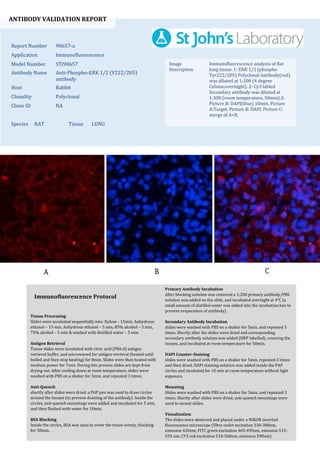
1. ANTIBODY VALIDATION REPORT
Report Number 90657-a
Application Immunofluorescence
Model Number STJ90657
Antibody Name Anti-Phospho-ERK 1/2 (Y222/205)
antibody
Host Rabbit
Clonality Polyclonal
Clone ID NA
Species RAT Tissue LUNG
Image
Description
Immunofluorescence analysis of Rat
lung tissue. 1: ERK 1/2 (phospho
Tyr222/205) Polyclonal Antibody(red)
was diluted at 1:200 (4 degree
Celsius,overnight). 2: Cy3 labled
Secondary antibody was diluted at
1:300 (room temperature, 50min).3:
Picture B: DAPI(blue) 10min. Picture
A:Target. Picture B: DAPI. Picture C:
merge of A+B.
Primary Antibody Incubation
After blocking solution was removed a 1:200 primary antibody/PBS
solution was added on the slide, and incubated overnight at 4°C (a
small amount of distilled water was added into the incubation box to
prevent evaporation of antibody).
Secondary Antibody Incubation
slides were washed with PBS on a shaker for 5min, and repeated 3
times. Shortly after the slides were dried and corresponding
secondary antibody solution was added (HRP labelled), covering the
tissues, and incubated at room temperature for 50min.
DAPI Counter-Staining
slides were washed with PBS on a shaker for 5min, repeated 3 times
and then dried. DAPI staining solution was added inside the PAP
circles and incubated for 10 min at room temperature without light
exposure.
Mounting
Slides were washed with PBS on a shaker for 5min, and repeated 3
times. Shortly after slides were dried, anti-quench mountings were
used to mount slides.
Visualization
The slides were observed and placed under a NIKON inverted
fluorescence microscope (Ultra violet excitation 330-380nm,
emission 420nm; FITC green excitation 465-495nm, emission 515-
555 nm; CY3 red excitation 510-560nm, emission 590nm)
Immunofluorescence Protocol
Tissue Processing
Slides were incubated sequentially into: Xylene - 15min, Anhydrous
ethanol – 15 min, Anhydrous ethanol – 5 min, 85% alcohol – 5 min,
75% alcohol – 5 min & washed with distilled water – 5 min.
Antigen Retrieval
Tissue slides were incubated with citric acid (PH6.0) antigen
retrieval buffer, and microwaved for antigen retrieval (heated until
boiled and then stop heating) for 8min. Slides were then heated with
medium power for 7min. During this process slides are kept from
drying out. After cooling down at room temperature, slides were
washed with PBS on a shaker for 5min, and repeated 3 times.
Anti-Quench
shortly after slides were dried, a PAP pen was used to draw circles
around the tissues (to prevent draining of the antibody). Inside the
circles, anti-quench mountings were added and incubated for 5 min,
and then flushed with water for 10min.
BSA Blocking
Inside the circles, BSA was used to cover the tissue evenly, blocking
for 30min.
2. ANTIBODY VALIDATION REPORT
Report Number 90657-b
Application Immunofluorescence
Model Number STJ90657
Antibody Name Anti-Phospho-ERK 1/2 (Y222/205)
antibody
Host Rabbit
Clonality Polyclonal
Clone ID NA
Species RAT Tissue LUNG
Image
Description
Immunofluorescence analysis of Rat
lung tissue. 1: ERK 1/2 (phospho
Tyr222/205) Polyclonal Antibody(red)
was diluted at 1:200 (4 degree
Celsius,overnight). 2: Cy3 labled
Secondary antibody was diluted at
1:300 (room temperature, 50min).3:
Picture B: DAPI(blue) 10min. Picture
A:Target. Picture B: DAPI. Picture C:
merge of A+B.
Primary Antibody Incubation
After blocking solution was removed a 1:200 primary antibody/PBS
solution was added on the slide, and incubated overnight at 4°C (a
small amount of distilled water was added into the incubation box to
prevent evaporation of antibody).
Secondary Antibody Incubation
slides were washed with PBS on a shaker for 5min, and repeated 3
times. Shortly after the slides were dried and corresponding
secondary antibody solution was added (HRP labelled), covering the
tissues, and incubated at room temperature for 50min.
DAPI Counter-Staining
slides were washed with PBS on a shaker for 5min, repeated 3 times
and then dried. DAPI staining solution was added inside the PAP
circles and incubated for 10 min at room temperature without light
exposure.
Mounting
Slides were washed with PBS on a shaker for 5min, and repeated 3
times. Shortly after slides were dried, anti-quench mountings were
used to mount slides.
Visualization
The slides were observed and placed under a NIKON inverted
fluorescence microscope (Ultra violet excitation 330-380nm,
emission 420nm; FITC green excitation 465-495nm, emission 515-
555 nm; CY3 red excitation 510-560nm, emission 590nm)
Immunofluorescence Protocol
Tissue Processing
Slides were incubated sequentially into: Xylene - 15min, Anhydrous
ethanol – 15 min, Anhydrous ethanol – 5 min, 85% alcohol – 5 min,
75% alcohol – 5 min & washed with distilled water – 5 min.
Antigen Retrieval
Tissue slides were incubated with citric acid (PH6.0) antigen
retrieval buffer, and microwaved for antigen retrieval (heated until
boiled and then stop heating) for 8min. Slides were then heated with
medium power for 7min. During this process slides are kept from
drying out. After cooling down at room temperature, slides were
washed with PBS on a shaker for 5min, and repeated 3 times.
Anti-Quench
shortly after slides were dried, a PAP pen was used to draw circles
around the tissues (to prevent draining of the antibody). Inside the
circles, anti-quench mountings were added and incubated for 5 min,
and then flushed with water for 10min.
BSA Blocking
Inside the circles, BSA was used to cover the tissue evenly, blocking
for 30min.
3. ANTIBODY VALIDATION REPORT
Report Number 90657-c
Application Immunofluorescence
Model Number STJ90657
Antibody Name Anti-Phospho-ERK 1/2 (Y222/205)
antibody
Host Rabbit
Clonality Polyclonal
Clone ID NA
Species RAT Tissue SPLEEN
Image
Description
Immunofluorescence analysis of Rat
spleen tissue. 1: ERK 1/2 (phospho
Tyr222/205) Polyclonal Antibody(red)
was diluted at 1:200 (4 degree
Celsius,overnight). 2: Cy3 labled
Secondary antibody was diluted at
1:300 (room temperature, 50min).3:
Picture B: DAPI(blue) 10min. Picture
A:Target. Picture B: DAPI. Picture C:
merge of A+B.
Primary Antibody Incubation
After blocking solution was removed a 1:200 primary antibody/PBS
solution was added on the slide, and incubated overnight at 4°C (a
small amount of distilled water was added into the incubation box to
prevent evaporation of antibody).
Secondary Antibody Incubation
slides were washed with PBS on a shaker for 5min, and repeated 3
times. Shortly after the slides were dried and corresponding
secondary antibody solution was added (HRP labelled), covering the
tissues, and incubated at room temperature for 50min.
DAPI Counter-Staining
slides were washed with PBS on a shaker for 5min, repeated 3 times
and then dried. DAPI staining solution was added inside the PAP
circles and incubated for 10 min at room temperature without light
exposure.
Mounting
Slides were washed with PBS on a shaker for 5min, and repeated 3
times. Shortly after slides were dried, anti-quench mountings were
used to mount slides.
Visualization
The slides were observed and placed under a NIKON inverted
fluorescence microscope (Ultra violet excitation 330-380nm,
emission 420nm; FITC green excitation 465-495nm, emission 515-
555 nm; CY3 red excitation 510-560nm, emission 590nm)
Immunofluorescence Protocol
Tissue Processing
Slides were incubated sequentially into: Xylene - 15min, Anhydrous
ethanol – 15 min, Anhydrous ethanol – 5 min, 85% alcohol – 5 min,
75% alcohol – 5 min & washed with distilled water – 5 min.
Antigen Retrieval
Tissue slides were incubated with citric acid (PH6.0) antigen
retrieval buffer, and microwaved for antigen retrieval (heated until
boiled and then stop heating) for 8min. Slides were then heated with
medium power for 7min. During this process slides are kept from
drying out. After cooling down at room temperature, slides were
washed with PBS on a shaker for 5min, and repeated 3 times.
Anti-Quench
shortly after slides were dried, a PAP pen was used to draw circles
around the tissues (to prevent draining of the antibody). Inside the
circles, anti-quench mountings were added and incubated for 5 min,
and then flushed with water for 10min.
BSA Blocking
Inside the circles, BSA was used to cover the tissue evenly, blocking
for 30min.
4. ANTIBODY VALIDATION REPORT
Report Number 90657-d
Application Immunofluorescence
Model Number STJ90657
Antibody Name Anti-Phospho-ERK 1/2 (Y222/205)
antibody
Host Rabbit
Clonality Polyclonal
Clone ID NA
Species RAT Tissue SPLEEN
Image
Description
Immunofluorescence analysis of Rat
spleen tissue. 1: ERK 1/2 (phospho
Tyr222/205) Polyclonal Antibody(red)
was diluted at 1:200 (4 degree
Celsius,overnight). 2: Cy3 labled
Secondary antibody was diluted at
1:300 (room temperature, 50min).3:
Picture B: DAPI(blue) 10min. Picture
A:Target. Picture B: DAPI. Picture C:
merge of A+B.
Primary Antibody Incubation
After blocking solution was removed a 1:200 primary antibody/PBS
solution was added on the slide, and incubated overnight at 4°C (a
small amount of distilled water was added into the incubation box to
prevent evaporation of antibody).
Secondary Antibody Incubation
slides were washed with PBS on a shaker for 5min, and repeated 3
times. Shortly after the slides were dried and corresponding
secondary antibody solution was added (HRP labelled), covering the
tissues, and incubated at room temperature for 50min.
DAPI Counter-Staining
slides were washed with PBS on a shaker for 5min, repeated 3 times
and then dried. DAPI staining solution was added inside the PAP
circles and incubated for 10 min at room temperature without light
exposure.
Mounting
Slides were washed with PBS on a shaker for 5min, and repeated 3
times. Shortly after slides were dried, anti-quench mountings were
used to mount slides.
Visualization
The slides were observed and placed under a NIKON inverted
fluorescence microscope (Ultra violet excitation 330-380nm,
emission 420nm; FITC green excitation 465-495nm, emission 515-
555 nm; CY3 red excitation 510-560nm, emission 590nm)
Immunofluorescence Protocol
Tissue Processing
Slides were incubated sequentially into: Xylene - 15min, Anhydrous
ethanol – 15 min, Anhydrous ethanol – 5 min, 85% alcohol – 5 min,
75% alcohol – 5 min & washed with distilled water – 5 min.
Antigen Retrieval
Tissue slides were incubated with citric acid (PH6.0) antigen
retrieval buffer, and microwaved for antigen retrieval (heated until
boiled and then stop heating) for 8min. Slides were then heated with
medium power for 7min. During this process slides are kept from
drying out. After cooling down at room temperature, slides were
washed with PBS on a shaker for 5min, and repeated 3 times.
Anti-Quench
shortly after slides were dried, a PAP pen was used to draw circles
around the tissues (to prevent draining of the antibody). Inside the
circles, anti-quench mountings were added and incubated for 5 min,
and then flushed with water for 10min.
BSA Blocking
Inside the circles, BSA was used to cover the tissue evenly, blocking
for 30min.