This report summarizes the validation of an anti-CD45 monoclonal antibody (clone 12A9) for use in immunofluorescence analysis of human cancer tissues. The antibody was tested on liver, lung, and kidney cancer tissues and showed specific staining of CD45 targets. The report details the immunofluorescence protocol used, including tissue processing, antigen retrieval, antibody incubations, counterstaining, mounting, and visualization under a fluorescence microscope.
Involved in DNA non-homologous end joining (NHEJ) required for double-strand break repair and V(D)J recombination. Binds to DNA and to DNA ligase IV (LIG4). The LIG4-XRCC4 complex is responsible for the NHEJ ligation step, and XRCC4 enhances the joining activity of LIG4. Binding of the LIG4-XRCC4 complex to DNA ends is dependent on the assembly of the DNA-dependent protein kinase complex DNA-PK to these DNA ends.
Anti-XRCC4 -http://www.stjohnslabs.com/xrcc4-antibody-p-98623
Join our Antibody Validation Project - http://www.stjohnslabs.com/services/antibody-validation
CD23, also known as Fc epsilon RII, or FcεRII, is the "low-affinity" receptor for IgE, an antibody isotype involved in allergy and resistance to parasites, and is important in regulation of IgE levels. Unlike many of the antibody receptors, CD23 is a C-type lectin. It is found on mature B cells, activated macrophages, eosinophils, follicular dendritic cells, and platelets.
There are two forms of CD23: CD23a and CD23b. CD23a is present on follicular B cells, whereas CD23b requires IL-4 to be expressed on T-cells, monocytes, Langerhans cells, eosinophils, and macrophages. CD23 is known to have a role of transportation in antibody feedback regulation. Antigens which enter the blood stream can be captured by antigen specific IgE antibodies. The IgE immune complexes that are formed bind to CD23 molecules on B cells, and are transported to the B cell follicles of the spleen. The antigen is then transferred from CD23+ B cells to CD11c+ antigen presenting cells. The CD11c+ cells in turn present the antigen to CD4+ T cells, which can lead to an enhanced antibody response.
Anti-CD23 -http://www.stjohnslabs.com/cd23-antibody-p-98586
Join our Antibody Validation Project - http://www.stjohnslabs.com/services/antibody-validation
Could play a role in phagocytic activities of tissue macrophages, both in intracellular lysosomal metabolism and extracellular cell-cell and cell-pathogen interactions. Binds to tissue- and organ-specific lectins or selectins, allowing homing of macrophage subsets to particular sites. Rapid recirculation of CD68 from endosomes and lysosomes to the plasma membrane may allow macrophages to crawl over selectin-bearing substrates or other cells.
Anti-CD68 -http://www.stjohnslabs.com/cd68-antibody-p-98587
Join our Antibody Validation Project - http://www.stjohnslabs.com/services/antibody-validation
Involved in the base excision repair (BER) pathway, by catalyzing the poly(ADP-ribosyl)ation of a limited number of acceptor proteins involved in chromatin architecture and in DNA metabolism. This modification follows DNA damages and appears as an obligatory step in a detection/signaling pathway leading to the reparation of DNA strand breaks. Mediates the poly(ADP-ribosyl)ation of APLF and CHFR. Positively regulates the transcription of MTUS1 and negatively regulates the transcription of MTUS2/TIP150. With EEF1A1 and TXK, forms a complex that acts as a T-helper 1 (Th1) cell-specific transcription factor and binds the promoter of IFN-gamma to directly regulate its transcription, and is thus involved importantly in Th1 cytokine production. Required for PARP9 and DTX3L recruitment to DNA damage sites. PARP1-dependent PARP9-DTX3L-mediated ubiquitination promotes the rapid and specific recruitment of 53BP1/TP53BP1, UIMC1/RAP80, and BRCA1 to DNA damage sites. / NAD+ + (ADP-D-ribosyl)(n)-acceptor = nicotinamide + (ADP-D-ribosyl)(n+1)-acceptor.
Anti-Cleaved PARP -http://www.stjohnslabs.com/cleaved-parp-
antibody-p-98682
Join our Antibody Validation Project - http://www.stjohnslabs.com/services/antibody-validation
Immunofluorescence Antibody Validation Report for Anti-CA IX Antibody (STJ96978)St John's Laboratory Ltd
Reversible hydration of carbon dioxide. Participates in pH regulation. May be involved in the control of cell proliferation and transformation. Appears to be a novel specific biomarker for a cervical neoplasia.
Anti-CA IX -http://www.stjohnslabs.com/ca-ix-antibody-p-98613
Join our Antibody Validation Project - http://www.stjohnslabs.com/services/antibody-validation
Accessory protein for MHC class-II antigen/T-cell receptor interaction. May regulate T-cell activation. Induces the aggregation of lipid rafts. / (Microbial infection) Acts as a receptor for human immunodeficiency virus-1 . Down-regulated by HIV-1 Vpu . Acts as a receptor for Human Herpes virus 7/HHV-7.
Anti-CD4 -http://www.stjohnslabs.com/cd4-antibody-p-98607
Join our Antibody Validation Project - http://www.stjohnslabs.com/services/antibody-validation
CD1 (cluster of differentiation 1) is a family of glycoproteins expressed on the surface of various human antigen-presenting cells. They are related to the class I MHC molecules, and are involved in the presentation of lipid antigens to T cells. However their precise function is unknown.
Anti-CD1 -http://www.stjohnslabs.com/cd1-antibody-p-98605
Join our Antibody Validation Project - http://www.stjohnslabs.com/services/antibody-validation
Keratin 16 is a protein that in humans is encoded by the KRT16 gene.
Keratin 16 is a type I cytokeratin. It is paired with keratin 6 in a number of epithelial tissues, including nail bed, esophagus, tongue, and hair follicles. Mutations in the gene encoding this protein are associated with the genetic skin disorders pachyonychia congenita, non-epidermolytic palmoplantar keratoderma and unilateral palmoplantar verrucous nevus.
Anti-CK16 -http://www.stjohnslabs.com/ck16-antibody-p-98593
Join our Antibody Validation Project - http://www.stjohnslabs.com/services/antibody-validation
Type I keratin involved in the formation and maintenance of various skin appendages, specifically in determining shape and orientation of hair (By similarity). Required for the correct growth of hair follicles, in particular for the persistence of the anagen (growth) state (By similarity). Modulates the function of TNF-alpha in the specific context of hair cycling. Regulates protein synthesis and epithelial cell growth through binding to the adapter protein SFN and by stimulating Akt/mTOR pathway (By similarity). Involved in tissue repair. May be a marker of basal cell differentiation in complex epithelia and therefore indicative of a certain type of epithelial "stem cells". Acts as a promoter of epithelial proliferation by acting a regulator of immune response in skin: promotes Th1/Th17-dominated immune environment contributing to the development of basaloid skin tumors (By similarity). May act as an autoantigen in the immunopathogenesis of psoriasis, with certain peptide regions being a major target for autoreactive T-cells and hence causing their proliferation.
Anti-CK17 -http://www.stjohnslabs.com/ck17-antibody-p-98618
Join our Antibody Validation Project - http://www.stjohnslabs.com/services/antibody-validation
Collagen alpha-2(I) chain is a protein that in humans is encoded by the COL1A2 gene.
This gene encodes one of the chains for type I collagen, the fibrillar collagen found in most connective tissues. Mutations in this gene are associated with osteogenesis imperfecta, Ehlers-Danlos syndrome, idiopathic osteoporosis, and atypical Marfan syndrome. Symptoms associated with mutations in this gene, however, tend to be less severe than mutations in the gene for alpha-1 type I collagen since alpha-2 is less abundant. Multiple messages for this gene result from multiple polyadenylation signals, a feature shared by most of the other collagen genes.
Anti-COL1A2 -http://www.stjohnslabs.com/col1a2-antibody
Join our Antibody Validation Project - http://www.stjohnslabs.com/services/antibody-validation
Claudins are a family of proteins that are the most important components of the tight junctions, where they establish the paracellular barrier that controls the flow of molecules in the intercellular space between the cells of an epithelium. They have four transmembrane domains, with the N-terminus and the C-terminus in the cytoplasm.
Anti-Claudin-5 -http://www.stjohnslabs.com/claudin-5-antibody-p-95192
Join our Antibody Validation Project - http://www.stjohnslabs.com/services/antibody-validation
Plays a role in the cellular breakdown of insulin, IAPP, glucagon, bradykinin, kallidin and other peptides, and thereby plays a role in intercellular peptide signaling. Degrades amyloid formed by APP and IAPP. May play a role in the degradation and clearance of naturally secreted amyloid beta-protein by neurons and microglia. / (Microbial infection) The membrane-associated isoform acts as an entry receptor for varicella-zoster virus (VZV). / Degradation of insulin, glucagon and other polypeptides. No action on proteins. / Zn2+ / Activated by small peptides (By similarity). Activated by ATP and GTP, and to a lesser extent by CTP, TTP and PPPi. Inhibited by bacitracin. Inhibited by S-nitrosylation and oxidation agents.
Anti-IDE -http://www.stjohnslabs.com/ide-antibody-p-98620
Join our Antibody Validation Project - http://www.stjohnslabs.com/services/antibody-validation
Functions as cell surface receptor for TIMP1 and plays a role in the activation of cellular signaling cascades. Plays a role in the activation of ITGB1 and integrin signaling, leading to the activation of AKT, FAK/PTK2 and MAP kinases. Promotes cell survival, reorganization of the actin cytoskeleton, cell adhesion, spreading and migration, via its role in the activation of AKT and FAK/PTK2. Plays a role in VEGFA signaling via its role in regulating the internalization of KDR/VEGFR2. Plays a role in intracellular vesicular transport processes, and is required for normal trafficking of the PMEL luminal domain that is essential for the development and maturation of melanocytes. Plays a role in the adhesion of leukocytes onto endothelial cells via its role in the regulation of SELP trafficking. May play a role in mast cell degranulation in response to Ms4a2/FceRI stimulation, but not in mast cell degranulation in response to other stimuli.
Anti-CD63 -http://www.stjohnslabs.com/cd63-antibody-p-98525
Join our Antibody Validation Project - http://www.stjohnslabs.com/services/antibody-validation
Transferrins are iron binding transport proteins which can bind two Fe3+ ions in association with the binding of an anion, usually bicarbonate. It is responsible for the transport of iron from sites of absorption and heme degradation to those of storage and utilization. Serum transferrin may also have a further role in stimulating cell proliferation.
Anti-Transferrin -http://www.stjohnslabs.com/transferrin-antibody-p-98689
Join our Antibody Validation Project - http://www.stjohnslabs.com/services/antibody-validation
Drug efflux transporter present in a number of stem cells that acts as a regulator of cellular differentiation. Able to mediate efflux from cells of the rhodamine dye and of the therapeutic drug doxorubicin. Specifically present in limbal stem cells, where it plays a key role in corneal development and repair.
Anti-ABCB5 -http://www.stjohnslabs.com/abcb5-antibody-p-98611
Join our Antibody Validation Project - http://www.stjohnslabs.com/services/antibody-validation
Aquaporin 4 also known as AQP4 is a protein which in humans is encoded by the AQP4 gene. AQP4 belongs to the aquaporin family of integral membrane proteins that conduct water through the cell membrane.
AQP4 is constitutively expressed in the basolateral cell membrane of principal collecting duct cells in the kidney and provide a pathway for water to exit these cells.
AQP4 is also expressed in astrocytes and is upregulated by direct insult to the central nervous system.
Anti-Aquaporin 4 -http://www.stjohnslabs.com/aquaporin-4-antibody-p-98599
Join our Antibody Validation Project - http://www.stjohnslabs.com/services/antibody-validation
Transcription factor that binds DNA in a non-specific manner, yet also specifically recognizes the core sequence 5'-CAC[GA]TG-3'. Activates the transcription of growth-related genes.
Anti-c-Myc -http://www.stjohnslabs.com/c-myc-antibody-p-91755
Join our Antibody Validation Project - http://www.stjohnslabs.com/services/antibody-validation
Thermolysin-like specificity, but is almost confined on acting on polypeptides of up to 30 amino acids . Biologically important in the destruction of opioid peptides such as Met- and Leu-enkephalins by cleavage of a Gly-Phe bond. Able to cleave angiotensin-1, angiotensin-2 and angiotensin 1-9. Involved in the degradation of atrial natriuretic factor (ANF). Displays UV-inducible elastase activity toward skin preelastic and elastic fibers.
Anti-CD10 -http://www.stjohnslabs.com/cd10-antibody-p-98609
Join our Antibody Validation Project - http://www.stjohnslabs.com/services/antibody-validation
Receptor for TNFSF6/FASLG. The adapter molecule FADD recruits caspase-8 to the activated receptor. The resulting death-inducing signaling complex (DISC) performs caspase-8 proteolytic activation which initiates the subsequent cascade of caspases (aspartate-specific cysteine proteases) mediating apoptosis. FAS-mediated apoptosis may have a role in the induction of peripheral tolerance, in the antigen-stimulated suicide of mature T-cells, or both. The secreted isoforms 2 to 6 block apoptosis (in vitro).
Anti-FAS -http://www.stjohnslabs.com/fas-antibody-p-92276
Join our Antibody Validation Project - http://www.stjohnslabs.com/services/antibody-validation
Auxiliary protein of DNA polymerase delta and is involved in the control of eukaryotic DNA replication by increasing the polymerase's processibility during elongation of the leading strand. Induces a robust stimulatory effect on the 3'-5' exonuclease and 3'-phosphodiesterase, but not apurinic-apyrimidinic (AP) endonuclease, APEX2 activities. Has to be loaded onto DNA in order to be able to stimulate APEX2. Plays a key role in DNA damage response (DDR) by being conveniently positioned at the replication fork to coordinate DNA replication with DNA repair and DNA damage tolerance pathways.
Anti-PCNA -http://www.stjohnslabs.com/pcna-antibody-p-98568
Join our Antibody Validation Project - http://www.stjohnslabs.com/services/antibody-validation
Epidermis-specific type I keratin involved in wound healing. Involved in the activation of follicular keratinocytes after wounding, while it does not play a major role in keratinocyte proliferation or migration. Participates in the regulation of epithelial migration by inhibiting the activity of SRC during wound repair.
Anti-Cytokeratin 6 -http://www.stjohnslabs.com/cytokeratin-6-antibody-p-98617
Join our Antibody Validation Project - http://www.stjohnslabs.com/services/antibody-validation
Has 2-hydroxyacid oxidase activity. Most active on the 2-carbon substrate glycolate, but is also active on 2-hydroxy fatty acids, with high activity towards 2-hydroxy palmitate and 2-hydroxy octanoate. / (S)-2-hydroxy acid + O2 = 2-oxo acid + H2O2.
Anti-HAO1 -http://www.stjohnslabs.com/hao1-antibody-p-99046
Join our Antibody Validation Project - http://www.stjohnslabs.com/services/antibody-validation
Keratin-7 is a member of the keratin gene family. The type II cytokeratins consist of basic or neutral proteins which are arranged in pairs of heterotypic keratin chains coexpressed during differentiation of simple and stratified epithelial tissues. This type II cytokeratin is specifically expressed in the simple epithelia lining the cavities of the internal organs and in the gland ducts and blood vessels. The genes encoding the type II cytokeratins are clustered in a region of chromosome 12q12-q13. Alternative splicing may result in several transcript variants; however, not all variants have been fully described.
Anti-CK7 -http://www.stjohnslabs.com/ck7-antibody-p-98591
Join our Antibody Validation Project - http://www.stjohnslabs.com/services/antibody-validation
Non-receptor protein-tyrosine kinase that plays an essential role in regulating cell migration, adhesion, spreading, reorganization of the actin cytoskeleton, formation and disassembly of focal adhesions and cell protrusions, cell cycle progression, cell proliferation and apoptosis. Required for early embryonic development and placenta development. Required for embryonic angiogenesis, normal cardiomyocyte migration and proliferation, and normal heart development. Regulates axon growth and neuronal cell migration, axon branching and synapse formation; required for normal development of the nervous system. Plays a role in osteogenesis and differentiation of osteoblasts.
Anti-FAK -http://www.stjohnslabs.com/fak-antibody-p-92262
Join our Antibody Validation Project - http://www.stjohnslabs.com/services/antibody-validation
In mammalian cells, the enzyme is responsible for converting (2R)-methylacyl-CoA esters to their (2S)-methylacyl-CoA epimers and known substrates, including coenzyme A esters of pristanic acid (mostly derived from phytanic acid, a 3-methyl branched-chain fatty acid that is abundant in the diet) and bile acids derived from cholesterol. This transformation is required in order to degrade (2R)-methylacyl-CoA esters by β-oxidation, which process requires the (2S)-epimer. The enzyme is known to be localised in peroxisomes and mitochondria, both of which are known to β-oxidize 2-methylacyl-CoA esters.
Anti-AMACR -http://www.stjohnslabs.com/amacr-antibody-p-98584
Join our Antibody Validation Project - http://www.stjohnslabs.com/services/antibody-validation
Involved in the activation cascade of caspases responsible for apoptosis execution. Binding of caspase-9 to Apaf-1 leads to activation of the protease which then cleaves and activates caspase-3. Promotes DNA damage-induced apoptosis in a ABL1/c-Abl-dependent manner. Proteolytically cleaves poly(ADP-ribose) polymerase (PARP). Isoform 2 lacks activity is an dominant-negative inhibitor of caspase-9. Strict requirement for an Asp residue at position P1 and with a marked preference for His at position P2. It has a preferred cleavage sequence of Leu-Gly-His-Asp-|-Xaa. Inhibited by the effector protein NleF that is produced by pathogenic E.coli; this inhibits apoptosis.
Anti-Caspase 9 -http://www.stjohnslabs.com/caspase-9-antibody-p-98614
Join our Antibody Validation Project - http://www.stjohnslabs.com/services/antibody-validation
Occurs in almost all aerobically respiring organisms and serves to protect cells from the toxic effects of hydrogen peroxide. Promotes growth of cells including T-cells, B-cells, myeloid leukemia cells, melanoma cells, mastocytoma cells and normal and transformed fibroblast cells. / 2 H2O2 = O2 + 2 H2O. / heme / NADP+.
Anti-Catalase -http://www.stjohnslabs.com/catalase-antibody-p-94943
Join our Antibody Validation Project - http://www.stjohnslabs.com/services/antibody-validation
Tubulin in molecular biology can refer either to the tubulin protein superfamily of globular proteins, or one of the member proteins of that superfamily. α- and β-tubulins polymerize into microtubules, a major component of the eukaryotic cytoskeleton. Microtubules function in many essential cellular processes, including mitosis. Tubulin-binding drugs kill cancerous cells by inhibiting microtubule dynamics, which are required for DNA segregation and therefore cell division. In eukaryotes there are six members of the tubulin superfamily, although not all are present in all species (see below).[2][3] Both α and β tubulins have a mass of around 50 kDa and are thus in a similar range compared to actin with ~42 kDa. In contrast, tubulin polymers (microtubules) tend to be much bigger than actin filaments due to their cylindrical nature.
Anti-α-tubulin -http://www.stjohnslabs.com/a-tubulin-antibody-p-98572
Join our Antibody Validation Project - http://www.stjohnslabs.com/services/antibody-validation
Has both glyceraldehyde-3-phosphate dehydrogenase and nitrosylase activities, thereby playing a role in glycolysis and nuclear functions, respectively. Participates in nuclear events including transcription, RNA transport, DNA replication and apoptosis. Nuclear functions are probably due to the nitrosylase activity that mediates cysteine S-nitrosylation of nuclear target proteins such as SIRT1, HDAC2 and PRKDC. Modulates the organization and assembly of the cytoskeleton. Facilitates the CHP1-dependent microtubule and membrane associations through its ability to stimulate the binding of CHP1 to microtubules.
Anti-GAPDH -http://www.stjohnslabs.com/gapdh-antibody-p-98566
Join our Antibody Validation Project - http://www.stjohnslabs.com/services/antibody-validation
Protein tyrosine-protein phosphatase required for T-cell activation through the antigen receptor. Acts as a positive regulator of T-cell coactivation upon binding to DPP4. The first PTPase domain has enzymatic activity, while the second one seems to affect the substrate specificity of the first one. Upon T-cell activation, recruits and dephosphorylates SKAP1 and FYN. Dephosphorylates LYN, and thereby modulates LYN activity (By similarity).
Anti-CD45-http://www.stjohnslabs.com/cd45-antibody-p-98583
Join our Antibody Validation Project - http://www.stjohnslabs.com/services/antibody-validation
Tubulin in molecular biology can refer either to the tubulin protein superfamily of globular proteins, or one of the member proteins of that superfamily. α- and β-tubulins polymerize into microtubules, a major component of the eukaryotic cytoskeleton. Microtubules function in many essential cellular processes, including mitosis. Tubulin-binding drugs kill cancerous cells by inhibiting microtubule dynamics, which are required for DNA segregation and therefore cell division. In eukaryotes there are six members of the tubulin superfamily, although not all are present in all species (see below).[2][3] Both α and β tubulins have a mass of around 50 kDa and are thus in a similar range compared to actin with ~42 kDa. In contrast, tubulin polymers (microtubules) tend to be much bigger than actin filaments due to their cylindrical nature.
Anti-α-tubulin -http://www.stjohnslabs.com/a-tubulin-antibody-p-98572
Join our Antibody Validation Project - http://www.stjohnslabs.com/services/antibody-validation
Immunocytochemistry Antibody Customer Review for Anti β-actin Antibody (STJ96...St John's Laboratory Ltd
Beta-actin (human gene and protein symbol ACTB/ACTB) is one of six different actin isoforms which have been identified in humans. This is one of the two nonmuscle cytoskeletal actins. Actins are highly conserved proteins that are involved in cell motility, structure and integrity. Alpha actins are a major constituent of the contractile apparatus.
Beta actin is usually used as a loading control, for among others, the integrity of cells, protein degradation, in PCR and Western blotting. Its molecular weight is approximately 42 kDa.
Anti β-actin - http://www.stjohnslabs.com/b-actin-antibody-p-98565?filter_name=STJ96930
Join our Antibody Validation Project - http://www.stjohnslabs.com/services/antibody-validation
Beta-actin (human gene and protein symbol ACTB/ACTB) is one of six different actin isoforms which have been identified in humans. This is one of the two nonmuscle cytoskeletal actins. Actins are highly conserved proteins[3][4] that are involved in cell motility, structure and integrity. Alpha actins are a major constituent of the contractile apparatus.
Beta actin is usually used as a loading control, for among others, the integrity of cells, protein degradation, in PCR and Western blotting. Its molecular weight is approximately 42 kDa.
Anti-β-actin -http://www.stjohnslabs.com/b-actin-antibody-p-98565
Join our Antibody Validation Project - http://www.stjohnslabs.com/services/antibody-validation
Has both glyceraldehyde-3-phosphate dehydrogenase and nitrosylase activities, thereby playing a role in glycolysis and nuclear functions, respectively. Participates in nuclear events including transcription, RNA transport, DNA replication and apoptosis. Nuclear functions are probably due to the nitrosylase activity that mediates cysteine S-nitrosylation of nuclear target proteins such as SIRT1, HDAC2 and PRKDC. Modulates the organization and assembly of the cytoskeleton. Facilitates the CHP1-dependent microtubule and membrane associations through its ability to stimulate the binding of CHP1 to microtubules (By similarity). Glyceraldehyde-3-phosphate dehydrogenase is a key enzyme in glycolysis that catalyzes the first step of the pathway by converting D-glyceraldehyde 3-phosphate (G3P) into 3-phospho-D-glyceroyl phosphate. Component of the GAIT (gamma interferon-activated inhibitor of translation) complex which mediates interferon-gamma-induced transcript-selective translation inhibition in inflammation processes. Upon interferon-gamma treatment assembles into the GAIT complex which binds to stem loop-containing GAIT elements in the 3'-UTR of diverse inflammatory mRNAs (such as ceruplasmin) and suppresses their translation.
Anti-GAPDH-http://www.stjohnslabs.com/gapdh-antibody-p-98566
Join our Antibody Validation Project - http://www.stjohnslabs.com/services/antibody-validation
Has both glyceraldehyde-3-phosphate dehydrogenase and nitrosylase activities, thereby playing a role in glycolysis and nuclear functions, respectively. Participates in nuclear events including transcription, RNA transport, DNA replication and apoptosis. Nuclear functions are probably due to the nitrosylase activity that mediates cysteine S-nitrosylation of nuclear target proteins such as SIRT1, HDAC2 and PRKDC. Modulates the organization and assembly of the cytoskeleton. Facilitates the CHP1-dependent microtubule and membrane associations through its ability to stimulate the binding of CHP1 to microtubules (By similarity). Glyceraldehyde-3-phosphate dehydrogenase is a key enzyme in glycolysis that catalyzes the first step of the pathway by converting D-glyceraldehyde 3-phosphate (G3P) into 3-phospho-D-glyceroyl phosphate. Component of the GAIT (gamma interferon-activated inhibitor of translation) complex which mediates interferon-gamma-induced transcript-selective translation inhibition in inflammation processes. Upon interferon-gamma treatment assembles into the GAIT complex which binds to stem loop-containing GAIT elements in the 3'-UTR of diverse inflammatory mRNAs (such as ceruplasmin) and suppresses their translation.
To purchase this antibody, use the following link: http://www.stjohnslabs.com/gapdh-antibody-p-94882?filter_name=STJ96417
Customer Review For Tubulin-beta Polyclonal Antibody- IF Application (STJ31562)St John's Laboratory Ltd
Tubulin is the major constituent of microtubules. It binds two moles of GTP, one at an exchangeable site on the beta chain and one at a non-exchangeable site on the alpha chain. TUBB3 plays a critical role in proper axon guidance and maintenance.
To purchase this antibody use the following link: http://www.stjohnslabs.com/tubulin-beta-antibody?filter_name=STJ31562
Tubulin is the major constituent of microtubules. It binds two molecules of GTP, one at an exchangeable site on the beta chain and one at a non-exchangeable site on the alpha chain. TUBB3 plays a critical role in proper axon guidance and mantainance.
To purchase this antibody, use the following link: http://www.stjohnslabs.com/tubulin-beta-antibody?filter_name=STJ31562
Immunofluorescence Antibody Validation Report for Anti-NFκB-p105/p50 Antibody...St John's Laboratory Ltd
NF-kappa-B is a pleiotropic transcription factor present in almost all cell types and is the endpoint of a series of signal transduction events that are initiated by a vast array of stimuli related to many biological processes such as inflammation, immunity, differentiation, cell growth, tumorigenesis and apoptosis. NF-kappa-B is a homo- or heterodimeric complex formed by the Rel-like domain-containing proteins RELA/p65, RELB, NFKB1/p105, NFKB1/p50, REL and NFKB2/p52 and the heterodimeric p65-p50 complex appears to be most abundant one. The dimers bind at kappa-B sites in the DNA of their target genes and the individual dimers have distinct preferences for different kappa-B sites that they can bind with distinguishable affinity and specificity.
Anti-NFκB-p105/p50 -http://www.stjohnslabs.com/nfkb-p105p50-antibody
Join our Antibody Validation Project - http://www.stjohnslabs.com/services/antibody-validation
Antigen KI-67 also known as Ki-67 or MKI67 is a protein that in humans is encoded by the MKI67 gene (antigen identified by monoclonal antibody Ki-67). Antigen KI-67 is a nuclear protein that is associated with and may be necessary for cellular proliferation. Furthermore, it is associated with ribosomal RNA transcription.[5] Inactivation of antigen KI-67 leads to inhibition of ribosomal RNA synthesis.
Anti-Ki 67 -http://www.stjohnslabs.com/ki-67-antibody-p-98601
Join our Antibody Validation Project - http://www.stjohnslabs.com/services/antibody-validation
Complement receptor type 2 (CR2), also known as complement C3d receptor, Epstein-Barr virus receptor, and CD21 (cluster of differentiation 21), is a protein that in humans is encoded by the CR2 gene. CR2 is involved in the complement system. It binds to iC3b (inactive derivative of C3b), C3dg, or C3d. B cells have CR2 receptors on their surfaces, allowing the complement system to play a role in B-cell activation and maturation.
Anti-CD21 -http://www.stjohnslabs.com/cd21-antibody-p-98597
Join our Antibody Validation Project - http://www.stjohnslabs.com/services/antibody-validation
Alpha-fetoprotein (AFP, α-fetoprotein; also sometimes called alpha-1-fetoprotein, alpha-fetoglobulin, or alpha fetal protein) is a protein that in humans is encoded by the AFP gene. The AFP gene is located on the q arm of chromosome 4 (4q25). AFP is a major plasma protein produced by the yolk sac and the liver during fetal development. It is thought to be the fetal form of serum albumin. AFP binds to copper, nickel, fatty acids and bilirubin[6] and is found in monomeric, dimeric and trimeric forms.
Anti-AFP alpha 1 Fetoprotein -http://www.stjohnslabs.com/afp-alpha-1-fetoprotein-antibody
Join our Antibody Validation Project - http://www.stjohnslabs.com/services/antibody-validation
Regulatory subunit of lactose synthase, changes the substrate specificity of galactosyltransferase in the mammary gland making glucose a good acceptor substrate for this enzyme. This enables LS to synthesize lactose, the major carbohydrate component of milk. In other tissues, galactosyltransferase transfers galactose onto the N-acetylglucosamine of the oligosaccharide chains in glycoproteins.
Anti-alpha Lactalbumin -http://www.stjohnslabs.com/alpha-lactalbumin-antibody-p-98621
Join our Antibody Validation Project - http://www.stjohnslabs.com/services/antibody-validation
CD5 is a cluster of differentiation expressed on the surface of T cells (various species) and in a subset of murine B cells known as B-1a. The expression of this receptor in human B cells has been a controversial topic and up to date there is no consensus regarding the role of this receptor as a marker of human B cells. B-1 cells have limited diversity of their B-cell receptor due to their lack of the enzyme terminal deoxynucleotidyl transferase (TdT) and are potentially self-reactive. CD5 serves to mitigate activating signals from the BCR so that the B-1 cells can only be activated by very strong stimuli (such as bacterial proteins) and not by normal tissue proteins. CD5 was used as a T-cell marker until monoclonal antibodies against CD3 were developed.
Anti-CD5 -http://www.stjohnslabs.com/cd5-antibody-p-98608
Join our Antibody Validation Project - http://www.stjohnslabs.com/services/antibody-validation
Multifunctional redox sensitive protein with various roles in different cellular compartments. In the nucleus is one of the major chromatin-associated non-histone proteins and acts as a DNA chaperone involved in replication, transcription, chromatin remodeling, V(D)J recombination, DNA repair and genome stability. Proposed to be an universal biosensor for nucleic acids. Promotes host inflammatory response to sterile and infectious signals and is involved in the coordination and integration of innate and adaptive immune responses. In the cytoplasm functions as sensor and/or chaperone for immunogenic nucleic acids implicating the activation of TLR9-mediated immune responses, and mediates autophagy.
Anti-HMG-1 -http://www.stjohnslabs.com/hmg-1-antibody-p-
98502
Join our Antibody Validation Project - http://www.stjohnslabs.com/services/antibody-validation
Transcription activator that binds to antioxidant response (ARE) elements in the promoter regions of target genes. Important for the coordinated up-regulation of genes in response to oxidative stress. May be involved in the transcriptional activation of genes of the beta-globin cluster by mediating enhancer activity of hypersensitive site 2 of the beta-globin locus control region.
Anti-Nrf2 -http://www.stjohnslabs.com/nrf2-antibody-p-95061
Join our Antibody Validation Project - http://www.stjohnslabs.com/services/antibody-validation
Electron carrier protein. The oxidized form of the cytochrome c heme group can accept an electron from the heme group of the cytochrome c1 subunit of cytochrome reductase. Cytochrome c then transfers this electron to the cytochrome oxidase complex, the final protein carrier in the mitochondrial electron-transport chain. / Plays a role in apoptosis. Suppression of the anti-apoptotic members or activation of the pro-apoptotic members of the Bcl-2 family leads to altered mitochondrial membrane permeability resulting in release of cytochrome c into the cytosol. Binding of cytochrome c to Apaf-1 triggers the activation of caspase-9, which then accelerates apoptosis by activating other caspases.
Anti-CYCS -http://www.stjohnslabs.com/cycs-antibody-p-99070
Join our Antibody Validation Project - http://www.stjohnslabs.com/services/antibody-validation
Tubulin is the major constituent of microtubules. The gamma chain is found at microtubule organizing centers (MTOC) such as the spindle poles or the centrosome. Pericentriolar matrix component that regulates alpha/beta chain minus-end nucleation, centrosome duplication and spindle formation.
Anti-Gamma Tubulin -http://www.stjohnslabs.com/gamma-tubulin-antibody
Join our Antibody Validation Project - http://www.stjohnslabs.com/services/antibody-validation
Lamin-B1 is a protein that in humans is encoded by the LMNB1 gene.
The nuclear lamina consists of a two-dimensional matrix of proteins located next to the inner nuclear membrane. The lamin family of proteins make up the matrix and are highly conserved in evolution. During mitosis, the lamina matrix is reversibly disassembled as the lamin proteins are phosphorylated. Lamin proteins are thought to be involved in nuclear stability, chromatin structure, and gene expression. Vertebrate lamins consist of two types, A and B. This gene encodes one of the two B type proteins, B1. Lamin B, along with heterochromatin, is anchored to the inner surface of the nuclear membrane by the lamin B receptor.
Anti-Lamin B1 -http://www.stjohnslabs.com/lamin-b1-antibody-p-98573
Join our Antibody Validation Project - http://www.stjohnslabs.com/services/antibody-validation
The steroid hormones and their receptors are involved in the regulation of eukaryotic gene expression and affect cellular proliferation and differentiation in target tissues. Progesterone receptor isoform B (PRB) is involved activation of c-SRC/MAPK signaling on hormone stimulation. / Isoform A: inactive in stimulating c-Src/MAPK signaling on hormone stimulation. / Isoform 4: Increases mitochondrial membrane potential and cellular respiration upon stimulation by progesterone.
Anti-PR -http://www.stjohnslabs.com/pr-antibody-p-99016
Join our Antibody Validation Project - http://www.stjohnslabs.com/services/antibody-validation
IL-8 is a chemotactic factor that attracts neutrophils, basophils, and T-cells, but not monocytes. It is also involved in neutrophil activation. It is released from several cell types in response to an inflammatory stimulus.
Anti-IL8 -http://www.stjohnslabs.com/il-8-antibody-p-94941
Join our Antibody Validation Project - http://www.stjohnslabs.com/services/antibody-validation
High-capacity urate exporter functioning in both renal and extrarenal urate excretion. Plays a role in porphyrin homeostasis as it is able to mediates the export of protoporhyrin IX (PPIX) both from mitochondria to cytosol and from cytosol to extracellular space, and cellular export of hemin, and heme. Xenobiotic transporter that may play an important role in the exclusion of xenobiotics from the brain. Appears to play a major role in the multidrug resistance phenotype of several cancer cell lines. Implicated in the efflux of numerous drugs and xenobiotics: mitoxantrone, the photosensitizer pheophorbide, camptothecin, methotrexate, azidothymidine (AZT), and the anthracyclines daunorubicin and doxorubicin.
Anti-ABCG2 -http://www.stjohnslabs.com/abcg2-antibody-p-98888
Join our Antibody Validation Project - http://www.stjohnslabs.com/services/antibody-validation
Cadherins are calcium-dependent cell adhesion proteins. They preferentially interact with themselves in a homophilic manner in connecting cells; cadherins may thus contribute to the sorting of heterogeneous cell types. This cadherin may play a important role in endothelial cell biology through control of the cohesion and organization of the intercellular junctions. It associates with alpha-catenin forming a link to the cytoskeleton. Acts in concert with KRIT1 to establish and maintain correct endothelial cell polarity and vascular lumen. These effects are mediated by recruitment and activation of the Par polarity complex and RAP1B. Required for activation of PRKCZ and for the localization of phosphorylated PRKCZ, PARD3, TIAM1 and RAP1B to the cell junction.
Anti-VE-Cadherin -http://www.stjohnslabs.com/ve-cadherin-antibody-p-98941
Join our Antibody Validation Project - http://www.stjohnslabs.com/services/antibody-validation
Immunofluorescence Antibody Validation Report for Anti-Collagen IV Antibody (...St John's Laboratory Ltd
Type IV collagen is the major structural component of glomerular basement membranes (GBM), forming a 'chicken-wire' meshwork together with laminins, proteoglycans and entactin/nidogen.; Arresten, comprising the C-terminal NC1 domain, inhibits angiogenesis and tumor formation. The C-terminal half is found to possess the anti-angiogenic activity. Specifically inhibits endothelial cell proliferation, migration and tube formation. Inhibits expression of hypoxia-inducible factor 1alpha and ERK1/2 and p38 MAPK activation. Ligand for alpha1/beta1 integrin.
Anti-Collagen IV - http://www.stjohnslabs.com/anti-collagen-iv-antibody?filter_name=STJ98907
Join our Antibody Validation Project - http://www.stjohnslabs.com/services/antibody-validation
Ubiquitin: Exists either covalently attached to another protein, or free (unanchored). When covalently bound, it is conjugated to target proteins via an isopeptide bond either as a monomer (monoubiquitin), a polymer linked via different Lys residues of the ubiquitin (polyubiquitin chains) or a linear polymer linked via the initiator Met of the ubiquitin (linear polyubiquitin chains). Polyubiquitin chains, when attached to a target protein, have different functions depending on the Lys residue of the ubiquitin that is linked: Lys-6-linked may be involved in DNA repair; Lys-11-linked is involved in ERAD (endoplasmic reticulum-associated degradation) and in cell-cycle regulation; Lys-29-linked is involved in lysosomal degradation; Lys-33-linked is involved in kinase modification; Lys-48-linked is involved in protein degradation via the proteasome; Lys-63-linked is involved in endocytosis, DNA-damage responses as well as in signaling processes leading to activation of the transcription factor NF-kappa-B.
Anti-Ub -http://www.stjohnslabs.com/ub-antibody-p-98871
Join our Antibody Validation Project - http://www.stjohnslabs.com/services/antibody-validation
Microtubules of the eukaryotic cytoskeleton perform essential and diverse functions and are composed of a heterodimer of alpha and beta tubulins. The genes encoding these microtubule constituents belong to the tubulin superfamily, which is composed of six distinct families. Genes from the alpha, beta and gamma tubulin families are found in all eukaryotes. The alpha and beta tubulins represent the major components of microtubules, while gamma tubulin plays a critical role in the nucleation of microtubule assembly. There are multiple alpha and beta tubulin genes, which are highly conserved among species. This gene encodes alpha tubulin and is highly similar to mouse and rat Tuba1 gene. Northern blotting studies have shown that the gene expression is predominantly found in morphologically differentiated neurologic cells.
Anti-Tubulin α -http://www.stjohnslabs.com/tubulin-a-antibody
Immunofluorescence Antibody Validation Report for Anti-β I tubulin Antibody (...St John's Laboratory Ltd
Tubulin in molecular biology can refer either to the tubulin protein superfamily of globular proteins, or one of the member proteins of that superfamily. α- and β-tubulins polymerize into microtubules, a major component of the eukaryotic cytoskeleton. Microtubules function in many essential cellular processes, including mitosis. Tubulin-binding drugs kill cancerous cells by inhibiting microtubule dynamics, which are required for DNA segregation and therefore cell division. In eukaryotes there are six members of the tubulin superfamily, although not all are present in all species (see below).[2][3] Both α and β tubulins have a mass of around 50 kDa and are thus in a similar range compared to actin with ~42 kDa. In contrast, tubulin polymers (microtubules) tend to be much bigger than actin filaments due to their cylindrical nature.
Anti-β I tubulin -http://www.stjohnslabs.com/b-i-tubulin-antibody-p-98574
Join our Antibody Validation Project - http://www.stjohnslabs.com/services/antibody-validation
Transcription factor that binds to the octamer motif (5'-ATTTGCAT-3') and activates the promoters of the genes for some small nuclear RNAs (snRNA) and of genes such as those for histone H2B and immunoglobulins. Modulates transcription transactivation by NR3C1, AR and PGR (By similarity). In case of human herpes simplex virus (HSV) infection, POU2F1 forms a multiprotein-DNA complex with the viral transactivator protein VP16 and HCFC1 thereby enabling the transcription of the viral immediate early genes.
Anti-OCT1 -http://www.stjohnslabs.com/oct1-antibody-p-98626
Join our Antibody Validation Project - http://www.stjohnslabs.com/services/antibody-validation
Inhibits NF-kappa-B by complexing with and trapping it in the cytoplasm. However, the unphosphorylated form resynthesized after cell stimulation is able to bind NF-kappa-B allowing its transport to the nucleus and protecting it to further NFKBIA-dependent inactivation. Association with inhibitor kappa B-interacting NKIRAS1 and NKIRAS2 prevent its phosphorylation rendering it more resistant to degradation, explaining its slower degradation.
Anti-IκB β -http://www.stjohnslabs.com/ikb-b-antibody-1f3
Join our Antibody Validation Project - http://www.stjohnslabs.com/services/antibody-validation
Involved in the activation cascade of caspases responsible for apoptosis execution. At the onset of apoptosis it proteolytically cleaves poly(ADP-ribose) polymerase (PARP) at a '216-Asp-|-Gly-217' bond. Cleaves and activates sterol regulatory element binding proteins (SREBPs) between the basic helix-loop-helix leucine zipper domain and the membrane attachment domain. Cleaves and activates caspase-6, -7 and -9. Involved in the cleavage of huntingtin. Triggers cell adhesion in sympathetic neurons through RET cleavage. / Strict requirement for an Asp residue at positions P1 and P4. It has a preferred cleavage sequence of Asp-Xaa-Xaa-Asp-|- with a hydrophobic amino-acid residue at P2 and a hydrophilic amino-acid residue at P3, although Val or Ala are also accepted at this position. / Inhibited by isatin sulfonamides.
Anti-Cleaved-Caspase-3 p17 (D175) -http://www.stjohnslabs.com/cleaved-caspase-3-p17-d175-antibody-p-89637
Join our Antibody Validation Project - http://www.stjohnslabs.com/services/antibody-validation
Metalloproteinase inhibitor that functions by forming one to one complexes with target metalloproteinases, such as collagenases, and irreversibly inactivates them by binding to their catalytic zinc cofactor. Acts on MMP1, MMP2, MMP3, MMP7, MMP8, MMP9, MMP10, MMP11, MMP12, MMP13 and MMP16. Does not act on MMP14. Also functions as a growth factor that regulates cell differentiation, migration and cell death and activates cellular signaling cascades via CD63 and ITGB1. Plays a role in integrin signaling. Mediates erythropoiesis in vitro; but, unlike IL3, it is species-specific, stimulating the growth and differentiation of only human and murine thyroid progenitors.
Anti-TIMP-1 -http://www.stjohnslabs.com/timp-1-antibody
Join our Antibody Validation Project - http://www.stjohnslabs.com/services/antibody-validation
Transthyretin (TTR) is a transport protein in the serum and cerebrospinal fluid that carries the thyroid hormone thyroxine (T4) and retinol-binding protein bound to retinol. This is how transthyretin gained its name: transports thyroxine and retinol. The liver secretes transthyretin into the blood, and the choroid plexus secretes TTR into the cerebrospinal fluid.
TTR was originally called prealbumin(or thyroxine-binding prealbumin) because it ran faster than albumin on electrophoresis gels.
Anti-TTR - http://www.stjohnslabs.com/anti-ttr-antibody?filter_name=STJ98876
Join our Antibody Validation Project - http://www.stjohnslabs.com/services/antibody-validation
Ubiquitin-like modifier involved in formation of autophagosomal vacuoles (autophagosomes) . Whereas LC3s are involved in elongation of the phagophore membrane, the GABARAP/GATE-16 subfamily is essential for a later stage in autophagosome maturation.
Anti-LC3A -http://www.stjohnslabs.com/lc3a-antibody
Join our Antibody Validation Project - http://www.stjohnslabs.com/services/antibody-validation
Similar to Immunofluorescence Antibody Validation Report for Anti-CD45 Antibody (STJ96948) (17)
G protein-coupled receptor that probably associates with the patched protein (PTCH) to transduce the hedgehog's proteins signal. Binding of sonic hedgehog (SHH) to its receptor patched is thought to prevent normal inhibition by patched of smoothened (SMO). Required for the accumulation of KIF7 and GLI3 in the cilia.
Anti-Smo antibody (STJ95710): http://www.stjohnslabs.com/smo-antibody-p-94371?filter_name=STJ95710
Join our Antibody Validation Project - http://www.stjohnslabs.com/services/antibody-validation
Western Blot Customer Review Anti Glucocorticoid Receptor antibody (STJ97101)St John's Laboratory Ltd
Receptor for glucocorticoids (GC). Has a dual mode of action: as a transcription factor that binds to glucocorticoid response elements (GRE), both for nuclear and mitochondrial DNA, and as a modulator of other transcription factors. Affects inflammatory responses, cellular proliferation and differentiation in target tissues. Involved in chromatin remodeling . Plays a role in rapid mRNA degradation by binding to the 5' UTR of target mRNAs and interacting with PNRC2 in a ligand-dependent manner. Could act as a coactivator for STAT5-dependent transcription upon growth hormone (GH) stimulation and could reveal an essential role of hepatic GR in the control of body growth (By similarity). Has transcriptional activation and repression activity . Mediates glucocorticoid-induced apoptosis . Promotes accurate chromosome segregation during mitosis . May act as a tumor suppressor . May play a negative role in adipogenesis through the regulation of lipolytic and antilipogenic gene expression (By similarity). / Isoform Beta: Acts as a dominant negative inhibitor of isoform Alpha . Has intrinsic transcriptional activity independent of isoform Alpha when both isoforms are coexpressed.
Join our Antibody Validation Project: http://www.stjohnslabs.com/services/antibody-validation
Anti glucocorticoid receptor antibody (STJ97101):
http://www.stjohnslabs.com/glucocorticoid-receptor-antibody-p-98736?filter_name=STJ97101
Western Blot Customer Review Anti-Phospho-Cofilin (S3) Antibody (STJ90230)St John's Laboratory Ltd
Binds to F-actin and exhibits pH-sensitive F-actin depolymerizing activity. Regulates actin cytoskeleton dynamics. Important for normal progress through mitosis and normal cytokinesis. Plays a role in the regulation of cell morphology and cytoskeletal organization. Required for the up-regulation of atypical chemokine receptor ACKR2 from endosomal compartment to cell membrane, increasing its efficiency in chemokine uptake and degradation.
Anti-Phospho-Cofilin (S3) -http://www.stjohnslabs.com/phospho-cofilin-s3-antibody
Join our Antibody Validation Project - http://www.stjohnslabs.com/services/antibody-validation
This June, Dr. Byron Baron from the University of Malta, Malta, is our Scientist of the Month! He's shared with us his research highlights, his current projects and some comments on the biotechnology industry.
Want to be our Scientist of the Month? Contact info@stjohnslabs.com
Downstream effector molecule involved in the transmission of signals from tyrosine kinase receptors and small GTPases to the actin cytoskeleton. Promotes formation of actin filaments. Part of the WAVE complex that regulates lamellipodia formation. The WAVE complex regulates actin filament reorganization via its interaction with the Arp2/3 complex.
Anti-WAVE2 -http://www.stjohnslabs.com/wave2-antibody
Join our Antibody Validation Project - http://www.stjohnslabs.com/services/antibody-validation
Implicated in synaptic vesicle endocytosis. May recruit other proteins to membranes with high curvature.
Brain, mostly in frontal cortex. Expressed at high level in fetal cerebellum.
Anti-Endophilin I -http://www.stjohnslabs.com/endophilin-i-antibody
Join our Antibody Validation Project - http://www.stjohnslabs.com/services/antibody-validation
Tubulin is the major constituent of microtubules. It binds two moles of GTP, one at an exchangeable site on the beta chain and one at a non-exchangeable site on the alpha chain. TUBB3 plays a critical role in proper axon guidance and mantainance.
Anti-β-tubulin -http://www.stjohnslabs.com/b-tubulin-antibody-p-98672
Join our Antibody Validation Project - http://www.stjohnslabs.com/services/antibody-validation
Most upstream protease of the activation cascade of caspases responsible for the TNFRSF6/FAS mediated and TNFRSF1A induced cell death. Binding to the adapter molecule FADD recruits it to either receptor. The resulting aggregate called death-inducing signaling complex (DISC) performs CASP8 proteolytic activation. The active dimeric enzyme is then liberated from the DISC and free to activate downstream apoptotic proteases. Proteolytic fragments of the N-terminal propeptide (termed CAP3, CAP5 and CAP6) are likely retained in the DISC. Cleaves and activates CASP3, CASP4, CASP6, CASP7, CASP9 and CASP10. May participate in the GZMB apoptotic pathways. Cleaves ADPRT. Hydrolyzes the small-molecule substrate, Ac-Asp-Glu-Val-Asp-|-AMC. Likely target for the cowpox virus CRMA death inhibitory protein. Isoform 5, isoform 6, isoform 7 and isoform 8 lack the catalytic site and may interfere with the pro-apoptotic activity of the complex. / Strict requirement for Asp at position P1 and has a preferred cleavage sequence of (Leu/Asp/Val)-Glu-Thr-Asp-|-(Gly/Ser/Ala). / Inhibited by the effector protein NleF that is produced by pathogenic E.coli; this inhibits apoptosis.
Anti-Caspase-8-http://www.stjohnslabs.com/caspase-8-antibody-p-99045
Join our Antibody Validation Project - http://www.stjohnslabs.com/services/antibody-validation
Tubulin is the major constituent of microtubules. The gamma chain is found at microtubule organizing centers (MTOC) such as the spindle poles or the centrosome. Pericentriolar matrix component that regulates alpha/beta chain minus-end nucleation, centrosome duplication and spindle formation.
Anti-Gamma Tubulin-http://www.stjohnslabs.com/gamma-tubulin-antibody
Join our Antibody Validation Project - http://www.stjohnslabs.com/services/antibody-validation
Tubulin in molecular biology can refer either to the tubulin protein superfamily of globular proteins, or one of the member proteins of that superfamily. α- and β-tubulins polymerize into microtubules, a major component of the eukaryotic cytoskeleton. Microtubules function in many essential cellular processes, including mitosis. Tubulin-binding drugs kill cancerous cells by inhibiting microtubule dynamics, which are required for DNA segregation and therefore cell division. In eukaryotes there are six members of the tubulin superfamily, although not all are present in all species (see below). Both α and β tubulins have a mass of around 50 kDa and are thus in a similar range compared to actin with ~42 kDa. In contrast, tubulin polymers (microtubules) tend to be much bigger than actin filaments due to their cylindrical nature. Tubulin was long thought to be specific to eukaryotes. More recently, however, several prokaryotic proteins have been shown to be related to tubulin.
Anti-Epsilon Tubulin -http://www.stjohnslabs.com/epsilon-tubulin-antibody-2
Join our Antibody Validation Project - http://www.stjohnslabs.com/services/antibody-validation
Ubiquitin-like modifier involved in formation of autophagosomal vacuoles (autophagosomes) . Whereas LC3s are involved in elongation of the phagophore membrane, the GABARAP/GATE-16 subfamily is essential for a later stage in autophagosome maturation .
Anti-LC3A-http://www.stjohnslabs.com/lc3a-antibody
Join our Antibody Validation Project - http://www.stjohnslabs.com/services/antibody-validation
Multifunctional transcription factor in ER stress response. Plays an essential role in the response to a wide variety of cell stresses and induces cell cycle arrest and apoptosis in response to ER stress. Plays a dual role both as an inhibitor of CCAAT/enhancer-binding protein (C/EBP) function and as an activator of other genes. Acts as a dominant-negative regulator of C/EBP-induced transcription: dimerizes with members of the C/EBP family, impairs their association with C/EBP binding sites in the promoter regions, and inhibits the expression of C/EBP regulated genes. Positively regulates the transcription of TRIB3, IL6, IL8, IL23, TNFRSF10B/DR5, PPP1R15A/GADD34, BBC3/PUMA, BCL2L11/BIM and ERO1L. Negatively regulates; expression of BCL2 and MYOD1, ATF4-dependent transcriptional activation of asparagine synthetase (ASNS), CEBPA-dependent transcriptional activation of hepcidin (HAMP) and CEBPB-mediated expression of peroxisome proliferator-activated receptor gamma (PPARG).
Anti-CHOP-http://www.stjohnslabs.com/chop-antibody-2
Join our Antibody Validation Project - http://www.stjohnslabs.com/services/antibody-validation
Pseudokinase that plays a key role in TNF-induced necroptosis, a programmed cell death process. Activated following phosphorylation by RIPK3, leading to homotrimerization, localization to the plasma membrane and execution of programmed necrosis characterized by calcium influx and plasma membrane damage. Does not have protein kinase activity.
Anti-phospho-MLKL (S358)-http://www.stjohnslabs.com/phospho-mlkl-s358-antibody-1
Join our Antibody Validation Project - http://www.stjohnslabs.com/services/antibody-validation
Serine/threonine kinase which acts as an essential component of the MAP kinase signal transduction pathway. MAPK1/ERK2 and MAPK3/ERK1 are the 2 MAPKs which play an important role in the MAPK/ERK cascade. They participate also in a signaling cascade initiated by activated KIT and KITLG/SCF. Depending on the cellular context, the MAPK/ERK cascade mediates diverse biological functions such as cell growth, adhesion, survival and differentiation through the regulation of transcription, translation, cytoskeletal rearrangements. The MAPK/ERK cascade plays also a role in initiation and regulation of meiosis, mitosis, and postmitotic functions in differentiated cells by phosphorylating a number of transcription factors. About 160 substrates have already been discovered for ERKs. Many of these substrates are localized in the nucleus, and seem to participate in the regulation of transcription upon stimulation.
Anti-ERK1-http://www.stjohnslabs.com/erk1-antibody-3
Join our Antibody Validation Project - http://www.stjohnslabs.com/services/antibody-validation
Tyrosine-protein kinase that acts as a cell-surface receptor for PDGFA, PDGFB and PDGFC and plays an essential role in the regulation of embryonic development, cell proliferation, survival and chemotaxis. Depending on the context, promotes or inhibits cell proliferation and cell migration. Plays an important role in the differentiation of bone marrow-derived mesenchymal stem cells. Required for normal skeleton development and cephalic closure during embryonic development. Required for normal development of the mucosa lining the gastrointestinal tract, and for recruitment of mesenchymal cells and normal development of intestinal villi. Plays a role in cell migration and chemotaxis in wound healing. Plays a role in platelet activation, secretion of agonists from platelet granules, and in thrombin-induced platelet aggregation.
Anti-PDGFRα-http://www.stjohnslabs.com/pdgfra-antibody-2
Join our Antibody Validation Project - http://www.stjohnslabs.com/services/antibody-validation
Thiol protease that cleaves IL-1 beta between an Asp and an Ala, releasing the mature cytokine which is involved in a variety of inflammatory processes. Important for defense against pathogens. Cleaves and activates sterol regulatory element binding proteins (SREBPs). Can also promote apoptosis. / Strict requirement for an Asp residue at position P1 and has a preferred cleavage sequence of Tyr-Val-Ala-Asp-|-. / Specifically inhibited by the cowpox virus Crma protein.
Anti-Caspase-1-http://www.stjohnslabs.com/caspase-1-antibody-1
Join our Antibody Validation Project - http://www.stjohnslabs.com/services/antibody-validation
Functions as a cell surface receptor and performs physiological functions on the surface of neurons relevant to neurite growth, neuronal adhesion and axonogenesis. Involved in cell mobility and transcription regulation through protein-protein interactions. Can promote transcription activation through binding to APBB1-KAT5 and inhibits Notch signaling through interaction with Numb. Couples to apoptosis-inducing pathways such as those mediated by G(O) and JIP. Inhibits G(o) alpha ATPase activity (By similarity). Acts as a kinesin I membrane receptor, mediating the axonal transport of beta-secretase and presenilin 1. Involved in copper homeostasis/oxidative stress through copper ion reduction. In vitro, copper-metallated APP induces neuronal death directly or is potentiated through Cu2+-mediated low-density lipoprotein oxidation.
Anti-Amyloid-β-http://www.stjohnslabs.com/amyloid-v-antibody
Join our Antibody Validation Project - http://www.stjohnslabs.com/services/antibody-validation
Suppresses apoptosis in a variety of cell systems including factor-dependent lymphohematopoietic and neural cells. Regulates cell death by controlling the mitochondrial membrane permeability. Appears to function in a feedback loop system with caspases. Inhibits caspase activity either by preventing the release of cytochrome c from the mitochondria and/or by binding to the apoptosis-activating factor (APAF-1). May attenuate inflammation by impairing NLRP1-inflammasome activation, hence CASP1 activation and IL1B release .
Anti-Bcl-2-http://www.stjohnslabs.com/bcl-2-antibody-1
Join our Antibody Validation Project - http://www.stjohnslabs.com/services/antibody-validation
Alpha-actin-2 also known as actin, aortic smooth muscle or alpha smooth muscle actin (α-SMA, SMactin, alpha-SM-actin, ASMA) is a protein that in humans is encoded by the ACTA2 gene located on 10q22-q24. Actin alpha 2, the human aortic smooth muscle actin gene, is one of six different actin isoforms which have been identified. Actins are highly conserved proteins that are involved in cell motility, structure and integrity. Alpha actins are a major constituent of the contractile apparatus. Alpha-smooth muscle actin (α-SMA) is commonly used as a marker of myofibroblast formation.
Anti-α-SMA -http://www.stjohnslabs.com/a-sma-antibody-1
Join our Antibody Validation Project - http://www.stjohnslabs.com/services/antibody-validation
Cancer cell metabolism: special Reference to Lactate PathwayAADYARAJPANDEY1
Normal Cell Metabolism:
Cellular respiration describes the series of steps that cells use to break down sugar and other chemicals to get the energy we need to function.
Energy is stored in the bonds of glucose and when glucose is broken down, much of that energy is released.
Cell utilize energy in the form of ATP.
The first step of respiration is called glycolysis. In a series of steps, glycolysis breaks glucose into two smaller molecules - a chemical called pyruvate. A small amount of ATP is formed during this process.
Most healthy cells continue the breakdown in a second process, called the Kreb's cycle. The Kreb's cycle allows cells to “burn” the pyruvates made in glycolysis to get more ATP.
The last step in the breakdown of glucose is called oxidative phosphorylation (Ox-Phos).
It takes place in specialized cell structures called mitochondria. This process produces a large amount of ATP. Importantly, cells need oxygen to complete oxidative phosphorylation.
If a cell completes only glycolysis, only 2 molecules of ATP are made per glucose. However, if the cell completes the entire respiration process (glycolysis - Kreb's - oxidative phosphorylation), about 36 molecules of ATP are created, giving it much more energy to use.
IN CANCER CELL:
Unlike healthy cells that "burn" the entire molecule of sugar to capture a large amount of energy as ATP, cancer cells are wasteful.
Cancer cells only partially break down sugar molecules. They overuse the first step of respiration, glycolysis. They frequently do not complete the second step, oxidative phosphorylation.
This results in only 2 molecules of ATP per each glucose molecule instead of the 36 or so ATPs healthy cells gain. As a result, cancer cells need to use a lot more sugar molecules to get enough energy to survive.
introduction to WARBERG PHENOMENA:
WARBURG EFFECT Usually, cancer cells are highly glycolytic (glucose addiction) and take up more glucose than do normal cells from outside.
Otto Heinrich Warburg (; 8 October 1883 – 1 August 1970) In 1931 was awarded the Nobel Prize in Physiology for his "discovery of the nature and mode of action of the respiratory enzyme.
WARNBURG EFFECT : cancer cells under aerobic (well-oxygenated) conditions to metabolize glucose to lactate (aerobic glycolysis) is known as the Warburg effect. Warburg made the observation that tumor slices consume glucose and secrete lactate at a higher rate than normal tissues.
Seminar of U.V. Spectroscopy by SAMIR PANDASAMIR PANDA
Spectroscopy is a branch of science dealing the study of interaction of electromagnetic radiation with matter.
Ultraviolet-visible spectroscopy refers to absorption spectroscopy or reflect spectroscopy in the UV-VIS spectral region.
Ultraviolet-visible spectroscopy is an analytical method that can measure the amount of light received by the analyte.
Deep Behavioral Phenotyping in Systems Neuroscience for Functional Atlasing a...Ana Luísa Pinho
Functional Magnetic Resonance Imaging (fMRI) provides means to characterize brain activations in response to behavior. However, cognitive neuroscience has been limited to group-level effects referring to the performance of specific tasks. To obtain the functional profile of elementary cognitive mechanisms, the combination of brain responses to many tasks is required. Yet, to date, both structural atlases and parcellation-based activations do not fully account for cognitive function and still present several limitations. Further, they do not adapt overall to individual characteristics. In this talk, I will give an account of deep-behavioral phenotyping strategies, namely data-driven methods in large task-fMRI datasets, to optimize functional brain-data collection and improve inference of effects-of-interest related to mental processes. Key to this approach is the employment of fast multi-functional paradigms rich on features that can be well parametrized and, consequently, facilitate the creation of psycho-physiological constructs to be modelled with imaging data. Particular emphasis will be given to music stimuli when studying high-order cognitive mechanisms, due to their ecological nature and quality to enable complex behavior compounded by discrete entities. I will also discuss how deep-behavioral phenotyping and individualized models applied to neuroimaging data can better account for the subject-specific organization of domain-general cognitive systems in the human brain. Finally, the accumulation of functional brain signatures brings the possibility to clarify relationships among tasks and create a univocal link between brain systems and mental functions through: (1) the development of ontologies proposing an organization of cognitive processes; and (2) brain-network taxonomies describing functional specialization. To this end, tools to improve commensurability in cognitive science are necessary, such as public repositories, ontology-based platforms and automated meta-analysis tools. I will thus discuss some brain-atlasing resources currently under development, and their applicability in cognitive as well as clinical neuroscience.
Comparing Evolved Extractive Text Summary Scores of Bidirectional Encoder Rep...University of Maribor
Slides from:
11th International Conference on Electrical, Electronics and Computer Engineering (IcETRAN), Niš, 3-6 June 2024
Track: Artificial Intelligence
https://www.etran.rs/2024/en/home-english/
A brief information about the SCOP protein database used in bioinformatics.
The Structural Classification of Proteins (SCOP) database is a comprehensive and authoritative resource for the structural and evolutionary relationships of proteins. It provides a detailed and curated classification of protein structures, grouping them into families, superfamilies, and folds based on their structural and sequence similarities.
This presentation explores a brief idea about the structural and functional attributes of nucleotides, the structure and function of genetic materials along with the impact of UV rays and pH upon them.
THE IMPORTANCE OF MARTIAN ATMOSPHERE SAMPLE RETURN.Sérgio Sacani
The return of a sample of near-surface atmosphere from Mars would facilitate answers to several first-order science questions surrounding the formation and evolution of the planet. One of the important aspects of terrestrial planet formation in general is the role that primary atmospheres played in influencing the chemistry and structure of the planets and their antecedents. Studies of the martian atmosphere can be used to investigate the role of a primary atmosphere in its history. Atmosphere samples would also inform our understanding of the near-surface chemistry of the planet, and ultimately the prospects for life. High-precision isotopic analyses of constituent gases are needed to address these questions, requiring that the analyses are made on returned samples rather than in situ.
THE IMPORTANCE OF MARTIAN ATMOSPHERE SAMPLE RETURN.
Immunofluorescence Antibody Validation Report for Anti-CD45 Antibody (STJ96948)
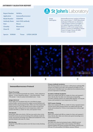
1. ANTIBODY VALIDATION REPORT
Report Number 96948-a
Application Immunofluorescence
Model Number STJ96948
Antibody Name Anti-CD45 antibody
Host Mouse
Clonality Monoclonal
Clone ID 12A9
Species HUMAN Tissue LIVER CANCER
Image
Description
Immunofluorescence analysis of Human
liver cancer tissue. 1: CD45 Monoclonal
Antibody(12A9)(red) was diluted at
1:200 (4 degree Celsius,overnight). 2:
Cy3 labled Secondary antibody was
diluted at 1:300 (room temperature,
50min).3: Picture B: DAPI(blue) 10min.
Picture A:Target. Picture B: DAPI.
Picture C: merge of A+B.
Primary Antibody Incubation
After blocking solution was removed a 1:200 primary antibody/PBS
solution was added on the slide, and incubated overnight at 4°C (a
small amount of distilled water was added into the incubation box to
prevent evaporation of antibody).
Secondary Antibody Incubation
slides were washed with PBS on a shaker for 5min, and repeated 3
times. Shortly after the slides were dried and corresponding
secondary antibody solution was added (HRP labelled), covering the
tissues, and incubated at room temperature for 50min.
DAPI Counter-Staining
slides were washed with PBS on a shaker for 5min, repeated 3 times
and then dried. DAPI staining solution was added inside the PAP
circles and incubated for 10 min at room temperature without light
exposure.
Mounting
Slides were washed with PBS on a shaker for 5min, and repeated 3
times. Shortly after slides were dried, anti-quench mountings were
used to mount slides.
Visualization
The slides were observed and placed under a NIKON inverted
fluorescence microscope (Ultra violet excitation 330-380nm,
emission 420nm; FITC green excitation 465-495nm, emission 515-
555 nm; CY3 red excitation 510-560nm, emission 590nm)
Immunofluorescence Protocol
Tissue Processing
Slides were incubated sequentially into: Xylene - 15min, Anhydrous
ethanol – 15 min, Anhydrous ethanol – 5 min, 85% alcohol – 5 min,
75% alcohol – 5 min & washed with distilled water – 5 min.
Antigen Retrieval
Tissue slides were incubated with citric acid (PH6.0) antigen
retrieval buffer, and microwaved for antigen retrieval (heated until
boiled and then stop heating) for 8min. Slides were then heated with
medium power for 7min. During this process slides are kept from
drying out. After cooling down at room temperature, slides were
washed with PBS on a shaker for 5min, and repeated 3 times.
Anti-Quench
shortly after slides were dried, a PAP pen was used to draw circles
around the tissues (to prevent draining of the antibody). Inside the
circles, anti-quench mountings were added and incubated for 5 min,
and then flushed with water for 10min.
BSA Blocking
Inside the circles, BSA was used to cover the tissue evenly, blocking
for 30min.
St John's Laboratory Ltd.
www.stjohnslabs.com
2. ANTIBODY VALIDATION REPORT
Report Number 96948-b
Application Immunofluorescence
Model Number STJ96948
Antibody Name Anti-CD45 antibody
Host Mouse
Clonality Monoclonal
Clone ID 12A9
Species HUMAN Tissue LIVER CANCER
Image
Description
Immunofluorescence analysis of Human
liver cancer tissue. 1: CD45 Monoclonal
Antibody(12A9)(red) was diluted at
1:200 (4 degree Celsius,overnight). 2:
Cy3 labled Secondary antibody was
diluted at 1:300 (room temperature,
50min).3: Picture B: DAPI(blue) 10min.
Picture A:Target. Picture B: DAPI.
Picture C: merge of A+B.
Primary Antibody Incubation
After blocking solution was removed a 1:200 primary antibody/PBS
solution was added on the slide, and incubated overnight at 4°C (a
small amount of distilled water was added into the incubation box to
prevent evaporation of antibody).
Secondary Antibody Incubation
slides were washed with PBS on a shaker for 5min, and repeated 3
times. Shortly after the slides were dried and corresponding
secondary antibody solution was added (HRP labelled), covering the
tissues, and incubated at room temperature for 50min.
DAPI Counter-Staining
slides were washed with PBS on a shaker for 5min, repeated 3 times
and then dried. DAPI staining solution was added inside the PAP
circles and incubated for 10 min at room temperature without light
exposure.
Mounting
Slides were washed with PBS on a shaker for 5min, and repeated 3
times. Shortly after slides were dried, anti-quench mountings were
used to mount slides.
Visualization
The slides were observed and placed under a NIKON inverted
fluorescence microscope (Ultra violet excitation 330-380nm,
emission 420nm; FITC green excitation 465-495nm, emission 515-
555 nm; CY3 red excitation 510-560nm, emission 590nm)
Immunofluorescence Protocol
Tissue Processing
Slides were incubated sequentially into: Xylene - 15min, Anhydrous
ethanol – 15 min, Anhydrous ethanol – 5 min, 85% alcohol – 5 min,
75% alcohol – 5 min & washed with distilled water – 5 min.
Antigen Retrieval
Tissue slides were incubated with citric acid (PH6.0) antigen
retrieval buffer, and microwaved for antigen retrieval (heated until
boiled and then stop heating) for 8min. Slides were then heated with
medium power for 7min. During this process slides are kept from
drying out. After cooling down at room temperature, slides were
washed with PBS on a shaker for 5min, and repeated 3 times.
Anti-Quench
shortly after slides were dried, a PAP pen was used to draw circles
around the tissues (to prevent draining of the antibody). Inside the
circles, anti-quench mountings were added and incubated for 5 min,
and then flushed with water for 10min.
BSA Blocking
Inside the circles, BSA was used to cover the tissue evenly, blocking
for 30min.
St John's Laboratory Ltd.
www.stjohnslabs.com
3. ANTIBODY VALIDATION REPORT
Report Number 96948-c
Application Immunofluorescence
Model Number STJ96948
Antibody Name Anti-CD45 antibody
Host Mouse
Clonality Monoclonal
Clone ID 12A9
Species HUMAN Tissue LUNG CANCER
Image
Description
Immunofluorescence analysis of Human
lung cancer tissue. 1: CD45 Monoclonal
Antibody(12A9)(red) was diluted at
1:200 (4 degree Celsius,overnight). 2:
Cy3 labled Secondary antibody was
diluted at 1:300 (room temperature,
50min).3: Picture B: DAPI(blue) 10min.
Picture A:Target. Picture B: DAPI.
Picture C: merge of A+B.
Primary Antibody Incubation
After blocking solution was removed a 1:200 primary antibody/PBS
solution was added on the slide, and incubated overnight at 4°C (a
small amount of distilled water was added into the incubation box to
prevent evaporation of antibody).
Secondary Antibody Incubation
slides were washed with PBS on a shaker for 5min, and repeated 3
times. Shortly after the slides were dried and corresponding
secondary antibody solution was added (HRP labelled), covering the
tissues, and incubated at room temperature for 50min.
DAPI Counter-Staining
slides were washed with PBS on a shaker for 5min, repeated 3 times
and then dried. DAPI staining solution was added inside the PAP
circles and incubated for 10 min at room temperature without light
exposure.
Mounting
Slides were washed with PBS on a shaker for 5min, and repeated 3
times. Shortly after slides were dried, anti-quench mountings were
used to mount slides.
Visualization
The slides were observed and placed under a NIKON inverted
fluorescence microscope (Ultra violet excitation 330-380nm,
emission 420nm; FITC green excitation 465-495nm, emission 515-
555 nm; CY3 red excitation 510-560nm, emission 590nm)
Immunofluorescence Protocol
Tissue Processing
Slides were incubated sequentially into: Xylene - 15min, Anhydrous
ethanol – 15 min, Anhydrous ethanol – 5 min, 85% alcohol – 5 min,
75% alcohol – 5 min & washed with distilled water – 5 min.
Antigen Retrieval
Tissue slides were incubated with citric acid (PH6.0) antigen
retrieval buffer, and microwaved for antigen retrieval (heated until
boiled and then stop heating) for 8min. Slides were then heated with
medium power for 7min. During this process slides are kept from
drying out. After cooling down at room temperature, slides were
washed with PBS on a shaker for 5min, and repeated 3 times.
Anti-Quench
shortly after slides were dried, a PAP pen was used to draw circles
around the tissues (to prevent draining of the antibody). Inside the
circles, anti-quench mountings were added and incubated for 5 min,
and then flushed with water for 10min.
BSA Blocking
Inside the circles, BSA was used to cover the tissue evenly, blocking
for 30min.
St John's Laboratory Ltd.
www.stjohnslabs.com
4. ANTIBODY VALIDATION REPORT
Report Number 96948-d
Application Immunofluorescence
Model Number STJ96948
Antibody Name Anti-CD45 antibody
Host Mouse
Clonality Monoclonal
Clone ID 12A9
Species HUMAN Tissue LUNG CANCER
Image
Description
Immunofluorescence analysis of Human
lung cancer tissue. 1: CD45 Monoclonal
Antibody(12A9)(red) was diluted at
1:200 (4 degree Celsius,overnight). 2:
Cy3 labled Secondary antibody was
diluted at 1:300 (room temperature,
50min).3: Picture B: DAPI(blue) 10min.
Picture A:Target. Picture B: DAPI.
Picture C: merge of A+B.
Primary Antibody Incubation
After blocking solution was removed a 1:200 primary antibody/PBS
solution was added on the slide, and incubated overnight at 4°C (a
small amount of distilled water was added into the incubation box to
prevent evaporation of antibody).
Secondary Antibody Incubation
slides were washed with PBS on a shaker for 5min, and repeated 3
times. Shortly after the slides were dried and corresponding
secondary antibody solution was added (HRP labelled), covering the
tissues, and incubated at room temperature for 50min.
DAPI Counter-Staining
slides were washed with PBS on a shaker for 5min, repeated 3 times
and then dried. DAPI staining solution was added inside the PAP
circles and incubated for 10 min at room temperature without light
exposure.
Mounting
Slides were washed with PBS on a shaker for 5min, and repeated 3
times. Shortly after slides were dried, anti-quench mountings were
used to mount slides.
Visualization
The slides were observed and placed under a NIKON inverted
fluorescence microscope (Ultra violet excitation 330-380nm,
emission 420nm; FITC green excitation 465-495nm, emission 515-
555 nm; CY3 red excitation 510-560nm, emission 590nm)
Immunofluorescence Protocol
Tissue Processing
Slides were incubated sequentially into: Xylene - 15min, Anhydrous
ethanol – 15 min, Anhydrous ethanol – 5 min, 85% alcohol – 5 min,
75% alcohol – 5 min & washed with distilled water – 5 min.
Antigen Retrieval
Tissue slides were incubated with citric acid (PH6.0) antigen
retrieval buffer, and microwaved for antigen retrieval (heated until
boiled and then stop heating) for 8min. Slides were then heated with
medium power for 7min. During this process slides are kept from
drying out. After cooling down at room temperature, slides were
washed with PBS on a shaker for 5min, and repeated 3 times.
Anti-Quench
shortly after slides were dried, a PAP pen was used to draw circles
around the tissues (to prevent draining of the antibody). Inside the
circles, anti-quench mountings were added and incubated for 5 min,
and then flushed with water for 10min.
BSA Blocking
Inside the circles, BSA was used to cover the tissue evenly, blocking
for 30min.
St John's Laboratory Ltd.
www.stjohnslabs.com
5. ANTIBODY VALIDATION REPORT
Report Number 96948-e
Application Immunofluorescence
Model Number STJ96948
Antibody Name Anti-CD45 antibody
Host Mouse
Clonality Monoclonal
Clone ID 12A9
Species HUMAN Tissue KIDNEY
Image
Description
Immunofluorescence analysis of Human
kidney cancer tissue. 1: CD45
Monoclonal Antibody(12A9)(red) was
diluted at 1:200 (4 degree
Celsius,overnight). 2: Cy3 labled
Secondary antibody was diluted at
1:300 (room temperature, 50min).3:
Picture B: DAPI(blue) 10min. Picture
A:Target. Picture B: DAPI. Picture C:
merge of A+B.
Primary Antibody Incubation
After blocking solution was removed a 1:200 primary antibody/PBS
solution was added on the slide, and incubated overnight at 4°C (a
small amount of distilled water was added into the incubation box to
prevent evaporation of antibody).
Secondary Antibody Incubation
slides were washed with PBS on a shaker for 5min, and repeated 3
times. Shortly after the slides were dried and corresponding
secondary antibody solution was added (HRP labelled), covering the
tissues, and incubated at room temperature for 50min.
DAPI Counter-Staining
slides were washed with PBS on a shaker for 5min, repeated 3 times
and then dried. DAPI staining solution was added inside the PAP
circles and incubated for 10 min at room temperature without light
exposure.
Mounting
Slides were washed with PBS on a shaker for 5min, and repeated 3
times. Shortly after slides were dried, anti-quench mountings were
used to mount slides.
Visualization
The slides were observed and placed under a NIKON inverted
fluorescence microscope (Ultra violet excitation 330-380nm,
emission 420nm; FITC green excitation 465-495nm, emission 515-
555 nm; CY3 red excitation 510-560nm, emission 590nm)
Immunofluorescence Protocol
Tissue Processing
Slides were incubated sequentially into: Xylene - 15min, Anhydrous
ethanol – 15 min, Anhydrous ethanol – 5 min, 85% alcohol – 5 min,
75% alcohol – 5 min & washed with distilled water – 5 min.
Antigen Retrieval
Tissue slides were incubated with citric acid (PH6.0) antigen
retrieval buffer, and microwaved for antigen retrieval (heated until
boiled and then stop heating) for 8min. Slides were then heated with
medium power for 7min. During this process slides are kept from
drying out. After cooling down at room temperature, slides were
washed with PBS on a shaker for 5min, and repeated 3 times.
Anti-Quench
shortly after slides were dried, a PAP pen was used to draw circles
around the tissues (to prevent draining of the antibody). Inside the
circles, anti-quench mountings were added and incubated for 5 min,
and then flushed with water for 10min.
BSA Blocking
Inside the circles, BSA was used to cover the tissue evenly, blocking
for 30min.
St John's Laboratory Ltd.
www.stjohnslabs.com
6. ANTIBODY VALIDATION REPORT
Report Number 96948-f
Application Immunofluorescence
Model Number STJ96948
Antibody Name Anti-CD45 antibody
Host Mouse
Clonality Monoclonal
Clone ID 12A9
Species HUMAN Tissue KIDNEY
Image
Description
Immunofluorescence analysis of Human
kidney cancer tissue. 1: CD45
Monoclonal Antibody(12A9)(red) was
diluted at 1:200 (4 degree
Celsius,overnight). 2: Cy3 labled
Secondary antibody was diluted at
1:300 (room temperature, 50min).3:
Picture B: DAPI(blue) 10min. Picture
A:Target. Picture B: DAPI. Picture C:
merge of A+B.
Primary Antibody Incubation
After blocking solution was removed a 1:200 primary antibody/PBS
solution was added on the slide, and incubated overnight at 4°C (a
small amount of distilled water was added into the incubation box to
prevent evaporation of antibody).
Secondary Antibody Incubation
slides were washed with PBS on a shaker for 5min, and repeated 3
times. Shortly after the slides were dried and corresponding
secondary antibody solution was added (HRP labelled), covering the
tissues, and incubated at room temperature for 50min.
DAPI Counter-Staining
slides were washed with PBS on a shaker for 5min, repeated 3 times
and then dried. DAPI staining solution was added inside the PAP
circles and incubated for 10 min at room temperature without light
exposure.
Mounting
Slides were washed with PBS on a shaker for 5min, and repeated 3
times. Shortly after slides were dried, anti-quench mountings were
used to mount slides.
Visualization
The slides were observed and placed under a NIKON inverted
fluorescence microscope (Ultra violet excitation 330-380nm,
emission 420nm; FITC green excitation 465-495nm, emission 515-
555 nm; CY3 red excitation 510-560nm, emission 590nm)
Immunofluorescence Protocol
Tissue Processing
Slides were incubated sequentially into: Xylene - 15min, Anhydrous
ethanol – 15 min, Anhydrous ethanol – 5 min, 85% alcohol – 5 min,
75% alcohol – 5 min & washed with distilled water – 5 min.
Antigen Retrieval
Tissue slides were incubated with citric acid (PH6.0) antigen
retrieval buffer, and microwaved for antigen retrieval (heated until
boiled and then stop heating) for 8min. Slides were then heated with
medium power for 7min. During this process slides are kept from
drying out. After cooling down at room temperature, slides were
washed with PBS on a shaker for 5min, and repeated 3 times.
Anti-Quench
shortly after slides were dried, a PAP pen was used to draw circles
around the tissues (to prevent draining of the antibody). Inside the
circles, anti-quench mountings were added and incubated for 5 min,
and then flushed with water for 10min.
BSA Blocking
Inside the circles, BSA was used to cover the tissue evenly, blocking
for 30min.
St John's Laboratory Ltd.
www.stjohnslabs.com