SlideShare a Scribd company logo
Figure:
Immunohistochemical
analysis of paraffin
embedded Human
uterus tissue. 1: PCNA
Monoclonal
Antibody(12D10) was
diluted at 1:200 (4
degree
Celsius,overnight). 2:
Sodium citrate pH 6.0
was used for antibody
retrieval (>98 degree
Celsius,20min). 3:
Secondary antibody was
diluted at 1:200 (room
temperature, 30min).
Negative control was
used by secondary
antibody only.
Report Number 96933-a Host Mouse
Application IHC-P Clonality Monoclonal
Model Number STJ96933 Clone ID 12D10
Antibody Name Anti-PCNA antibody
Testing Species HUMAN Testing Tissue UTERUS
ANTIBODY VALIDATION REPORT
a. (A small amount of distilled water was added into the incubation
box to prevent evaporation of antibody).
128. Secondary antibody incubation
a. Slides were washed 3 times, with PBS on a shaker for 5min.
Shortly after the slides were dried the corresponding secondary
antibody solution was added (HRP labelled), covering the
tissues, and incubated at room temperature for 30min.
b.
129. DAB staining
a. Slides were washed 3 times, with PBS on a shaker for 5min.
b. Shortly after, the slides were dried and fresh DAB staining buffer
was added inside the circles. The staining time was adjusted
under a microscope. Yellow-brown colour represented a positive
result. Slides were washed with water to stop the staining.
c.
130. Haematoxylin staining
a. Haematoxylin was used to counter-staining for 1min, and then
the slides were washed with water. 1% Hydrochloric acid and
alcohol was added for several seconds and then washed with
water. Ammonia was used to reveal blue colour, and then
flushed with water.
b.
131. Desolation and Clearing
i. Slides were incubated sequentially into: 75% alcohol 5min, 85%
alcohol 5min, Anhydrous ethanol - 5min, Anhydrous ethanol -
5min & Xylene - 5min. Shortly after slides were dried and neutral
gum was used to seal the slides.
ii.
132. Visualization
a. Results were validated with microscope, and the slides were
scanned.
Paraffin-Embedded
Immunohistochemistry Protocol
122.
123. Tissue processing
a. Slides were incubated sequentially into Xylene; 15min –
Xylene, 15min - Anhydrous ethanol, 5min - Anhydrous
ethanol, 5min - 85% alcohol, 5min - 75% alcohol & 5min –
wash in distilled water.
b.
124. Antigen retrieval
a. Tissue slides were incubated with citric acid (PH6.0) antigen
retrieval buffer and microwaved for antigen retrieval (heated
until boiled and then stopped heating) for 8min. Slides were
then heated with medium power for 7min. During this
process slides were kept from drying out. After cooling down
at room temperature, slides were washed with PBS on
shaker for 5min, repeated for 3 times.
b.
125. Inhibition of endogenous peroxidase
a. Slides were placed in 3% Hydrogen peroxide solution, and
incubated for 10 min at room temperature without light
exposure. Slides were then washed 3 times with PBS on a
shaker for 5mins.
b.
126. BSA Blocking
a. Shortly after slides were dried, a PAP pen was used to draw
circles around the tissue sections (and to prevent draining of
the antibody solution). Inside the circles, BSA was used to
cover the tissue evenly, blocking for 30min.
b.
127. Primary antibody incubation
After blocking solution was removed a 1:200 solution of
primary antibody/PBS was added on the slide, and incubated
overnight at 4°C.
St John's Laboratory Ltd.
www.stjohnslabs.com
Figure:
Immunohistochemical
analysis of paraffin
embedded Human
uterus cancer tissue. 1:
PCNA Monoclonal
Antibody(12D10) was
diluted at 1:200 (4
degree
Celsius,overnight). 2:
Sodium citrate pH 6.0
was used for antibody
retrieval (>98 degree
Celsius,20min). 3:
Secondary antibody was
diluted at 1:200 (room
temperature, 30min).
Negative control was
used by secondary
antibody only.
Report Number 96933-b Host Mouse
Application IHC-P Clonality Monoclonal
Model Number STJ96933 Clone ID 12D10
Antibody Name Anti-PCNA antibody
Testing Species HUMAN Testing Tissue UTERUS CANCER
ANTIBODY VALIDATION REPORT
a. (A small amount of distilled water was added into the incubation
box to prevent evaporation of antibody).
117. Secondary antibody incubation
a. Slides were washed 3 times, with PBS on a shaker for 5min.
Shortly after the slides were dried the corresponding secondary
antibody solution was added (HRP labelled), covering the
tissues, and incubated at room temperature for 30min.
b.
118. DAB staining
a. Slides were washed 3 times, with PBS on a shaker for 5min.
b. Shortly after, the slides were dried and fresh DAB staining buffer
was added inside the circles. The staining time was adjusted
under a microscope. Yellow-brown colour represented a positive
result. Slides were washed with water to stop the staining.
c.
119. Haematoxylin staining
a. Haematoxylin was used to counter-staining for 1min, and then
the slides were washed with water. 1% Hydrochloric acid and
alcohol was added for several seconds and then washed with
water. Ammonia was used to reveal blue colour, and then
flushed with water.
b.
120. Desolation and Clearing
i. Slides were incubated sequentially into: 75% alcohol 5min, 85%
alcohol 5min, Anhydrous ethanol - 5min, Anhydrous ethanol -
5min & Xylene - 5min. Shortly after slides were dried and neutral
gum was used to seal the slides.
ii.
121. Visualization
a. Results were validated with microscope, and the slides were
scanned.
Paraffin-Embedded
Immunohistochemistry Protocol
111.
112. Tissue processing
a. Slides were incubated sequentially into Xylene; 15min –
Xylene, 15min - Anhydrous ethanol, 5min - Anhydrous
ethanol, 5min - 85% alcohol, 5min - 75% alcohol & 5min –
wash in distilled water.
b.
113. Antigen retrieval
a. Tissue slides were incubated with citric acid (PH6.0) antigen
retrieval buffer and microwaved for antigen retrieval (heated
until boiled and then stopped heating) for 8min. Slides were
then heated with medium power for 7min. During this
process slides were kept from drying out. After cooling down
at room temperature, slides were washed with PBS on
shaker for 5min, repeated for 3 times.
b.
114. Inhibition of endogenous peroxidase
a. Slides were placed in 3% Hydrogen peroxide solution, and
incubated for 10 min at room temperature without light
exposure. Slides were then washed 3 times with PBS on a
shaker for 5mins.
b.
115. BSA Blocking
a. Shortly after slides were dried, a PAP pen was used to draw
circles around the tissue sections (and to prevent draining of
the antibody solution). Inside the circles, BSA was used to
cover the tissue evenly, blocking for 30min.
b.
116. Primary antibody incubation
After blocking solution was removed a 1:200 solution of
primary antibody/PBS was added on the slide, and incubated
overnight at 4°C.
St John's Laboratory Ltd.
www.stjohnslabs.com
Figure:
Immunohistochemical
analysis of paraffin
embedded Human
Tonsil tissue. 1: PCNA
Monoclonal
Antibody(12D10) was
diluted at 1:200 (4
degree
Celsius,overnight). 2:
Sodium citrate pH 6.0
was used for antibody
retrieval (>98 degree
Celsius,20min). 3:
Secondary antibody was
diluted at 1:200 (room
temperature, 30min).
Negative control was
used by secondary
antibody only.
Report Number 96933-c Host Mouse
Application IHC-P Clonality Monoclonal
Model Number STJ96933 Clone ID 12D10
Antibody Name Anti-PCNA antibody
Testing Species HUMAN Testing Tissue TONSIL
ANTIBODY VALIDATION REPORT
a. (A small amount of distilled water was added into the incubation
box to prevent evaporation of antibody).
106. Secondary antibody incubation
a. Slides were washed 3 times, with PBS on a shaker for 5min.
Shortly after the slides were dried the corresponding secondary
antibody solution was added (HRP labelled), covering the
tissues, and incubated at room temperature for 30min.
b.
107. DAB staining
a. Slides were washed 3 times, with PBS on a shaker for 5min.
b. Shortly after, the slides were dried and fresh DAB staining buffer
was added inside the circles. The staining time was adjusted
under a microscope. Yellow-brown colour represented a positive
result. Slides were washed with water to stop the staining.
c.
108. Haematoxylin staining
a. Haematoxylin was used to counter-staining for 1min, and then
the slides were washed with water. 1% Hydrochloric acid and
alcohol was added for several seconds and then washed with
water. Ammonia was used to reveal blue colour, and then
flushed with water.
b.
109. Desolation and Clearing
i. Slides were incubated sequentially into: 75% alcohol 5min, 85%
alcohol 5min, Anhydrous ethanol - 5min, Anhydrous ethanol -
5min & Xylene - 5min. Shortly after slides were dried and neutral
gum was used to seal the slides.
ii.
110. Visualization
a. Results were validated with microscope, and the slides were
scanned.
Paraffin-Embedded
Immunohistochemistry Protocol
100.
101. Tissue processing
a. Slides were incubated sequentially into Xylene; 15min –
Xylene, 15min - Anhydrous ethanol, 5min - Anhydrous
ethanol, 5min - 85% alcohol, 5min - 75% alcohol & 5min –
wash in distilled water.
b.
102. Antigen retrieval
a. Tissue slides were incubated with citric acid (PH6.0) antigen
retrieval buffer and microwaved for antigen retrieval (heated
until boiled and then stopped heating) for 8min. Slides were
then heated with medium power for 7min. During this
process slides were kept from drying out. After cooling down
at room temperature, slides were washed with PBS on
shaker for 5min, repeated for 3 times.
b.
103. Inhibition of endogenous peroxidase
a. Slides were placed in 3% Hydrogen peroxide solution, and
incubated for 10 min at room temperature without light
exposure. Slides were then washed 3 times with PBS on a
shaker for 5mins.
b.
104. BSA Blocking
a. Shortly after slides were dried, a PAP pen was used to draw
circles around the tissue sections (and to prevent draining of
the antibody solution). Inside the circles, BSA was used to
cover the tissue evenly, blocking for 30min.
b.
105. Primary antibody incubation
After blocking solution was removed a 1:200 solution of
primary antibody/PBS was added on the slide, and incubated
overnight at 4°C.
St John's Laboratory Ltd.
www.stjohnslabs.com
Figure:
Immunohistochemical
analysis of paraffin
embedded Human colon
tissue. 1: PCNA
Monoclonal
Antibody(12D10) was
diluted at 1:200 (4
degree
Celsius,overnight). 2:
Sodium citrate pH 6.0
was used for antibody
retrieval (>98 degree
Celsius,20min). 3:
Secondary antibody was
diluted at 1:200 (room
temperature, 30min).
Negative control was
used by secondary
antibody only.
Report Number 96933-d Host Mouse
Application IHC-P Clonality Monoclonal
Model Number STJ96933 Clone ID 12D10
Antibody Name Anti-PCNA antibody
Testing Species HUMAN Testing Tissue COLON
ANTIBODY VALIDATION REPORT
a. (A small amount of distilled water was added into the incubation
box to prevent evaporation of antibody).
95. Secondary antibody incubation
a. Slides were washed 3 times, with PBS on a shaker for 5min.
Shortly after the slides were dried the corresponding secondary
antibody solution was added (HRP labelled), covering the
tissues, and incubated at room temperature for 30min.
b.
96. DAB staining
a. Slides were washed 3 times, with PBS on a shaker for 5min.
b. Shortly after, the slides were dried and fresh DAB staining buffer
was added inside the circles. The staining time was adjusted
under a microscope. Yellow-brown colour represented a positive
result. Slides were washed with water to stop the staining.
c.
97. Haematoxylin staining
a. Haematoxylin was used to counter-staining for 1min, and then
the slides were washed with water. 1% Hydrochloric acid and
alcohol was added for several seconds and then washed with
water. Ammonia was used to reveal blue colour, and then
flushed with water.
b.
98. Desolation and Clearing
i. Slides were incubated sequentially into: 75% alcohol 5min, 85%
alcohol 5min, Anhydrous ethanol - 5min, Anhydrous ethanol -
5min & Xylene - 5min. Shortly after slides were dried and neutral
gum was used to seal the slides.
ii.
99. Visualization
a. Results were validated with microscope, and the slides were
scanned.
Paraffin-Embedded
Immunohistochemistry Protocol
89.
90. Tissue processing
a. Slides were incubated sequentially into Xylene; 15min –
Xylene, 15min - Anhydrous ethanol, 5min - Anhydrous
ethanol, 5min - 85% alcohol, 5min - 75% alcohol & 5min –
wash in distilled water.
b.
91. Antigen retrieval
a. Tissue slides were incubated with citric acid (PH6.0) antigen
retrieval buffer and microwaved for antigen retrieval (heated
until boiled and then stopped heating) for 8min. Slides were
then heated with medium power for 7min. During this
process slides were kept from drying out. After cooling down
at room temperature, slides were washed with PBS on
shaker for 5min, repeated for 3 times.
b.
92. Inhibition of endogenous peroxidase
a. Slides were placed in 3% Hydrogen peroxide solution, and
incubated for 10 min at room temperature without light
exposure. Slides were then washed 3 times with PBS on a
shaker for 5mins.
b.
93. BSA Blocking
a. Shortly after slides were dried, a PAP pen was used to draw
circles around the tissue sections (and to prevent draining of
the antibody solution). Inside the circles, BSA was used to
cover the tissue evenly, blocking for 30min.
b.
94. Primary antibody incubation
After blocking solution was removed a 1:200 solution of
primary antibody/PBS was added on the slide, and incubated
overnight at 4°C.
St John's Laboratory Ltd.
www.stjohnslabs.com
Figure:
Immunohistochemical
analysis of paraffin
embedded Human liver
tissue. 1: PCNA
Monoclonal
Antibody(12D10) was
diluted at 1:200 (4
degree
Celsius,overnight). 2:
Sodium citrate pH 6.0
was used for antibody
retrieval (>98 degree
Celsius,20min). 3:
Secondary antibody was
diluted at 1:200 (room
temperature, 30min).
Negative control was
used by secondary
antibody only.
Report Number 96933-e Host Mouse
Application IHC-P Clonality Monoclonal
Model Number STJ96933 Clone ID 12D10
Antibody Name Anti-PCNA antibody
Testing Species HUMAN Testing Tissue LIVER
ANTIBODY VALIDATION REPORT
a. (A small amount of distilled water was added into the incubation
box to prevent evaporation of antibody).
84. Secondary antibody incubation
a. Slides were washed 3 times, with PBS on a shaker for 5min.
Shortly after the slides were dried the corresponding secondary
antibody solution was added (HRP labelled), covering the
tissues, and incubated at room temperature for 30min.
b.
85. DAB staining
a. Slides were washed 3 times, with PBS on a shaker for 5min.
b. Shortly after, the slides were dried and fresh DAB staining buffer
was added inside the circles. The staining time was adjusted
under a microscope. Yellow-brown colour represented a positive
result. Slides were washed with water to stop the staining.
c.
86. Haematoxylin staining
a. Haematoxylin was used to counter-staining for 1min, and then
the slides were washed with water. 1% Hydrochloric acid and
alcohol was added for several seconds and then washed with
water. Ammonia was used to reveal blue colour, and then
flushed with water.
b.
87. Desolation and Clearing
i. Slides were incubated sequentially into: 75% alcohol 5min, 85%
alcohol 5min, Anhydrous ethanol - 5min, Anhydrous ethanol -
5min & Xylene - 5min. Shortly after slides were dried and neutral
gum was used to seal the slides.
ii.
88. Visualization
a. Results were validated with microscope, and the slides were
scanned.
Paraffin-Embedded
Immunohistochemistry Protocol
78.
79. Tissue processing
a. Slides were incubated sequentially into Xylene; 15min –
Xylene, 15min - Anhydrous ethanol, 5min - Anhydrous
ethanol, 5min - 85% alcohol, 5min - 75% alcohol & 5min –
wash in distilled water.
b.
80. Antigen retrieval
a. Tissue slides were incubated with citric acid (PH6.0) antigen
retrieval buffer and microwaved for antigen retrieval (heated
until boiled and then stopped heating) for 8min. Slides were
then heated with medium power for 7min. During this
process slides were kept from drying out. After cooling down
at room temperature, slides were washed with PBS on
shaker for 5min, repeated for 3 times.
b.
81. Inhibition of endogenous peroxidase
a. Slides were placed in 3% Hydrogen peroxide solution, and
incubated for 10 min at room temperature without light
exposure. Slides were then washed 3 times with PBS on a
shaker for 5mins.
b.
82. BSA Blocking
a. Shortly after slides were dried, a PAP pen was used to draw
circles around the tissue sections (and to prevent draining of
the antibody solution). Inside the circles, BSA was used to
cover the tissue evenly, blocking for 30min.
b.
83. Primary antibody incubation
After blocking solution was removed a 1:200 solution of
primary antibody/PBS was added on the slide, and incubated
overnight at 4°C.
St John's Laboratory Ltd.
www.stjohnslabs.com
Figure:
Immunohistochemical
analysis of paraffin
embedded Human liver
cancer tissue. 1: PCNA
Monoclonal
Antibody(12D10) was
diluted at 1:200 (4
degree
Celsius,overnight). 2:
Sodium citrate pH 6.0
was used for antibody
retrieval (>98 degree
Celsius,20min). 3:
Secondary antibody was
diluted at 1:200 (room
temperature, 30min).
Negative control was
used by secondary
antibody only.
Report Number 96933-f Host Mouse
Application IHC-P Clonality Monoclonal
Model Number STJ96933 Clone ID 12D10
Antibody Name Anti-PCNA antibody
Testing Species HUMAN Testing Tissue LIVER CANCER
ANTIBODY VALIDATION REPORT
a. (A small amount of distilled water was added into the incubation
box to prevent evaporation of antibody).
73. Secondary antibody incubation
a. Slides were washed 3 times, with PBS on a shaker for 5min.
Shortly after the slides were dried the corresponding secondary
antibody solution was added (HRP labelled), covering the
tissues, and incubated at room temperature for 30min.
b.
74. DAB staining
a. Slides were washed 3 times, with PBS on a shaker for 5min.
b. Shortly after, the slides were dried and fresh DAB staining buffer
was added inside the circles. The staining time was adjusted
under a microscope. Yellow-brown colour represented a positive
result. Slides were washed with water to stop the staining.
c.
75. Haematoxylin staining
a. Haematoxylin was used to counter-staining for 1min, and then
the slides were washed with water. 1% Hydrochloric acid and
alcohol was added for several seconds and then washed with
water. Ammonia was used to reveal blue colour, and then
flushed with water.
b.
76. Desolation and Clearing
i. Slides were incubated sequentially into: 75% alcohol 5min, 85%
alcohol 5min, Anhydrous ethanol - 5min, Anhydrous ethanol -
5min & Xylene - 5min. Shortly after slides were dried and neutral
gum was used to seal the slides.
ii.
77. Visualization
a. Results were validated with microscope, and the slides were
scanned.
Paraffin-Embedded
Immunohistochemistry Protocol
67.
68. Tissue processing
a. Slides were incubated sequentially into Xylene; 15min –
Xylene, 15min - Anhydrous ethanol, 5min - Anhydrous
ethanol, 5min - 85% alcohol, 5min - 75% alcohol & 5min –
wash in distilled water.
b.
69. Antigen retrieval
a. Tissue slides were incubated with citric acid (PH6.0) antigen
retrieval buffer and microwaved for antigen retrieval (heated
until boiled and then stopped heating) for 8min. Slides were
then heated with medium power for 7min. During this
process slides were kept from drying out. After cooling down
at room temperature, slides were washed with PBS on
shaker for 5min, repeated for 3 times.
b.
70. Inhibition of endogenous peroxidase
a. Slides were placed in 3% Hydrogen peroxide solution, and
incubated for 10 min at room temperature without light
exposure. Slides were then washed 3 times with PBS on a
shaker for 5mins.
b.
71. BSA Blocking
a. Shortly after slides were dried, a PAP pen was used to draw
circles around the tissue sections (and to prevent draining of
the antibody solution). Inside the circles, BSA was used to
cover the tissue evenly, blocking for 30min.
b.
72. Primary antibody incubation
After blocking solution was removed a 1:200 solution of
primary antibody/PBS was added on the slide, and incubated
overnight at 4°C.
St John's Laboratory Ltd.
www.stjohnslabs.com
Figure:
Immunohistochemical
analysis of paraffin
embedded Human lung
tissue. 1: PCNA
Monoclonal
Antibody(12D10) was
diluted at 1:200 (4
degree
Celsius,overnight). 2:
Sodium citrate pH 6.0
was used for antibody
retrieval (>98 degree
Celsius,20min). 3:
Secondary antibody was
diluted at 1:200 (room
temperature, 30min).
Negative control was
used by secondary
antibody only.
Report Number 96933-g Host Mouse
Application IHC-P Clonality Monoclonal
Model Number STJ96933 Clone ID 12D10
Antibody Name Anti-PCNA antibody
Testing Species HUMAN Testing Tissue LUNG
ANTIBODY VALIDATION REPORT
b. (A small amount of distilled water was added into the incubation
box to prevent evaporation of antibody).
62. Secondary antibody incubation
a. Slides were washed 3 times, with PBS on a shaker for 5min.
Shortly after the slides were dried the corresponding secondary
antibody solution was added (HRP labelled), covering the
tissues, and incubated at room temperature for 30min.
b.
63. DAB staining
a. Slides were washed 3 times, with PBS on a shaker for 5min.
b. Shortly after, the slides were dried and fresh DAB staining buffer
was added inside the circles. The staining time was adjusted
under a microscope. Yellow-brown colour represented a positive
result. Slides were washed with water to stop the staining.
c.
64. Haematoxylin staining
a. Haematoxylin was used to counter-staining for 1min, and then
the slides were washed with water. 1% Hydrochloric acid and
alcohol was added for several seconds and then washed with
water. Ammonia was used to reveal blue colour, and then
flushed with water.
b.
65. Desolation and Clearing
i. Slides were incubated sequentially into: 75% alcohol 5min, 85%
alcohol 5min, Anhydrous ethanol - 5min, Anhydrous ethanol -
5min & Xylene - 5min. Shortly after slides were dried and neutral
gum was used to seal the slides.
ii.
66. Visualization
a. Results were validated with microscope, and the slides were
scanned.
Paraffin-Embedded
Immunohistochemistry Protocol
56.
57. Tissue processing
a. Slides were incubated sequentially into Xylene; 15min –
Xylene, 15min - Anhydrous ethanol, 5min - Anhydrous
ethanol, 5min - 85% alcohol, 5min - 75% alcohol & 5min –
wash in distilled water.
b.
58. Antigen retrieval
a. Tissue slides were incubated with citric acid (PH6.0) antigen
retrieval buffer and microwaved for antigen retrieval (heated
until boiled and then stopped heating) for 8min. Slides were
then heated with medium power for 7min. During this
process slides were kept from drying out. After cooling down
at room temperature, slides were washed with PBS on
shaker for 5min, repeated for 3 times.
b.
59. Inhibition of endogenous peroxidase
a. Slides were placed in 3% Hydrogen peroxide solution, and
incubated for 10 min at room temperature without light
exposure. Slides were then washed 3 times with PBS on a
shaker for 5mins.
b.
60. BSA Blocking
a. Shortly after slides were dried, a PAP pen was used to draw
circles around the tissue sections (and to prevent draining of
the antibody solution). Inside the circles, BSA was used to
cover the tissue evenly, blocking for 30min.
b.
61. Primary antibody incubation
After blocking solution was removed a 1:200 solution of
primary antibody/PBS was added on the slide, and incubated
overnight at 4°C.
St John's Laboratory Ltd.
www.stjohnslabs.com
Figure:
Immunohistochemical
analysis of paraffin
embedded Human lung
cancer tissue. 1: PCNA
Monoclonal
Antibody(12D10) was
diluted at 1:200 (4
degree
Celsius,overnight). 2:
Sodium citrate pH 6.0
was used for antibody
retrieval (>98 degree
Celsius,20min). 3:
Secondary antibody was
diluted at 1:200 (room
temperature, 30min).
Negative control was
used by secondary
antibody only.
Report Number 96933-h Host Mouse
Application IHC-P Clonality Monoclonal
Model Number STJ96933 Clone ID 12D10
Antibody Name Anti-PCNA antibody
Testing Species HUMAN Testing Tissue LUNG CANCER
ANTIBODY VALIDATION REPORT
b. (A small amount of distilled water was added into the incubation
box to prevent evaporation of antibody).
51. Secondary antibody incubation
a. Slides were washed 3 times, with PBS on a shaker for 5min.
Shortly after the slides were dried the corresponding secondary
antibody solution was added (HRP labelled), covering the
tissues, and incubated at room temperature for 30min.
b.
52. DAB staining
a. Slides were washed 3 times, with PBS on a shaker for 5min.
b. Shortly after, the slides were dried and fresh DAB staining buffer
was added inside the circles. The staining time was adjusted
under a microscope. Yellow-brown colour represented a positive
result. Slides were washed with water to stop the staining.
c.
53. Haematoxylin staining
a. Haematoxylin was used to counter-staining for 1min, and then
the slides were washed with water. 1% Hydrochloric acid and
alcohol was added for several seconds and then washed with
water. Ammonia was used to reveal blue colour, and then
flushed with water.
b.
54. Desolation and Clearing
i. Slides were incubated sequentially into: 75% alcohol 5min, 85%
alcohol 5min, Anhydrous ethanol - 5min, Anhydrous ethanol -
5min & Xylene - 5min. Shortly after slides were dried and neutral
gum was used to seal the slides.
ii.
55. Visualization
a. Results were validated with microscope, and the slides were
scanned.
Paraffin-Embedded
Immunohistochemistry Protocol
45.
46. Tissue processing
a. Slides were incubated sequentially into Xylene; 15min –
Xylene, 15min - Anhydrous ethanol, 5min - Anhydrous
ethanol, 5min - 85% alcohol, 5min - 75% alcohol & 5min –
wash in distilled water.
b.
47. Antigen retrieval
a. Tissue slides were incubated with citric acid (PH6.0) antigen
retrieval buffer and microwaved for antigen retrieval (heated
until boiled and then stopped heating) for 8min. Slides were
then heated with medium power for 7min. During this
process slides were kept from drying out. After cooling down
at room temperature, slides were washed with PBS on
shaker for 5min, repeated for 3 times.
b.
48. Inhibition of endogenous peroxidase
a. Slides were placed in 3% Hydrogen peroxide solution, and
incubated for 10 min at room temperature without light
exposure. Slides were then washed 3 times with PBS on a
shaker for 5mins.
b.
49. BSA Blocking
a. Shortly after slides were dried, a PAP pen was used to draw
circles around the tissue sections (and to prevent draining of
the antibody solution). Inside the circles, BSA was used to
cover the tissue evenly, blocking for 30min.
b.
50. Primary antibody incubation
After blocking solution was removed a 1:200 solution of
primary antibody/PBS was added on the slide, and incubated
overnight at 4°C.
St John's Laboratory Ltd.
www.stjohnslabs.com
Figure:
Immunohistochemical
analysis of paraffin
embedded Human
stomach cancer tissue.
1: PCNA Monoclonal
Antibody(12D10) was
diluted at 1:200 (4
degree
Celsius,overnight). 2:
Sodium citrate pH 6.0
was used for antibody
retrieval (>98 degree
Celsius,20min). 3:
Secondary antibody was
diluted at 1:200 (room
temperature, 30min).
Negative control was
used by secondary
antibody only.
Report Number 96933-i Host Mouse
Application IHC-P Clonality Monoclonal
Model Number STJ96933 Clone ID 12D10
Antibody Name Anti-PCNA antibody
Testing Species HUMAN Testing Tissue STOMACH CANCER
ANTIBODY VALIDATION REPORT
b. (A small amount of distilled water was added into the incubation
box to prevent evaporation of antibody).
40. Secondary antibody incubation
a. Slides were washed 3 times, with PBS on a shaker for 5min.
Shortly after the slides were dried the corresponding secondary
antibody solution was added (HRP labelled), covering the
tissues, and incubated at room temperature for 30min.
b.
41. DAB staining
a. Slides were washed 3 times, with PBS on a shaker for 5min.
b. Shortly after, the slides were dried and fresh DAB staining buffer
was added inside the circles. The staining time was adjusted
under a microscope. Yellow-brown colour represented a positive
result. Slides were washed with water to stop the staining.
c.
42. Haematoxylin staining
a. Haematoxylin was used to counter-staining for 1min, and then
the slides were washed with water. 1% Hydrochloric acid and
alcohol was added for several seconds and then washed with
water. Ammonia was used to reveal blue colour, and then
flushed with water.
b.
43. Desolation and Clearing
i. Slides were incubated sequentially into: 75% alcohol 5min, 85%
alcohol 5min, Anhydrous ethanol - 5min, Anhydrous ethanol -
5min & Xylene - 5min. Shortly after slides were dried and neutral
gum was used to seal the slides.
ii.
44. Visualization
a. Results were validated with microscope, and the slides were
scanned.
Paraffin-Embedded
Immunohistochemistry Protocol
34.
35. Tissue processing
a. Slides were incubated sequentially into Xylene; 15min –
Xylene, 15min - Anhydrous ethanol, 5min - Anhydrous
ethanol, 5min - 85% alcohol, 5min - 75% alcohol & 5min –
wash in distilled water.
b.
36. Antigen retrieval
a. Tissue slides were incubated with citric acid (PH6.0) antigen
retrieval buffer and microwaved for antigen retrieval (heated
until boiled and then stopped heating) for 8min. Slides were
then heated with medium power for 7min. During this
process slides were kept from drying out. After cooling down
at room temperature, slides were washed with PBS on
shaker for 5min, repeated for 3 times.
b.
37. Inhibition of endogenous peroxidase
a. Slides were placed in 3% Hydrogen peroxide solution, and
incubated for 10 min at room temperature without light
exposure. Slides were then washed 3 times with PBS on a
shaker for 5mins.
b.
38. BSA Blocking
a. Shortly after slides were dried, a PAP pen was used to draw
circles around the tissue sections (and to prevent draining of
the antibody solution). Inside the circles, BSA was used to
cover the tissue evenly, blocking for 30min.
b.
39. Primary antibody incubation
After blocking solution was removed a 1:200 solution of
primary antibody/PBS was added on the slide, and incubated
overnight at 4°C.
St John's Laboratory Ltd.
www.stjohnslabs.com
Figure:
Immunohistochemical
analysis of paraffin
embedded Human
appendix tissue. 1:
PCNA Monoclonal
Antibody(12D10) was
diluted at 1:200 (4
degree
Celsius,overnight). 2:
Sodium citrate pH 6.0
was used for antibody
retrieval (>98 degree
Celsius,20min). 3:
Secondary antibody was
diluted at 1:200 (room
temperature, 30min).
Negative control was
used by secondary
antibody only.
Report Number 96933-j Host Mouse
Application IHC-P Clonality Monoclonal
Model Number STJ96933 Clone ID 12D10
Antibody Name Anti-PCNA antibody
Testing Species HUMAN Testing Tissue APPENDIX
ANTIBODY VALIDATION REPORT
b. (A small amount of distilled water was added into the incubation
box to prevent evaporation of antibody).
29. Secondary antibody incubation
a. Slides were washed 3 times, with PBS on a shaker for 5min.
Shortly after the slides were dried the corresponding secondary
antibody solution was added (HRP labelled), covering the
tissues, and incubated at room temperature for 30min.
b.
30. DAB staining
a. Slides were washed 3 times, with PBS on a shaker for 5min.
b. Shortly after, the slides were dried and fresh DAB staining buffer
was added inside the circles. The staining time was adjusted
under a microscope. Yellow-brown colour represented a positive
result. Slides were washed with water to stop the staining.
c.
31. Haematoxylin staining
a. Haematoxylin was used to counter-staining for 1min, and then
the slides were washed with water. 1% Hydrochloric acid and
alcohol was added for several seconds and then washed with
water. Ammonia was used to reveal blue colour, and then
flushed with water.
b.
32. Desolation and Clearing
i. Slides were incubated sequentially into: 75% alcohol 5min, 85%
alcohol 5min, Anhydrous ethanol - 5min, Anhydrous ethanol -
5min & Xylene - 5min. Shortly after slides were dried and neutral
gum was used to seal the slides.
ii.
33. Visualization
a. Results were validated with microscope, and the slides were
scanned.
Paraffin-Embedded
Immunohistochemistry Protocol
23.
24. Tissue processing
a. Slides were incubated sequentially into Xylene; 15min –
Xylene, 15min - Anhydrous ethanol, 5min - Anhydrous
ethanol, 5min - 85% alcohol, 5min - 75% alcohol & 5min –
wash in distilled water.
b.
25. Antigen retrieval
a. Tissue slides were incubated with citric acid (PH6.0) antigen
retrieval buffer and microwaved for antigen retrieval (heated
until boiled and then stopped heating) for 8min. Slides were
then heated with medium power for 7min. During this
process slides were kept from drying out. After cooling down
at room temperature, slides were washed with PBS on
shaker for 5min, repeated for 3 times.
b.
26. Inhibition of endogenous peroxidase
a. Slides were placed in 3% Hydrogen peroxide solution, and
incubated for 10 min at room temperature without light
exposure. Slides were then washed 3 times with PBS on a
shaker for 5mins.
b.
27. BSA Blocking
a. Shortly after slides were dried, a PAP pen was used to draw
circles around the tissue sections (and to prevent draining of
the antibody solution). Inside the circles, BSA was used to
cover the tissue evenly, blocking for 30min.
b.
28. Primary antibody incubation
After blocking solution was removed a 1:200 solution of
primary antibody/PBS was added on the slide, and incubated
overnight at 4°C.
St John's Laboratory Ltd.
www.stjohnslabs.com
Figure:
Immunohistochemical
analysis of paraffin
embedded Rat testis
tissue. 1: PCNA
Monoclonal
Antibody(12D10) was
diluted at 1:200 (4
degree
Celsius,overnight). 2:
Sodium citrate pH 6.0
was used for antibody
retrieval (>98 degree
Celsius,20min). 3:
Secondary antibody was
diluted at 1:200 (room
temperature, 30min).
Negative control was
used by secondary
antibody only.
Report Number 96933-k Host Mouse
Application IHC-P Clonality Monoclonal
Model Number STJ96933 Clone ID 12D10
Antibody Name Anti-PCNA antibody
Testing Species RAT Testing Tissue TESTIS
ANTIBODY VALIDATION REPORT
b. (A small amount of distilled water was added into the incubation
box to prevent evaporation of antibody).
18. Secondary antibody incubation
a. Slides were washed 3 times, with PBS on a shaker for 5min.
Shortly after the slides were dried the corresponding secondary
antibody solution was added (HRP labelled), covering the
tissues, and incubated at room temperature for 30min.
b.
19. DAB staining
a. Slides were washed 3 times, with PBS on a shaker for 5min.
b. Shortly after, the slides were dried and fresh DAB staining buffer
was added inside the circles. The staining time was adjusted
under a microscope. Yellow-brown colour represented a positive
result. Slides were washed with water to stop the staining.
c.
20. Haematoxylin staining
a. Haematoxylin was used to counter-staining for 1min, and then
the slides were washed with water. 1% Hydrochloric acid and
alcohol was added for several seconds and then washed with
water. Ammonia was used to reveal blue colour, and then
flushed with water.
b.
21. Desolation and Clearing
i. Slides were incubated sequentially into: 75% alcohol 5min, 85%
alcohol 5min, Anhydrous ethanol - 5min, Anhydrous ethanol -
5min & Xylene - 5min. Shortly after slides were dried and neutral
gum was used to seal the slides.
ii.
22. Visualization
a. Results were validated with microscope, and the slides were
scanned.
Paraffin-Embedded
Immunohistochemistry Protocol
12.
13. Tissue processing
a. Slides were incubated sequentially into Xylene; 15min –
Xylene, 15min - Anhydrous ethanol, 5min - Anhydrous
ethanol, 5min - 85% alcohol, 5min - 75% alcohol & 5min –
wash in distilled water.
b.
14. Antigen retrieval
a. Tissue slides were incubated with citric acid (PH6.0) antigen
retrieval buffer and microwaved for antigen retrieval (heated
until boiled and then stopped heating) for 8min. Slides were
then heated with medium power for 7min. During this
process slides were kept from drying out. After cooling down
at room temperature, slides were washed with PBS on
shaker for 5min, repeated for 3 times.
b.
15. Inhibition of endogenous peroxidase
a. Slides were placed in 3% Hydrogen peroxide solution, and
incubated for 10 min at room temperature without light
exposure. Slides were then washed 3 times with PBS on a
shaker for 5mins.
b.
16. BSA Blocking
a. Shortly after slides were dried, a PAP pen was used to draw
circles around the tissue sections (and to prevent draining of
the antibody solution). Inside the circles, BSA was used to
cover the tissue evenly, blocking for 30min.
b.
17. Primary antibody incubation
After blocking solution was removed a 1:200 solution of
primary antibody/PBS was added on the slide, and incubated
overnight at 4°C.
St John's Laboratory Ltd.
www.stjohnslabs.com
Figure:
Immunohistochemical
analysis of paraffin
embedded Rat liver
tissue. 1: PCNA
Monoclonal
Antibody(12D10) was
diluted at 1:200 (4
degree
Celsius,overnight). 2:
Sodium citrate pH 6.0
was used for antibody
retrieval (>98 degree
Celsius,20min). 3:
Secondary antibody was
diluted at 1:200 (room
temperature, 30min).
Negative control was
used by secondary
antibody only.
Report Number 96933-l Host Mouse
Application IHC-P Clonality Monoclonal
Model Number STJ96933 Clone ID 12D10
Antibody Name Anti-PCNA antibody
Testing Species RAT Testing Tissue LIVER
ANTIBODY VALIDATION REPORT
b. (A small amount of distilled water was added into the incubation
box to prevent evaporation of antibody).
7. Secondary antibody incubation
a. Slides were washed 3 times, with PBS on a shaker for 5min.
Shortly after the slides were dried the corresponding secondary
antibody solution was added (HRP labelled), covering the
tissues, and incubated at room temperature for 30min.
b.
8. DAB staining
a. Slides were washed 3 times, with PBS on a shaker for 5min.
b. Shortly after, the slides were dried and fresh DAB staining buffer
was added inside the circles. The staining time was adjusted
under a microscope. Yellow-brown colour represented a positive
result. Slides were washed with water to stop the staining.
c.
9. Haematoxylin staining
a. Haematoxylin was used to counter-staining for 1min, and then
the slides were washed with water. 1% Hydrochloric acid and
alcohol was added for several seconds and then washed with
water. Ammonia was used to reveal blue colour, and then
flushed with water.
b.
10. Desolation and Clearing
i. Slides were incubated sequentially into: 75% alcohol 5min, 85%
alcohol 5min, Anhydrous ethanol - 5min, Anhydrous ethanol -
5min & Xylene - 5min. Shortly after slides were dried and neutral
gum was used to seal the slides.
ii.
11. Visualization
a. Results were validated with microscope, and the slides were
scanned.
Paraffin-Embedded
Immunohistochemistry Protocol
1.
2. Tissue processing
a. Slides were incubated sequentially into Xylene; 15min –
Xylene, 15min - Anhydrous ethanol, 5min - Anhydrous
ethanol, 5min - 85% alcohol, 5min - 75% alcohol & 5min –
wash in distilled water.
b.
3. Antigen retrieval
a. Tissue slides were incubated with citric acid (PH6.0) antigen
retrieval buffer and microwaved for antigen retrieval (heated
until boiled and then stopped heating) for 8min. Slides were
then heated with medium power for 7min. During this
process slides were kept from drying out. After cooling down
at room temperature, slides were washed with PBS on
shaker for 5min, repeated for 3 times.
b.
4. Inhibition of endogenous peroxidase
a. Slides were placed in 3% Hydrogen peroxide solution, and
incubated for 10 min at room temperature without light
exposure. Slides were then washed 3 times with PBS on a
shaker for 5mins.
b.
5. BSA Blocking
a. Shortly after slides were dried, a PAP pen was used to draw
circles around the tissue sections (and to prevent draining of
the antibody solution). Inside the circles, BSA was used to
cover the tissue evenly, blocking for 30min.
b.
6. Primary antibody incubation
After blocking solution was removed a 1:200 solution of
primary antibody/PBS was added on the slide, and incubated
overnight at 4°C.
St John's Laboratory Ltd.
www.stjohnslabs.com
Figure:
Immunohistochemical
analysis of paraffin
embedded Rat brain
tissue. 1: PCNA
Monoclonal
Antibody(12D10) was
diluted at 1:200 (4
degree
Celsius,overnight). 2:
Sodium citrate pH 6.0
was used for antibody
retrieval (>98 degree
Celsius,20min). 3:
Secondary antibody was
diluted at 1:200 (room
temperature, 30min).
Negative control was
used by secondary
antibody only.
Report Number 96933-m Host Mouse
Application IHC-P Clonality Monoclonal
Model Number STJ96933 Clone ID 12D10
Antibody Name Anti-PCNA antibody
Testing Species RAT Testing Tissue BRAIN
ANTIBODY VALIDATION REPORT
c. (A small amount of distilled water was added into the incubation
box to prevent evaporation of antibody).
133. Secondary antibody incubation
a. Slides were washed 3 times, with PBS on a shaker for 5min.
Shortly after the slides were dried the corresponding secondary
antibody solution was added (HRP labelled), covering the
tissues, and incubated at room temperature for 30min.
b.
134. DAB staining
a. Slides were washed 3 times, with PBS on a shaker for 5min.
b. Shortly after, the slides were dried and fresh DAB staining buffer
was added inside the circles. The staining time was adjusted
under a microscope. Yellow-brown colour represented a positive
result. Slides were washed with water to stop the staining.
c.
135. Haematoxylin staining
a. Haematoxylin was used to counter-staining for 1min, and then
the slides were washed with water. 1% Hydrochloric acid and
alcohol was added for several seconds and then washed with
water. Ammonia was used to reveal blue colour, and then
flushed with water.
b.
136. Desolation and Clearing
i. Slides were incubated sequentially into: 75% alcohol 5min, 85%
alcohol 5min, Anhydrous ethanol - 5min, Anhydrous ethanol -
5min & Xylene - 5min. Shortly after slides were dried and neutral
gum was used to seal the slides.
ii.
137. Visualization
a. Results were validated with microscope, and the slides were
scanned.
Paraffin-Embedded
Immunohistochemistry Protocol
138.
139. Tissue processing
a. Slides were incubated sequentially into Xylene; 15min –
Xylene, 15min - Anhydrous ethanol, 5min - Anhydrous
ethanol, 5min - 85% alcohol, 5min - 75% alcohol & 5min –
wash in distilled water.
b.
140. Antigen retrieval
a. Tissue slides were incubated with citric acid (PH6.0) antigen
retrieval buffer and microwaved for antigen retrieval (heated
until boiled and then stopped heating) for 8min. Slides were
then heated with medium power for 7min. During this
process slides were kept from drying out. After cooling down
at room temperature, slides were washed with PBS on
shaker for 5min, repeated for 3 times.
b.
141. Inhibition of endogenous peroxidase
a. Slides were placed in 3% Hydrogen peroxide solution, and
incubated for 10 min at room temperature without light
exposure. Slides were then washed 3 times with PBS on a
shaker for 5mins.
b.
142. BSA Blocking
a. Shortly after slides were dried, a PAP pen was used to draw
circles around the tissue sections (and to prevent draining of
the antibody solution). Inside the circles, BSA was used to
cover the tissue evenly, blocking for 30min.
b.
143. Primary antibody incubation
After blocking solution was removed a 1:200 solution of
primary antibody/PBS was added on the slide, and incubated
overnight at 4°C.
St John's Laboratory Ltd.
www.stjohnslabs.com
Figure:
Immunohistochemical
analysis of paraffin
embedded Rat spleen
tissue. 1: PCNA
Monoclonal
Antibody(12D10) was
diluted at 1:200 (4
degree
Celsius,overnight). 2:
Sodium citrate pH 6.0
was used for antibody
retrieval (>98 degree
Celsius,20min). 3:
Secondary antibody was
diluted at 1:200 (room
temperature, 30min).
Negative control was
used by secondary
antibody only.
Report Number 96933-n Host Mouse
Application IHC-P Clonality Monoclonal
Model Number STJ96933 Clone ID 12D10
Antibody Name Anti-PCNA antibody
Testing Species RAT Testing Tissue SPLEEN
ANTIBODY VALIDATION REPORT
b. (A small amount of distilled water was added into the incubation
box to prevent evaporation of antibody).
144. Secondary antibody incubation
a. Slides were washed 3 times, with PBS on a shaker for 5min.
Shortly after the slides were dried the corresponding secondary
antibody solution was added (HRP labelled), covering the
tissues, and incubated at room temperature for 30min.
b.
145. DAB staining
a. Slides were washed 3 times, with PBS on a shaker for 5min.
b. Shortly after, the slides were dried and fresh DAB staining buffer
was added inside the circles. The staining time was adjusted
under a microscope. Yellow-brown colour represented a positive
result. Slides were washed with water to stop the staining.
c.
146. Haematoxylin staining
a. Haematoxylin was used to counter-staining for 1min, and then
the slides were washed with water. 1% Hydrochloric acid and
alcohol was added for several seconds and then washed with
water. Ammonia was used to reveal blue colour, and then
flushed with water.
b.
147. Desolation and Clearing
i. Slides were incubated sequentially into: 75% alcohol 5min, 85%
alcohol 5min, Anhydrous ethanol - 5min, Anhydrous ethanol -
5min & Xylene - 5min. Shortly after slides were dried and neutral
gum was used to seal the slides.
ii.
148. Visualization
a. Results were validated with microscope, and the slides were
scanned.
Paraffin-Embedded
Immunohistochemistry Protocol
149.
150. Tissue processing
a. Slides were incubated sequentially into Xylene; 15min –
Xylene, 15min - Anhydrous ethanol, 5min - Anhydrous
ethanol, 5min - 85% alcohol, 5min - 75% alcohol & 5min –
wash in distilled water.
b.
151. Antigen retrieval
a. Tissue slides were incubated with citric acid (PH6.0) antigen
retrieval buffer and microwaved for antigen retrieval (heated
until boiled and then stopped heating) for 8min. Slides were
then heated with medium power for 7min. During this
process slides were kept from drying out. After cooling down
at room temperature, slides were washed with PBS on
shaker for 5min, repeated for 3 times.
b.
152. Inhibition of endogenous peroxidase
a. Slides were placed in 3% Hydrogen peroxide solution, and
incubated for 10 min at room temperature without light
exposure. Slides were then washed 3 times with PBS on a
shaker for 5mins.
b.
153. BSA Blocking
a. Shortly after slides were dried, a PAP pen was used to draw
circles around the tissue sections (and to prevent draining of
the antibody solution). Inside the circles, BSA was used to
cover the tissue evenly, blocking for 30min.
b.
154. Primary antibody incubation
After blocking solution was removed a 1:200 solution of
primary antibody/PBS was added on the slide, and incubated
overnight at 4°C.
St John's Laboratory Ltd.
www.stjohnslabs.com
Figure:
Immunohistochemical
analysis of paraffin
embedded Mouse heart
tissue. 1: PCNA
Monoclonal
Antibody(12D10) was
diluted at 1:200 (4
degree
Celsius,overnight). 2:
Sodium citrate pH 6.0
was used for antibody
retrieval (>98 degree
Celsius,20min). 3:
Secondary antibody was
diluted at 1:200 (room
temperature, 30min).
Negative control was
used by secondary
antibody only.
Report Number 96933-o Host Mouse
Application IHC-P Clonality Monoclonal
Model Number STJ96933 Clone ID 12D10
Antibody Name Anti-PCNA antibody
Testing Species MOUSE Testing Tissue HEART
ANTIBODY VALIDATION REPORT
b. (A small amount of distilled water was added into the incubation
box to prevent evaporation of antibody).
155. Secondary antibody incubation
a. Slides were washed 3 times, with PBS on a shaker for 5min.
Shortly after the slides were dried the corresponding secondary
antibody solution was added (HRP labelled), covering the
tissues, and incubated at room temperature for 30min.
b.
156. DAB staining
a. Slides were washed 3 times, with PBS on a shaker for 5min.
b. Shortly after, the slides were dried and fresh DAB staining buffer
was added inside the circles. The staining time was adjusted
under a microscope. Yellow-brown colour represented a positive
result. Slides were washed with water to stop the staining.
c.
157. Haematoxylin staining
a. Haematoxylin was used to counter-staining for 1min, and then
the slides were washed with water. 1% Hydrochloric acid and
alcohol was added for several seconds and then washed with
water. Ammonia was used to reveal blue colour, and then
flushed with water.
b.
158. Desolation and Clearing
i. Slides were incubated sequentially into: 75% alcohol 5min, 85%
alcohol 5min, Anhydrous ethanol - 5min, Anhydrous ethanol -
5min & Xylene - 5min. Shortly after slides were dried and neutral
gum was used to seal the slides.
ii.
159. Visualization
a. Results were validated with microscope, and the slides were
scanned.
Paraffin-Embedded
Immunohistochemistry Protocol
160.
161. Tissue processing
a. Slides were incubated sequentially into Xylene; 15min –
Xylene, 15min - Anhydrous ethanol, 5min - Anhydrous
ethanol, 5min - 85% alcohol, 5min - 75% alcohol & 5min –
wash in distilled water.
b.
162. Antigen retrieval
a. Tissue slides were incubated with citric acid (PH6.0) antigen
retrieval buffer and microwaved for antigen retrieval (heated
until boiled and then stopped heating) for 8min. Slides were
then heated with medium power for 7min. During this
process slides were kept from drying out. After cooling down
at room temperature, slides were washed with PBS on
shaker for 5min, repeated for 3 times.
b.
163. Inhibition of endogenous peroxidase
a. Slides were placed in 3% Hydrogen peroxide solution, and
incubated for 10 min at room temperature without light
exposure. Slides were then washed 3 times with PBS on a
shaker for 5mins.
b.
164. BSA Blocking
a. Shortly after slides were dried, a PAP pen was used to draw
circles around the tissue sections (and to prevent draining of
the antibody solution). Inside the circles, BSA was used to
cover the tissue evenly, blocking for 30min.
b.
165. Primary antibody incubation
After blocking solution was removed a 1:200 solution of
primary antibody/PBS was added on the slide, and incubated
overnight at 4°C.
St John's Laboratory Ltd.
www.stjohnslabs.com
Figure:
Immunohistochemical
analysis of paraffin
embedded Mouse testis
tissue. 1: PCNA
Monoclonal
Antibody(12D10) was
diluted at 1:200 (4
degree
Celsius,overnight). 2:
Sodium citrate pH 6.0
was used for antibody
retrieval (>98 degree
Celsius,20min). 3:
Secondary antibody was
diluted at 1:200 (room
temperature, 30min).
Negative control was
used by secondary
antibody only.
Report Number 96933-p Host Mouse
Application IHC-P Clonality Monoclonal
Model Number STJ96933 Clone ID 12D10
Antibody Name Anti-PCNA antibody
Testing Species MOUSE Testing Tissue TESTIS
ANTIBODY VALIDATION REPORT
b. (A small amount of distilled water was added into the incubation
box to prevent evaporation of antibody).
166. Secondary antibody incubation
a. Slides were washed 3 times, with PBS on a shaker for 5min.
Shortly after the slides were dried the corresponding secondary
antibody solution was added (HRP labelled), covering the
tissues, and incubated at room temperature for 30min.
b.
167. DAB staining
a. Slides were washed 3 times, with PBS on a shaker for 5min.
b. Shortly after, the slides were dried and fresh DAB staining buffer
was added inside the circles. The staining time was adjusted
under a microscope. Yellow-brown colour represented a positive
result. Slides were washed with water to stop the staining.
c.
168. Haematoxylin staining
a. Haematoxylin was used to counter-staining for 1min, and then
the slides were washed with water. 1% Hydrochloric acid and
alcohol was added for several seconds and then washed with
water. Ammonia was used to reveal blue colour, and then
flushed with water.
b.
169. Desolation and Clearing
i. Slides were incubated sequentially into: 75% alcohol 5min, 85%
alcohol 5min, Anhydrous ethanol - 5min, Anhydrous ethanol -
5min & Xylene - 5min. Shortly after slides were dried and neutral
gum was used to seal the slides.
ii.
170. Visualization
a. Results were validated with microscope, and the slides were
scanned.
Paraffin-Embedded
Immunohistochemistry Protocol
171.
172. Tissue processing
a. Slides were incubated sequentially into Xylene; 15min –
Xylene, 15min - Anhydrous ethanol, 5min - Anhydrous
ethanol, 5min - 85% alcohol, 5min - 75% alcohol & 5min –
wash in distilled water.
b.
173. Antigen retrieval
a. Tissue slides were incubated with citric acid (PH6.0) antigen
retrieval buffer and microwaved for antigen retrieval (heated
until boiled and then stopped heating) for 8min. Slides were
then heated with medium power for 7min. During this
process slides were kept from drying out. After cooling down
at room temperature, slides were washed with PBS on
shaker for 5min, repeated for 3 times.
b.
174. Inhibition of endogenous peroxidase
a. Slides were placed in 3% Hydrogen peroxide solution, and
incubated for 10 min at room temperature without light
exposure. Slides were then washed 3 times with PBS on a
shaker for 5mins.
b.
175. BSA Blocking
a. Shortly after slides were dried, a PAP pen was used to draw
circles around the tissue sections (and to prevent draining of
the antibody solution). Inside the circles, BSA was used to
cover the tissue evenly, blocking for 30min.
b.
176. Primary antibody incubation
After blocking solution was removed a 1:200 solution of
primary antibody/PBS was added on the slide, and incubated
overnight at 4°C.
St John's Laboratory Ltd.
www.stjohnslabs.com
Figure:
Immunohistochemical
analysis of paraffin
embedded Mouse liver
tissue. 1: PCNA
Monoclonal
Antibody(12D10) was
diluted at 1:200 (4
degree
Celsius,overnight). 2:
Sodium citrate pH 6.0
was used for antibody
retrieval (>98 degree
Celsius,20min). 3:
Secondary antibody was
diluted at 1:200 (room
temperature, 30min).
Negative control was
used by secondary
antibody only.
Report Number 96933-q Host Mouse
Application IHC-P Clonality Monoclonal
Model Number STJ96933 Clone ID 12D10
Antibody Name Anti-PCNA antibody
Testing Species MOUSE Testing Tissue LIVER
ANTIBODY VALIDATION REPORT
b. (A small amount of distilled water was added into the incubation
box to prevent evaporation of antibody).
177. Secondary antibody incubation
a. Slides were washed 3 times, with PBS on a shaker for 5min.
Shortly after the slides were dried the corresponding secondary
antibody solution was added (HRP labelled), covering the
tissues, and incubated at room temperature for 30min.
b.
178. DAB staining
a. Slides were washed 3 times, with PBS on a shaker for 5min.
b. Shortly after, the slides were dried and fresh DAB staining buffer
was added inside the circles. The staining time was adjusted
under a microscope. Yellow-brown colour represented a positive
result. Slides were washed with water to stop the staining.
c.
179. Haematoxylin staining
a. Haematoxylin was used to counter-staining for 1min, and then
the slides were washed with water. 1% Hydrochloric acid and
alcohol was added for several seconds and then washed with
water. Ammonia was used to reveal blue colour, and then
flushed with water.
b.
180. Desolation and Clearing
i. Slides were incubated sequentially into: 75% alcohol 5min, 85%
alcohol 5min, Anhydrous ethanol - 5min, Anhydrous ethanol -
5min & Xylene - 5min. Shortly after slides were dried and neutral
gum was used to seal the slides.
ii.
181. Visualization
a. Results were validated with microscope, and the slides were
scanned.
Paraffin-Embedded
Immunohistochemistry Protocol
182.
183. Tissue processing
a. Slides were incubated sequentially into Xylene; 15min –
Xylene, 15min - Anhydrous ethanol, 5min - Anhydrous
ethanol, 5min - 85% alcohol, 5min - 75% alcohol & 5min –
wash in distilled water.
b.
184. Antigen retrieval
a. Tissue slides were incubated with citric acid (PH6.0) antigen
retrieval buffer and microwaved for antigen retrieval (heated
until boiled and then stopped heating) for 8min. Slides were
then heated with medium power for 7min. During this
process slides were kept from drying out. After cooling down
at room temperature, slides were washed with PBS on
shaker for 5min, repeated for 3 times.
b.
185. Inhibition of endogenous peroxidase
a. Slides were placed in 3% Hydrogen peroxide solution, and
incubated for 10 min at room temperature without light
exposure. Slides were then washed 3 times with PBS on a
shaker for 5mins.
b.
186. BSA Blocking
a. Shortly after slides were dried, a PAP pen was used to draw
circles around the tissue sections (and to prevent draining of
the antibody solution). Inside the circles, BSA was used to
cover the tissue evenly, blocking for 30min.
b.
187. Primary antibody incubation
After blocking solution was removed a 1:200 solution of
primary antibody/PBS was added on the slide, and incubated
overnight at 4°C.
St John's Laboratory Ltd.
www.stjohnslabs.com
Figure:
Immunohistochemical
analysis of paraffin
embedded Mouse lung
tissue. 1: PCNA
Monoclonal
Antibody(12D10) was
diluted at 1:200 (4
degree
Celsius,overnight). 2:
Sodium citrate pH 6.0
was used for antibody
retrieval (>98 degree
Celsius,20min). 3:
Secondary antibody was
diluted at 1:200 (room
temperature, 30min).
Negative control was
used by secondary
antibody only.
Report Number 96933-r Host Mouse
Application IHC-P Clonality Monoclonal
Model Number STJ96933 Clone ID 12D10
Antibody Name Anti-PCNA antibody
Testing Species MOUSE Testing Tissue LUNG
ANTIBODY VALIDATION REPORT
b. (A small amount of distilled water was added into the incubation
box to prevent evaporation of antibody).
188. Secondary antibody incubation
a. Slides were washed 3 times, with PBS on a shaker for 5min.
Shortly after the slides were dried the corresponding secondary
antibody solution was added (HRP labelled), covering the
tissues, and incubated at room temperature for 30min.
b.
189. DAB staining
a. Slides were washed 3 times, with PBS on a shaker for 5min.
b. Shortly after, the slides were dried and fresh DAB staining buffer
was added inside the circles. The staining time was adjusted
under a microscope. Yellow-brown colour represented a positive
result. Slides were washed with water to stop the staining.
c.
190. Haematoxylin staining
a. Haematoxylin was used to counter-staining for 1min, and then
the slides were washed with water. 1% Hydrochloric acid and
alcohol was added for several seconds and then washed with
water. Ammonia was used to reveal blue colour, and then
flushed with water.
b.
191. Desolation and Clearing
i. Slides were incubated sequentially into: 75% alcohol 5min, 85%
alcohol 5min, Anhydrous ethanol - 5min, Anhydrous ethanol -
5min & Xylene - 5min. Shortly after slides were dried and neutral
gum was used to seal the slides.
ii.
192. Visualization
a. Results were validated with microscope, and the slides were
scanned.
Paraffin-Embedded
Immunohistochemistry Protocol
193.
194. Tissue processing
a. Slides were incubated sequentially into Xylene; 15min –
Xylene, 15min - Anhydrous ethanol, 5min - Anhydrous
ethanol, 5min - 85% alcohol, 5min - 75% alcohol & 5min –
wash in distilled water.
b.
195. Antigen retrieval
a. Tissue slides were incubated with citric acid (PH6.0) antigen
retrieval buffer and microwaved for antigen retrieval (heated
until boiled and then stopped heating) for 8min. Slides were
then heated with medium power for 7min. During this
process slides were kept from drying out. After cooling down
at room temperature, slides were washed with PBS on
shaker for 5min, repeated for 3 times.
b.
196. Inhibition of endogenous peroxidase
a. Slides were placed in 3% Hydrogen peroxide solution, and
incubated for 10 min at room temperature without light
exposure. Slides were then washed 3 times with PBS on a
shaker for 5mins.
b.
197. BSA Blocking
a. Shortly after slides were dried, a PAP pen was used to draw
circles around the tissue sections (and to prevent draining of
the antibody solution). Inside the circles, BSA was used to
cover the tissue evenly, blocking for 30min.
b.
198. Primary antibody incubation
After blocking solution was removed a 1:200 solution of
primary antibody/PBS was added on the slide, and incubated
overnight at 4°C.
St John's Laboratory Ltd.
www.stjohnslabs.com
Figure:
Immunohistochemical
analysis of paraffin
embedded Mouse
kidney tissue. 1: PCNA
Monoclonal
Antibody(12D10) was
diluted at 1:200 (4
degree
Celsius,overnight). 2:
Sodium citrate pH 6.0
was used for antibody
retrieval (>98 degree
Celsius,20min). 3:
Secondary antibody was
diluted at 1:200 (room
temperature, 30min).
Negative control was
used by secondary
antibody only.
Report Number 96933-s Host Mouse
Application IHC-P Clonality Monoclonal
Model Number STJ96933 Clone ID 12D10
Antibody Name Anti-PCNA antibody
Testing Species MOUSE Testing Tissue KIDNEY
ANTIBODY VALIDATION REPORT
b. (A small amount of distilled water was added into the incubation
box to prevent evaporation of antibody).
199. Secondary antibody incubation
a. Slides were washed 3 times, with PBS on a shaker for 5min.
Shortly after the slides were dried the corresponding secondary
antibody solution was added (HRP labelled), covering the
tissues, and incubated at room temperature for 30min.
b.
200. DAB staining
a. Slides were washed 3 times, with PBS on a shaker for 5min.
b. Shortly after, the slides were dried and fresh DAB staining buffer
was added inside the circles. The staining time was adjusted
under a microscope. Yellow-brown colour represented a positive
result. Slides were washed with water to stop the staining.
c.
201. Haematoxylin staining
a. Haematoxylin was used to counter-staining for 1min, and then
the slides were washed with water. 1% Hydrochloric acid and
alcohol was added for several seconds and then washed with
water. Ammonia was used to reveal blue colour, and then
flushed with water.
b.
202. Desolation and Clearing
i. Slides were incubated sequentially into: 75% alcohol 5min, 85%
alcohol 5min, Anhydrous ethanol - 5min, Anhydrous ethanol -
5min & Xylene - 5min. Shortly after slides were dried and neutral
gum was used to seal the slides.
ii.
203. Visualization
a. Results were validated with microscope, and the slides were
scanned.
Paraffin-Embedded
Immunohistochemistry Protocol
204.
205. Tissue processing
a. Slides were incubated sequentially into Xylene; 15min –
Xylene, 15min - Anhydrous ethanol, 5min - Anhydrous
ethanol, 5min - 85% alcohol, 5min - 75% alcohol & 5min –
wash in distilled water.
b.
206. Antigen retrieval
a. Tissue slides were incubated with citric acid (PH6.0) antigen
retrieval buffer and microwaved for antigen retrieval (heated
until boiled and then stopped heating) for 8min. Slides were
then heated with medium power for 7min. During this
process slides were kept from drying out. After cooling down
at room temperature, slides were washed with PBS on
shaker for 5min, repeated for 3 times.
b.
207. Inhibition of endogenous peroxidase
a. Slides were placed in 3% Hydrogen peroxide solution, and
incubated for 10 min at room temperature without light
exposure. Slides were then washed 3 times with PBS on a
shaker for 5mins.
b.
208. BSA Blocking
a. Shortly after slides were dried, a PAP pen was used to draw
circles around the tissue sections (and to prevent draining of
the antibody solution). Inside the circles, BSA was used to
cover the tissue evenly, blocking for 30min.
b.
209. Primary antibody incubation
After blocking solution was removed a 1:200 solution of
primary antibody/PBS was added on the slide, and incubated
overnight at 4°C.
St John's Laboratory Ltd.
www.stjohnslabs.com

More Related Content

What's hot

Immunohistochemistry Antibody Validation Report for Anti-Calnexin Antibody (S...
Immunohistochemistry Antibody Validation Report for Anti-Calnexin Antibody (S...Immunohistochemistry Antibody Validation Report for Anti-Calnexin Antibody (S...
Immunohistochemistry Antibody Validation Report for Anti-Calnexin Antibody (S...
St John's Laboratory Ltd
 
Immunohistochemistry Antibody Validation Report for Anti-MIF Antibody (STJ94126)
Immunohistochemistry Antibody Validation Report for Anti-MIF Antibody (STJ94126)Immunohistochemistry Antibody Validation Report for Anti-MIF Antibody (STJ94126)
Immunohistochemistry Antibody Validation Report for Anti-MIF Antibody (STJ94126)
St John's Laboratory Ltd
 
Immunohistochemistry Antibody Validation Report for Anti-Caspase-8 Antibody (...
Immunohistochemistry Antibody Validation Report for Anti-Caspase-8 Antibody (...Immunohistochemistry Antibody Validation Report for Anti-Caspase-8 Antibody (...
Immunohistochemistry Antibody Validation Report for Anti-Caspase-8 Antibody (...
St John's Laboratory Ltd
 
Immunohistochemistry Antibody Validation Report for Anti-PPAR-γ Antibody (STJ...
Immunohistochemistry Antibody Validation Report for Anti-PPAR-γ Antibody (STJ...Immunohistochemistry Antibody Validation Report for Anti-PPAR-γ Antibody (STJ...
Immunohistochemistry Antibody Validation Report for Anti-PPAR-γ Antibody (STJ...
St John's Laboratory Ltd
 
Immunohistochemistry Antibody Validation Report for Anti-CD4 Antibody (STJ96972)
Immunohistochemistry Antibody Validation Report for Anti-CD4 Antibody (STJ96972)Immunohistochemistry Antibody Validation Report for Anti-CD4 Antibody (STJ96972)
Immunohistochemistry Antibody Validation Report for Anti-CD4 Antibody (STJ96972)
St John's Laboratory Ltd
 
Immunohistochemistry Antibody Validation Report for Anti-CD23 Antibody (STJ96...
Immunohistochemistry Antibody Validation Report for Anti-CD23 Antibody (STJ96...Immunohistochemistry Antibody Validation Report for Anti-CD23 Antibody (STJ96...
Immunohistochemistry Antibody Validation Report for Anti-CD23 Antibody (STJ96...
St John's Laboratory Ltd
 
Immunohistochemistry Antibody Validation Report for Anti-Catalase Antibody (S...
Immunohistochemistry Antibody Validation Report for Anti-Catalase Antibody (S...Immunohistochemistry Antibody Validation Report for Anti-Catalase Antibody (S...
Immunohistochemistry Antibody Validation Report for Anti-Catalase Antibody (S...
St John's Laboratory Ltd
 
Immunohistochemistry Antibody Validation Report for Anti-Cdk2 Antibody (STJ92...
Immunohistochemistry Antibody Validation Report for Anti-Cdk2 Antibody (STJ92...Immunohistochemistry Antibody Validation Report for Anti-Cdk2 Antibody (STJ92...
Immunohistochemistry Antibody Validation Report for Anti-Cdk2 Antibody (STJ92...
St John's Laboratory Ltd
 
Immunohistochemistry Antibody Validation Report for Anti-TTR Antibody (STJ98876)
Immunohistochemistry Antibody Validation Report for Anti-TTR Antibody (STJ98876)Immunohistochemistry Antibody Validation Report for Anti-TTR Antibody (STJ98876)
Immunohistochemistry Antibody Validation Report for Anti-TTR Antibody (STJ98876)
St John's Laboratory Ltd
 
Immunohistochemistry Antibody Validation Report for Anti-COL2A1 Antibody (STJ...
Immunohistochemistry Antibody Validation Report for Anti-COL2A1 Antibody (STJ...Immunohistochemistry Antibody Validation Report for Anti-COL2A1 Antibody (STJ...
Immunohistochemistry Antibody Validation Report for Anti-COL2A1 Antibody (STJ...
St John's Laboratory Ltd
 
Immunohistochemistry Antibody Validation Report for Anti-CD68 Antibody (STJ96...
Immunohistochemistry Antibody Validation Report for Anti-CD68 Antibody (STJ96...Immunohistochemistry Antibody Validation Report for Anti-CD68 Antibody (STJ96...
Immunohistochemistry Antibody Validation Report for Anti-CD68 Antibody (STJ96...
St John's Laboratory Ltd
 
Immunohistochemistry Antibody Validation Report for Anti-MDM2 Antibody (STJ96...
Immunohistochemistry Antibody Validation Report for Anti-MDM2 Antibody (STJ96...Immunohistochemistry Antibody Validation Report for Anti-MDM2 Antibody (STJ96...
Immunohistochemistry Antibody Validation Report for Anti-MDM2 Antibody (STJ96...
St John's Laboratory Ltd
 
Immunohistochemistry Antibody Validation Report for Anti-Smad2 Antibody (STJ9...
Immunohistochemistry Antibody Validation Report for Anti-Smad2 Antibody (STJ9...Immunohistochemistry Antibody Validation Report for Anti-Smad2 Antibody (STJ9...
Immunohistochemistry Antibody Validation Report for Anti-Smad2 Antibody (STJ9...
St John's Laboratory Ltd
 
Immunohistochemistry Antibody Validation Report for Anti-PPAR Delta Antibody ...
Immunohistochemistry Antibody Validation Report for Anti-PPAR Delta Antibody ...Immunohistochemistry Antibody Validation Report for Anti-PPAR Delta Antibody ...
Immunohistochemistry Antibody Validation Report for Anti-PPAR Delta Antibody ...
St John's Laboratory Ltd
 
Immunohistochemistry Antibody Validation Report for Anti-ERβ Antibody (STJ93002)
Immunohistochemistry Antibody Validation Report for Anti-ERβ Antibody (STJ93002)Immunohistochemistry Antibody Validation Report for Anti-ERβ Antibody (STJ93002)
Immunohistochemistry Antibody Validation Report for Anti-ERβ Antibody (STJ93002)
St John's Laboratory Ltd
 
Immunohistochemistry Antibody Validation Report for Anti-Cystatin C Antibody ...
Immunohistochemistry Antibody Validation Report for Anti-Cystatin C Antibody ...Immunohistochemistry Antibody Validation Report for Anti-Cystatin C Antibody ...
Immunohistochemistry Antibody Validation Report for Anti-Cystatin C Antibody ...
St John's Laboratory Ltd
 
Immunohistochemistry Antibody Validation Report for Anti-HSP90β Antibody (STJ...
Immunohistochemistry Antibody Validation Report for Anti-HSP90β Antibody (STJ...Immunohistochemistry Antibody Validation Report for Anti-HSP90β Antibody (STJ...
Immunohistochemistry Antibody Validation Report for Anti-HSP90β Antibody (STJ...
St John's Laboratory Ltd
 
Immunohistochemistry Antibody Validation Report for Anti-CD1 Antibody (STJ96970)
Immunohistochemistry Antibody Validation Report for Anti-CD1 Antibody (STJ96970)Immunohistochemistry Antibody Validation Report for Anti-CD1 Antibody (STJ96970)
Immunohistochemistry Antibody Validation Report for Anti-CD1 Antibody (STJ96970)
St John's Laboratory Ltd
 
Immunohistochemistry Antibody Validation Report for Anti-Cleaved PARP Antibod...
Immunohistochemistry Antibody Validation Report for Anti-Cleaved PARP Antibod...Immunohistochemistry Antibody Validation Report for Anti-Cleaved PARP Antibod...
Immunohistochemistry Antibody Validation Report for Anti-Cleaved PARP Antibod...
St John's Laboratory Ltd
 
Immunohistochemistry Antibody Validation Report for Anti-XRCC4 Antibody (STJ9...
Immunohistochemistry Antibody Validation Report for Anti-XRCC4 Antibody (STJ9...Immunohistochemistry Antibody Validation Report for Anti-XRCC4 Antibody (STJ9...
Immunohistochemistry Antibody Validation Report for Anti-XRCC4 Antibody (STJ9...
St John's Laboratory Ltd
 

What's hot (20)

Immunohistochemistry Antibody Validation Report for Anti-Calnexin Antibody (S...
Immunohistochemistry Antibody Validation Report for Anti-Calnexin Antibody (S...Immunohistochemistry Antibody Validation Report for Anti-Calnexin Antibody (S...
Immunohistochemistry Antibody Validation Report for Anti-Calnexin Antibody (S...
 
Immunohistochemistry Antibody Validation Report for Anti-MIF Antibody (STJ94126)
Immunohistochemistry Antibody Validation Report for Anti-MIF Antibody (STJ94126)Immunohistochemistry Antibody Validation Report for Anti-MIF Antibody (STJ94126)
Immunohistochemistry Antibody Validation Report for Anti-MIF Antibody (STJ94126)
 
Immunohistochemistry Antibody Validation Report for Anti-Caspase-8 Antibody (...
Immunohistochemistry Antibody Validation Report for Anti-Caspase-8 Antibody (...Immunohistochemistry Antibody Validation Report for Anti-Caspase-8 Antibody (...
Immunohistochemistry Antibody Validation Report for Anti-Caspase-8 Antibody (...
 
Immunohistochemistry Antibody Validation Report for Anti-PPAR-γ Antibody (STJ...
Immunohistochemistry Antibody Validation Report for Anti-PPAR-γ Antibody (STJ...Immunohistochemistry Antibody Validation Report for Anti-PPAR-γ Antibody (STJ...
Immunohistochemistry Antibody Validation Report for Anti-PPAR-γ Antibody (STJ...
 
Immunohistochemistry Antibody Validation Report for Anti-CD4 Antibody (STJ96972)
Immunohistochemistry Antibody Validation Report for Anti-CD4 Antibody (STJ96972)Immunohistochemistry Antibody Validation Report for Anti-CD4 Antibody (STJ96972)
Immunohistochemistry Antibody Validation Report for Anti-CD4 Antibody (STJ96972)
 
Immunohistochemistry Antibody Validation Report for Anti-CD23 Antibody (STJ96...
Immunohistochemistry Antibody Validation Report for Anti-CD23 Antibody (STJ96...Immunohistochemistry Antibody Validation Report for Anti-CD23 Antibody (STJ96...
Immunohistochemistry Antibody Validation Report for Anti-CD23 Antibody (STJ96...
 
Immunohistochemistry Antibody Validation Report for Anti-Catalase Antibody (S...
Immunohistochemistry Antibody Validation Report for Anti-Catalase Antibody (S...Immunohistochemistry Antibody Validation Report for Anti-Catalase Antibody (S...
Immunohistochemistry Antibody Validation Report for Anti-Catalase Antibody (S...
 
Immunohistochemistry Antibody Validation Report for Anti-Cdk2 Antibody (STJ92...
Immunohistochemistry Antibody Validation Report for Anti-Cdk2 Antibody (STJ92...Immunohistochemistry Antibody Validation Report for Anti-Cdk2 Antibody (STJ92...
Immunohistochemistry Antibody Validation Report for Anti-Cdk2 Antibody (STJ92...
 
Immunohistochemistry Antibody Validation Report for Anti-TTR Antibody (STJ98876)
Immunohistochemistry Antibody Validation Report for Anti-TTR Antibody (STJ98876)Immunohistochemistry Antibody Validation Report for Anti-TTR Antibody (STJ98876)
Immunohistochemistry Antibody Validation Report for Anti-TTR Antibody (STJ98876)
 
Immunohistochemistry Antibody Validation Report for Anti-COL2A1 Antibody (STJ...
Immunohistochemistry Antibody Validation Report for Anti-COL2A1 Antibody (STJ...Immunohistochemistry Antibody Validation Report for Anti-COL2A1 Antibody (STJ...
Immunohistochemistry Antibody Validation Report for Anti-COL2A1 Antibody (STJ...
 
Immunohistochemistry Antibody Validation Report for Anti-CD68 Antibody (STJ96...
Immunohistochemistry Antibody Validation Report for Anti-CD68 Antibody (STJ96...Immunohistochemistry Antibody Validation Report for Anti-CD68 Antibody (STJ96...
Immunohistochemistry Antibody Validation Report for Anti-CD68 Antibody (STJ96...
 
Immunohistochemistry Antibody Validation Report for Anti-MDM2 Antibody (STJ96...
Immunohistochemistry Antibody Validation Report for Anti-MDM2 Antibody (STJ96...Immunohistochemistry Antibody Validation Report for Anti-MDM2 Antibody (STJ96...
Immunohistochemistry Antibody Validation Report for Anti-MDM2 Antibody (STJ96...
 
Immunohistochemistry Antibody Validation Report for Anti-Smad2 Antibody (STJ9...
Immunohistochemistry Antibody Validation Report for Anti-Smad2 Antibody (STJ9...Immunohistochemistry Antibody Validation Report for Anti-Smad2 Antibody (STJ9...
Immunohistochemistry Antibody Validation Report for Anti-Smad2 Antibody (STJ9...
 
Immunohistochemistry Antibody Validation Report for Anti-PPAR Delta Antibody ...
Immunohistochemistry Antibody Validation Report for Anti-PPAR Delta Antibody ...Immunohistochemistry Antibody Validation Report for Anti-PPAR Delta Antibody ...
Immunohistochemistry Antibody Validation Report for Anti-PPAR Delta Antibody ...
 
Immunohistochemistry Antibody Validation Report for Anti-ERβ Antibody (STJ93002)
Immunohistochemistry Antibody Validation Report for Anti-ERβ Antibody (STJ93002)Immunohistochemistry Antibody Validation Report for Anti-ERβ Antibody (STJ93002)
Immunohistochemistry Antibody Validation Report for Anti-ERβ Antibody (STJ93002)
 
Immunohistochemistry Antibody Validation Report for Anti-Cystatin C Antibody ...
Immunohistochemistry Antibody Validation Report for Anti-Cystatin C Antibody ...Immunohistochemistry Antibody Validation Report for Anti-Cystatin C Antibody ...
Immunohistochemistry Antibody Validation Report for Anti-Cystatin C Antibody ...
 
Immunohistochemistry Antibody Validation Report for Anti-HSP90β Antibody (STJ...
Immunohistochemistry Antibody Validation Report for Anti-HSP90β Antibody (STJ...Immunohistochemistry Antibody Validation Report for Anti-HSP90β Antibody (STJ...
Immunohistochemistry Antibody Validation Report for Anti-HSP90β Antibody (STJ...
 
Immunohistochemistry Antibody Validation Report for Anti-CD1 Antibody (STJ96970)
Immunohistochemistry Antibody Validation Report for Anti-CD1 Antibody (STJ96970)Immunohistochemistry Antibody Validation Report for Anti-CD1 Antibody (STJ96970)
Immunohistochemistry Antibody Validation Report for Anti-CD1 Antibody (STJ96970)
 
Immunohistochemistry Antibody Validation Report for Anti-Cleaved PARP Antibod...
Immunohistochemistry Antibody Validation Report for Anti-Cleaved PARP Antibod...Immunohistochemistry Antibody Validation Report for Anti-Cleaved PARP Antibod...
Immunohistochemistry Antibody Validation Report for Anti-Cleaved PARP Antibod...
 
Immunohistochemistry Antibody Validation Report for Anti-XRCC4 Antibody (STJ9...
Immunohistochemistry Antibody Validation Report for Anti-XRCC4 Antibody (STJ9...Immunohistochemistry Antibody Validation Report for Anti-XRCC4 Antibody (STJ9...
Immunohistochemistry Antibody Validation Report for Anti-XRCC4 Antibody (STJ9...
 

Viewers also liked

Immunofluorescence Antibody Validation Report for Anti-Lamin B1 Antibody (STJ...
Immunofluorescence Antibody Validation Report for Anti-Lamin B1 Antibody (STJ...Immunofluorescence Antibody Validation Report for Anti-Lamin B1 Antibody (STJ...
Immunofluorescence Antibody Validation Report for Anti-Lamin B1 Antibody (STJ...
St John's Laboratory Ltd
 
Immunofluorescence Antibody Validation Report for Anti-PCNA Antibody (STJ96933)
Immunofluorescence Antibody Validation Report for Anti-PCNA Antibody (STJ96933)Immunofluorescence Antibody Validation Report for Anti-PCNA Antibody (STJ96933)
Immunofluorescence Antibody Validation Report for Anti-PCNA Antibody (STJ96933)
St John's Laboratory Ltd
 
Immunofluorescence Antibody Validation Report for Anti-Histone H3 Antibody (S...
Immunofluorescence Antibody Validation Report for Anti-Histone H3 Antibody (S...Immunofluorescence Antibody Validation Report for Anti-Histone H3 Antibody (S...
Immunofluorescence Antibody Validation Report for Anti-Histone H3 Antibody (S...
St John's Laboratory Ltd
 
Immunofluorescence Antibody Validation Report for Anti-Jagged1 Antibody (STJ9...
Immunofluorescence Antibody Validation Report for Anti-Jagged1 Antibody (STJ9...Immunofluorescence Antibody Validation Report for Anti-Jagged1 Antibody (STJ9...
Immunofluorescence Antibody Validation Report for Anti-Jagged1 Antibody (STJ9...
St John's Laboratory Ltd
 
Immunofluorescence Antibody Validation Report for Anti-GFAP Antibody (STJ96961)
Immunofluorescence Antibody Validation Report for Anti-GFAP Antibody (STJ96961)Immunofluorescence Antibody Validation Report for Anti-GFAP Antibody (STJ96961)
Immunofluorescence Antibody Validation Report for Anti-GFAP Antibody (STJ96961)
St John's Laboratory Ltd
 
Immunofluorescence Antibody Validation Report for Anti-β-Tubulin Antibody (ST...
Immunofluorescence Antibody Validation Report for Anti-β-Tubulin Antibody (ST...Immunofluorescence Antibody Validation Report for Anti-β-Tubulin Antibody (ST...
Immunofluorescence Antibody Validation Report for Anti-β-Tubulin Antibody (ST...
St John's Laboratory Ltd
 
Immunofluorescence Antibody Validation Report for Anti-GAPDH Antibody (STJ96931)
Immunofluorescence Antibody Validation Report for Anti-GAPDH Antibody (STJ96931)Immunofluorescence Antibody Validation Report for Anti-GAPDH Antibody (STJ96931)
Immunofluorescence Antibody Validation Report for Anti-GAPDH Antibody (STJ96931)
St John's Laboratory Ltd
 
Immunocytochemistry Antibody Customer Review for Anti β-actin Antibody (STJ96...
Immunocytochemistry Antibody Customer Review for Anti β-actin Antibody (STJ96...Immunocytochemistry Antibody Customer Review for Anti β-actin Antibody (STJ96...
Immunocytochemistry Antibody Customer Review for Anti β-actin Antibody (STJ96...
St John's Laboratory Ltd
 
Immunofluorescence Antibody Validation Report for Anti-β-actin Antibody (STJ9...
Immunofluorescence Antibody Validation Report for Anti-β-actin Antibody (STJ9...Immunofluorescence Antibody Validation Report for Anti-β-actin Antibody (STJ9...
Immunofluorescence Antibody Validation Report for Anti-β-actin Antibody (STJ9...
St John's Laboratory Ltd
 
Immunohistochemistry Antibody Validation Report for Anti-Lamin B1 Antibody (S...
Immunohistochemistry Antibody Validation Report for Anti-Lamin B1 Antibody (S...Immunohistochemistry Antibody Validation Report for Anti-Lamin B1 Antibody (S...
Immunohistochemistry Antibody Validation Report for Anti-Lamin B1 Antibody (S...
St John's Laboratory Ltd
 
Immunohistochemistry Antibody Validation Report for Anti-Histone H3 Antibody ...
Immunohistochemistry Antibody Validation Report for Anti-Histone H3 Antibody ...Immunohistochemistry Antibody Validation Report for Anti-Histone H3 Antibody ...
Immunohistochemistry Antibody Validation Report for Anti-Histone H3 Antibody ...
St John's Laboratory Ltd
 
Immunohistochemistry Antibody Validation Report for Anti-Bax Antibody (STJ97725)
Immunohistochemistry Antibody Validation Report for Anti-Bax Antibody (STJ97725)Immunohistochemistry Antibody Validation Report for Anti-Bax Antibody (STJ97725)
Immunohistochemistry Antibody Validation Report for Anti-Bax Antibody (STJ97725)
St John's Laboratory Ltd
 
Immunofluorescence Antibody Validation Report for Anti-Caspase-8 Antibody (ST...
Immunofluorescence Antibody Validation Report for Anti-Caspase-8 Antibody (ST...Immunofluorescence Antibody Validation Report for Anti-Caspase-8 Antibody (ST...
Immunofluorescence Antibody Validation Report for Anti-Caspase-8 Antibody (ST...
St John's Laboratory Ltd
 
Immunohistochemistry Antibody Validation Report for Anti-GFAP Antibody (STJ96...
Immunohistochemistry Antibody Validation Report for Anti-GFAP Antibody (STJ96...Immunohistochemistry Antibody Validation Report for Anti-GFAP Antibody (STJ96...
Immunohistochemistry Antibody Validation Report for Anti-GFAP Antibody (STJ96...
St John's Laboratory Ltd
 
Immunohistochemistry Antibody Validation Report for Anti-Jagged1 Antibody (ST...
Immunohistochemistry Antibody Validation Report for Anti-Jagged1 Antibody (ST...Immunohistochemistry Antibody Validation Report for Anti-Jagged1 Antibody (ST...
Immunohistochemistry Antibody Validation Report for Anti-Jagged1 Antibody (ST...
St John's Laboratory Ltd
 
Immunohistochemistry Antibody Validation Report for Anti-Collagen III Antibod...
Immunohistochemistry Antibody Validation Report for Anti-Collagen III Antibod...Immunohistochemistry Antibody Validation Report for Anti-Collagen III Antibod...
Immunohistochemistry Antibody Validation Report for Anti-Collagen III Antibod...
St John's Laboratory Ltd
 
Immunohistochemistry Antibody Validation Report for Anti-β-actin Antibody (ST...
Immunohistochemistry Antibody Validation Report for Anti-β-actin Antibody (ST...Immunohistochemistry Antibody Validation Report for Anti-β-actin Antibody (ST...
Immunohistochemistry Antibody Validation Report for Anti-β-actin Antibody (ST...
St John's Laboratory Ltd
 
Immunohistochemistry Antibody Validation Report for Anti-β-Tubulin Antibody (...
Immunohistochemistry Antibody Validation Report for Anti-β-Tubulin Antibody (...Immunohistochemistry Antibody Validation Report for Anti-β-Tubulin Antibody (...
Immunohistochemistry Antibody Validation Report for Anti-β-Tubulin Antibody (...
St John's Laboratory Ltd
 
Immunohistochemistry Antibody Validation Report for Anti-CHOP Antibody (STJ97...
Immunohistochemistry Antibody Validation Report for Anti-CHOP Antibody (STJ97...Immunohistochemistry Antibody Validation Report for Anti-CHOP Antibody (STJ97...
Immunohistochemistry Antibody Validation Report for Anti-CHOP Antibody (STJ97...
St John's Laboratory Ltd
 
Immunohistochemistry Antibody Validation Report for Anti-LC3A Antibody (STJ97...
Immunohistochemistry Antibody Validation Report for Anti-LC3A Antibody (STJ97...Immunohistochemistry Antibody Validation Report for Anti-LC3A Antibody (STJ97...
Immunohistochemistry Antibody Validation Report for Anti-LC3A Antibody (STJ97...
St John's Laboratory Ltd
 

Viewers also liked (20)

Immunofluorescence Antibody Validation Report for Anti-Lamin B1 Antibody (STJ...
Immunofluorescence Antibody Validation Report for Anti-Lamin B1 Antibody (STJ...Immunofluorescence Antibody Validation Report for Anti-Lamin B1 Antibody (STJ...
Immunofluorescence Antibody Validation Report for Anti-Lamin B1 Antibody (STJ...
 
Immunofluorescence Antibody Validation Report for Anti-PCNA Antibody (STJ96933)
Immunofluorescence Antibody Validation Report for Anti-PCNA Antibody (STJ96933)Immunofluorescence Antibody Validation Report for Anti-PCNA Antibody (STJ96933)
Immunofluorescence Antibody Validation Report for Anti-PCNA Antibody (STJ96933)
 
Immunofluorescence Antibody Validation Report for Anti-Histone H3 Antibody (S...
Immunofluorescence Antibody Validation Report for Anti-Histone H3 Antibody (S...Immunofluorescence Antibody Validation Report for Anti-Histone H3 Antibody (S...
Immunofluorescence Antibody Validation Report for Anti-Histone H3 Antibody (S...
 
Immunofluorescence Antibody Validation Report for Anti-Jagged1 Antibody (STJ9...
Immunofluorescence Antibody Validation Report for Anti-Jagged1 Antibody (STJ9...Immunofluorescence Antibody Validation Report for Anti-Jagged1 Antibody (STJ9...
Immunofluorescence Antibody Validation Report for Anti-Jagged1 Antibody (STJ9...
 
Immunofluorescence Antibody Validation Report for Anti-GFAP Antibody (STJ96961)
Immunofluorescence Antibody Validation Report for Anti-GFAP Antibody (STJ96961)Immunofluorescence Antibody Validation Report for Anti-GFAP Antibody (STJ96961)
Immunofluorescence Antibody Validation Report for Anti-GFAP Antibody (STJ96961)
 
Immunofluorescence Antibody Validation Report for Anti-β-Tubulin Antibody (ST...
Immunofluorescence Antibody Validation Report for Anti-β-Tubulin Antibody (ST...Immunofluorescence Antibody Validation Report for Anti-β-Tubulin Antibody (ST...
Immunofluorescence Antibody Validation Report for Anti-β-Tubulin Antibody (ST...
 
Immunofluorescence Antibody Validation Report for Anti-GAPDH Antibody (STJ96931)
Immunofluorescence Antibody Validation Report for Anti-GAPDH Antibody (STJ96931)Immunofluorescence Antibody Validation Report for Anti-GAPDH Antibody (STJ96931)
Immunofluorescence Antibody Validation Report for Anti-GAPDH Antibody (STJ96931)
 
Immunocytochemistry Antibody Customer Review for Anti β-actin Antibody (STJ96...
Immunocytochemistry Antibody Customer Review for Anti β-actin Antibody (STJ96...Immunocytochemistry Antibody Customer Review for Anti β-actin Antibody (STJ96...
Immunocytochemistry Antibody Customer Review for Anti β-actin Antibody (STJ96...
 
Immunofluorescence Antibody Validation Report for Anti-β-actin Antibody (STJ9...
Immunofluorescence Antibody Validation Report for Anti-β-actin Antibody (STJ9...Immunofluorescence Antibody Validation Report for Anti-β-actin Antibody (STJ9...
Immunofluorescence Antibody Validation Report for Anti-β-actin Antibody (STJ9...
 
Immunohistochemistry Antibody Validation Report for Anti-Lamin B1 Antibody (S...
Immunohistochemistry Antibody Validation Report for Anti-Lamin B1 Antibody (S...Immunohistochemistry Antibody Validation Report for Anti-Lamin B1 Antibody (S...
Immunohistochemistry Antibody Validation Report for Anti-Lamin B1 Antibody (S...
 
Immunohistochemistry Antibody Validation Report for Anti-Histone H3 Antibody ...
Immunohistochemistry Antibody Validation Report for Anti-Histone H3 Antibody ...Immunohistochemistry Antibody Validation Report for Anti-Histone H3 Antibody ...
Immunohistochemistry Antibody Validation Report for Anti-Histone H3 Antibody ...
 
Immunohistochemistry Antibody Validation Report for Anti-Bax Antibody (STJ97725)
Immunohistochemistry Antibody Validation Report for Anti-Bax Antibody (STJ97725)Immunohistochemistry Antibody Validation Report for Anti-Bax Antibody (STJ97725)
Immunohistochemistry Antibody Validation Report for Anti-Bax Antibody (STJ97725)
 
Immunofluorescence Antibody Validation Report for Anti-Caspase-8 Antibody (ST...
Immunofluorescence Antibody Validation Report for Anti-Caspase-8 Antibody (ST...Immunofluorescence Antibody Validation Report for Anti-Caspase-8 Antibody (ST...
Immunofluorescence Antibody Validation Report for Anti-Caspase-8 Antibody (ST...
 
Immunohistochemistry Antibody Validation Report for Anti-GFAP Antibody (STJ96...
Immunohistochemistry Antibody Validation Report for Anti-GFAP Antibody (STJ96...Immunohistochemistry Antibody Validation Report for Anti-GFAP Antibody (STJ96...
Immunohistochemistry Antibody Validation Report for Anti-GFAP Antibody (STJ96...
 
Immunohistochemistry Antibody Validation Report for Anti-Jagged1 Antibody (ST...
Immunohistochemistry Antibody Validation Report for Anti-Jagged1 Antibody (ST...Immunohistochemistry Antibody Validation Report for Anti-Jagged1 Antibody (ST...
Immunohistochemistry Antibody Validation Report for Anti-Jagged1 Antibody (ST...
 
Immunohistochemistry Antibody Validation Report for Anti-Collagen III Antibod...
Immunohistochemistry Antibody Validation Report for Anti-Collagen III Antibod...Immunohistochemistry Antibody Validation Report for Anti-Collagen III Antibod...
Immunohistochemistry Antibody Validation Report for Anti-Collagen III Antibod...
 
Immunohistochemistry Antibody Validation Report for Anti-β-actin Antibody (ST...
Immunohistochemistry Antibody Validation Report for Anti-β-actin Antibody (ST...Immunohistochemistry Antibody Validation Report for Anti-β-actin Antibody (ST...
Immunohistochemistry Antibody Validation Report for Anti-β-actin Antibody (ST...
 
Immunohistochemistry Antibody Validation Report for Anti-β-Tubulin Antibody (...
Immunohistochemistry Antibody Validation Report for Anti-β-Tubulin Antibody (...Immunohistochemistry Antibody Validation Report for Anti-β-Tubulin Antibody (...
Immunohistochemistry Antibody Validation Report for Anti-β-Tubulin Antibody (...
 
Immunohistochemistry Antibody Validation Report for Anti-CHOP Antibody (STJ97...
Immunohistochemistry Antibody Validation Report for Anti-CHOP Antibody (STJ97...Immunohistochemistry Antibody Validation Report for Anti-CHOP Antibody (STJ97...
Immunohistochemistry Antibody Validation Report for Anti-CHOP Antibody (STJ97...
 
Immunohistochemistry Antibody Validation Report for Anti-LC3A Antibody (STJ97...
Immunohistochemistry Antibody Validation Report for Anti-LC3A Antibody (STJ97...Immunohistochemistry Antibody Validation Report for Anti-LC3A Antibody (STJ97...
Immunohistochemistry Antibody Validation Report for Anti-LC3A Antibody (STJ97...
 

Similar to Immunohistochemistry Antibody Validation Report for Anti-PCNA Antibody (STJ96933)

Immunohistochemistry Antibody Validation Report for Anti-GSK3β Antibody (STJ9...
Immunohistochemistry Antibody Validation Report for Anti-GSK3β Antibody (STJ9...Immunohistochemistry Antibody Validation Report for Anti-GSK3β Antibody (STJ9...
Immunohistochemistry Antibody Validation Report for Anti-GSK3β Antibody (STJ9...
St John's Laboratory Ltd
 
Immunohistochemistry Antibody Validation Report for Anti-HIF-1α Antibody (STJ...
Immunohistochemistry Antibody Validation Report for Anti-HIF-1α Antibody (STJ...Immunohistochemistry Antibody Validation Report for Anti-HIF-1α Antibody (STJ...
Immunohistochemistry Antibody Validation Report for Anti-HIF-1α Antibody (STJ...
St John's Laboratory Ltd
 
Immunohistochemistry Antibody Validation Report for Anti-NSE Antibody (STJ96968)
Immunohistochemistry Antibody Validation Report for Anti-NSE Antibody (STJ96968)Immunohistochemistry Antibody Validation Report for Anti-NSE Antibody (STJ96968)
Immunohistochemistry Antibody Validation Report for Anti-NSE Antibody (STJ96968)
St John's Laboratory Ltd
 
Immunohistochemistry Antibody Validation Report for Anti-Dnmt3b Antibody (STJ...
Immunohistochemistry Antibody Validation Report for Anti-Dnmt3b Antibody (STJ...Immunohistochemistry Antibody Validation Report for Anti-Dnmt3b Antibody (STJ...
Immunohistochemistry Antibody Validation Report for Anti-Dnmt3b Antibody (STJ...
St John's Laboratory Ltd
 
Immunohistochemistry Antibody Validation Report for Anti-HSP70 Antibody (STJ9...
Immunohistochemistry Antibody Validation Report for Anti-HSP70 Antibody (STJ9...Immunohistochemistry Antibody Validation Report for Anti-HSP70 Antibody (STJ9...
Immunohistochemistry Antibody Validation Report for Anti-HSP70 Antibody (STJ9...
St John's Laboratory Ltd
 
Immunohistochemistry Antibody Validation Report for Anti-CD284 Antibody (STJ9...
Immunohistochemistry Antibody Validation Report for Anti-CD284 Antibody (STJ9...Immunohistochemistry Antibody Validation Report for Anti-CD284 Antibody (STJ9...
Immunohistochemistry Antibody Validation Report for Anti-CD284 Antibody (STJ9...
St John's Laboratory Ltd
 
Immunohistochemistry Antibody Validation Report for Anti-Desmin Antibody (STJ...
Immunohistochemistry Antibody Validation Report for Anti-Desmin Antibody (STJ...Immunohistochemistry Antibody Validation Report for Anti-Desmin Antibody (STJ...
Immunohistochemistry Antibody Validation Report for Anti-Desmin Antibody (STJ...
St John's Laboratory Ltd
 
Immunohistochemistry Antibody Validation Report for Anti-Claudin-5 Antibody (...
Immunohistochemistry Antibody Validation Report for Anti-Claudin-5 Antibody (...Immunohistochemistry Antibody Validation Report for Anti-Claudin-5 Antibody (...
Immunohistochemistry Antibody Validation Report for Anti-Claudin-5 Antibody (...
St John's Laboratory Ltd
 
Immunohistochemistry Antibody Validation Report for Anti-N-cadherin Antibody ...
Immunohistochemistry Antibody Validation Report for Anti-N-cadherin Antibody ...Immunohistochemistry Antibody Validation Report for Anti-N-cadherin Antibody ...
Immunohistochemistry Antibody Validation Report for Anti-N-cadherin Antibody ...
St John's Laboratory Ltd
 
Immunohistochemistry Antibody Validation Report for Anti-JAK1 Antibody (STJ93...
Immunohistochemistry Antibody Validation Report for Anti-JAK1 Antibody (STJ93...Immunohistochemistry Antibody Validation Report for Anti-JAK1 Antibody (STJ93...
Immunohistochemistry Antibody Validation Report for Anti-JAK1 Antibody (STJ93...
St John's Laboratory Ltd
 
Immunohistochemistry Antibody Validation Report for Anti-CK17 Antibody (STJ96...
Immunohistochemistry Antibody Validation Report for Anti-CK17 Antibody (STJ96...Immunohistochemistry Antibody Validation Report for Anti-CK17 Antibody (STJ96...
Immunohistochemistry Antibody Validation Report for Anti-CK17 Antibody (STJ96...
St John's Laboratory Ltd
 
Immunohistochemistry Antibody Validation Report for Anti-CD2 Antibody (STJ96971)
Immunohistochemistry Antibody Validation Report for Anti-CD2 Antibody (STJ96971)Immunohistochemistry Antibody Validation Report for Anti-CD2 Antibody (STJ96971)
Immunohistochemistry Antibody Validation Report for Anti-CD2 Antibody (STJ96971)
St John's Laboratory Ltd
 
Immunohistochemistry Antibody Validation Report for Anti-α-tubulin Antibody (...
Immunohistochemistry Antibody Validation Report for Anti-α-tubulin Antibody (...Immunohistochemistry Antibody Validation Report for Anti-α-tubulin Antibody (...
Immunohistochemistry Antibody Validation Report for Anti-α-tubulin Antibody (...
St John's Laboratory Ltd
 
Immunohistochemistry Antibody Validation Report for Anti-PI 3-kinase p110α An...
Immunohistochemistry Antibody Validation Report for Anti-PI 3-kinase p110α An...Immunohistochemistry Antibody Validation Report for Anti-PI 3-kinase p110α An...
Immunohistochemistry Antibody Validation Report for Anti-PI 3-kinase p110α An...
St John's Laboratory Ltd
 
Immunohistochemistry Antibody Validation Report for Anti-Smad3 Antibody (STJ9...
Immunohistochemistry Antibody Validation Report for Anti-Smad3 Antibody (STJ9...Immunohistochemistry Antibody Validation Report for Anti-Smad3 Antibody (STJ9...
Immunohistochemistry Antibody Validation Report for Anti-Smad3 Antibody (STJ9...
St John's Laboratory Ltd
 
Immunohistochemistry Antibody Validation Report for Anti-HER2 Antibody (STJ96...
Immunohistochemistry Antibody Validation Report for Anti-HER2 Antibody (STJ96...Immunohistochemistry Antibody Validation Report for Anti-HER2 Antibody (STJ96...
Immunohistochemistry Antibody Validation Report for Anti-HER2 Antibody (STJ96...
St John's Laboratory Ltd
 
Immunohistochemistry Antibody Validation Report for Anti-CA IX Antibody (STJ9...
Immunohistochemistry Antibody Validation Report for Anti-CA IX Antibody (STJ9...Immunohistochemistry Antibody Validation Report for Anti-CA IX Antibody (STJ9...
Immunohistochemistry Antibody Validation Report for Anti-CA IX Antibody (STJ9...
St John's Laboratory Ltd
 
Immunohistochemistry Antibody Validation Report for Anti-Survivin Antibody (S...
Immunohistochemistry Antibody Validation Report for Anti-Survivin Antibody (S...Immunohistochemistry Antibody Validation Report for Anti-Survivin Antibody (S...
Immunohistochemistry Antibody Validation Report for Anti-Survivin Antibody (S...
St John's Laboratory Ltd
 
Immunohistochemistry Antibody Validation Report for Anti-Bad Antibody (STJ91800)
Immunohistochemistry Antibody Validation Report for Anti-Bad Antibody (STJ91800)Immunohistochemistry Antibody Validation Report for Anti-Bad Antibody (STJ91800)
Immunohistochemistry Antibody Validation Report for Anti-Bad Antibody (STJ91800)
St John's Laboratory Ltd
 
Immunohistochemistry Antibody Validation Report for Anti-Akt1 Antibody (STJ91...
Immunohistochemistry Antibody Validation Report for Anti-Akt1 Antibody (STJ91...Immunohistochemistry Antibody Validation Report for Anti-Akt1 Antibody (STJ91...
Immunohistochemistry Antibody Validation Report for Anti-Akt1 Antibody (STJ91...
St John's Laboratory Ltd
 

Similar to Immunohistochemistry Antibody Validation Report for Anti-PCNA Antibody (STJ96933) (20)

Immunohistochemistry Antibody Validation Report for Anti-GSK3β Antibody (STJ9...
Immunohistochemistry Antibody Validation Report for Anti-GSK3β Antibody (STJ9...Immunohistochemistry Antibody Validation Report for Anti-GSK3β Antibody (STJ9...
Immunohistochemistry Antibody Validation Report for Anti-GSK3β Antibody (STJ9...
 
Immunohistochemistry Antibody Validation Report for Anti-HIF-1α Antibody (STJ...
Immunohistochemistry Antibody Validation Report for Anti-HIF-1α Antibody (STJ...Immunohistochemistry Antibody Validation Report for Anti-HIF-1α Antibody (STJ...
Immunohistochemistry Antibody Validation Report for Anti-HIF-1α Antibody (STJ...
 
Immunohistochemistry Antibody Validation Report for Anti-NSE Antibody (STJ96968)
Immunohistochemistry Antibody Validation Report for Anti-NSE Antibody (STJ96968)Immunohistochemistry Antibody Validation Report for Anti-NSE Antibody (STJ96968)
Immunohistochemistry Antibody Validation Report for Anti-NSE Antibody (STJ96968)
 
Immunohistochemistry Antibody Validation Report for Anti-Dnmt3b Antibody (STJ...
Immunohistochemistry Antibody Validation Report for Anti-Dnmt3b Antibody (STJ...Immunohistochemistry Antibody Validation Report for Anti-Dnmt3b Antibody (STJ...
Immunohistochemistry Antibody Validation Report for Anti-Dnmt3b Antibody (STJ...
 
Immunohistochemistry Antibody Validation Report for Anti-HSP70 Antibody (STJ9...
Immunohistochemistry Antibody Validation Report for Anti-HSP70 Antibody (STJ9...Immunohistochemistry Antibody Validation Report for Anti-HSP70 Antibody (STJ9...
Immunohistochemistry Antibody Validation Report for Anti-HSP70 Antibody (STJ9...
 
Immunohistochemistry Antibody Validation Report for Anti-CD284 Antibody (STJ9...
Immunohistochemistry Antibody Validation Report for Anti-CD284 Antibody (STJ9...Immunohistochemistry Antibody Validation Report for Anti-CD284 Antibody (STJ9...
Immunohistochemistry Antibody Validation Report for Anti-CD284 Antibody (STJ9...
 
Immunohistochemistry Antibody Validation Report for Anti-Desmin Antibody (STJ...
Immunohistochemistry Antibody Validation Report for Anti-Desmin Antibody (STJ...Immunohistochemistry Antibody Validation Report for Anti-Desmin Antibody (STJ...
Immunohistochemistry Antibody Validation Report for Anti-Desmin Antibody (STJ...
 
Immunohistochemistry Antibody Validation Report for Anti-Claudin-5 Antibody (...
Immunohistochemistry Antibody Validation Report for Anti-Claudin-5 Antibody (...Immunohistochemistry Antibody Validation Report for Anti-Claudin-5 Antibody (...
Immunohistochemistry Antibody Validation Report for Anti-Claudin-5 Antibody (...
 
Immunohistochemistry Antibody Validation Report for Anti-N-cadherin Antibody ...
Immunohistochemistry Antibody Validation Report for Anti-N-cadherin Antibody ...Immunohistochemistry Antibody Validation Report for Anti-N-cadherin Antibody ...
Immunohistochemistry Antibody Validation Report for Anti-N-cadherin Antibody ...
 
Immunohistochemistry Antibody Validation Report for Anti-JAK1 Antibody (STJ93...
Immunohistochemistry Antibody Validation Report for Anti-JAK1 Antibody (STJ93...Immunohistochemistry Antibody Validation Report for Anti-JAK1 Antibody (STJ93...
Immunohistochemistry Antibody Validation Report for Anti-JAK1 Antibody (STJ93...
 
Immunohistochemistry Antibody Validation Report for Anti-CK17 Antibody (STJ96...
Immunohistochemistry Antibody Validation Report for Anti-CK17 Antibody (STJ96...Immunohistochemistry Antibody Validation Report for Anti-CK17 Antibody (STJ96...
Immunohistochemistry Antibody Validation Report for Anti-CK17 Antibody (STJ96...
 
Immunohistochemistry Antibody Validation Report for Anti-CD2 Antibody (STJ96971)
Immunohistochemistry Antibody Validation Report for Anti-CD2 Antibody (STJ96971)Immunohistochemistry Antibody Validation Report for Anti-CD2 Antibody (STJ96971)
Immunohistochemistry Antibody Validation Report for Anti-CD2 Antibody (STJ96971)
 
Immunohistochemistry Antibody Validation Report for Anti-α-tubulin Antibody (...
Immunohistochemistry Antibody Validation Report for Anti-α-tubulin Antibody (...Immunohistochemistry Antibody Validation Report for Anti-α-tubulin Antibody (...
Immunohistochemistry Antibody Validation Report for Anti-α-tubulin Antibody (...
 
Immunohistochemistry Antibody Validation Report for Anti-PI 3-kinase p110α An...
Immunohistochemistry Antibody Validation Report for Anti-PI 3-kinase p110α An...Immunohistochemistry Antibody Validation Report for Anti-PI 3-kinase p110α An...
Immunohistochemistry Antibody Validation Report for Anti-PI 3-kinase p110α An...
 
Immunohistochemistry Antibody Validation Report for Anti-Smad3 Antibody (STJ9...
Immunohistochemistry Antibody Validation Report for Anti-Smad3 Antibody (STJ9...Immunohistochemistry Antibody Validation Report for Anti-Smad3 Antibody (STJ9...
Immunohistochemistry Antibody Validation Report for Anti-Smad3 Antibody (STJ9...
 
Immunohistochemistry Antibody Validation Report for Anti-HER2 Antibody (STJ96...
Immunohistochemistry Antibody Validation Report for Anti-HER2 Antibody (STJ96...Immunohistochemistry Antibody Validation Report for Anti-HER2 Antibody (STJ96...
Immunohistochemistry Antibody Validation Report for Anti-HER2 Antibody (STJ96...
 
Immunohistochemistry Antibody Validation Report for Anti-CA IX Antibody (STJ9...
Immunohistochemistry Antibody Validation Report for Anti-CA IX Antibody (STJ9...Immunohistochemistry Antibody Validation Report for Anti-CA IX Antibody (STJ9...
Immunohistochemistry Antibody Validation Report for Anti-CA IX Antibody (STJ9...
 
Immunohistochemistry Antibody Validation Report for Anti-Survivin Antibody (S...
Immunohistochemistry Antibody Validation Report for Anti-Survivin Antibody (S...Immunohistochemistry Antibody Validation Report for Anti-Survivin Antibody (S...
Immunohistochemistry Antibody Validation Report for Anti-Survivin Antibody (S...
 
Immunohistochemistry Antibody Validation Report for Anti-Bad Antibody (STJ91800)
Immunohistochemistry Antibody Validation Report for Anti-Bad Antibody (STJ91800)Immunohistochemistry Antibody Validation Report for Anti-Bad Antibody (STJ91800)
Immunohistochemistry Antibody Validation Report for Anti-Bad Antibody (STJ91800)
 
Immunohistochemistry Antibody Validation Report for Anti-Akt1 Antibody (STJ91...
Immunohistochemistry Antibody Validation Report for Anti-Akt1 Antibody (STJ91...Immunohistochemistry Antibody Validation Report for Anti-Akt1 Antibody (STJ91...
Immunohistochemistry Antibody Validation Report for Anti-Akt1 Antibody (STJ91...
 

More from St John's Laboratory Ltd

Immunohistochemistry Customer Review Anti-Smo antibody (STJ95710)
Immunohistochemistry Customer Review Anti-Smo antibody (STJ95710)Immunohistochemistry Customer Review Anti-Smo antibody (STJ95710)
Immunohistochemistry Customer Review Anti-Smo antibody (STJ95710)
St John's Laboratory Ltd
 
Western Blot Customer Review Anti Glucocorticoid Receptor antibody (STJ97101)
Western Blot Customer Review Anti Glucocorticoid Receptor antibody (STJ97101)Western Blot Customer Review Anti Glucocorticoid Receptor antibody (STJ97101)
Western Blot Customer Review Anti Glucocorticoid Receptor antibody (STJ97101)
St John's Laboratory Ltd
 
Western Blot Customer Review Anti-Phospho-Cofilin (S3) Antibody (STJ90230)
Western Blot Customer Review Anti-Phospho-Cofilin (S3) Antibody (STJ90230)Western Blot Customer Review Anti-Phospho-Cofilin (S3) Antibody (STJ90230)
Western Blot Customer Review Anti-Phospho-Cofilin (S3) Antibody (STJ90230)
St John's Laboratory Ltd
 
Scientist of the Month - May Byron Baron
Scientist of the Month - May Byron BaronScientist of the Month - May Byron Baron
Scientist of the Month - May Byron Baron
St John's Laboratory Ltd
 
Western Blot Customer Review Anti-WAVE2 Antibody (STJ96263)
Western Blot Customer Review Anti-WAVE2 Antibody (STJ96263)Western Blot Customer Review Anti-WAVE2 Antibody (STJ96263)
Western Blot Customer Review Anti-WAVE2 Antibody (STJ96263)
St John's Laboratory Ltd
 
Western Blot Customer Review Anti-Endophilin I Antibody (STJ92924)
Western Blot Customer Review Anti-Endophilin I Antibody (STJ92924)Western Blot Customer Review Anti-Endophilin I Antibody (STJ92924)
Western Blot Customer Review Anti-Endophilin I Antibody (STJ92924)
St John's Laboratory Ltd
 
Western Blot Customer Review Anti-β-tubulin Antibody (STJ97037)
Western Blot Customer Review Anti-β-tubulin Antibody (STJ97037)Western Blot Customer Review Anti-β-tubulin Antibody (STJ97037)
Western Blot Customer Review Anti-β-tubulin Antibody (STJ97037)
St John's Laboratory Ltd
 
Immunohistochemistry Antibody Validation Report for Anti-Gamma Tubulin Antibo...
Immunohistochemistry Antibody Validation Report for Anti-Gamma Tubulin Antibo...Immunohistochemistry Antibody Validation Report for Anti-Gamma Tubulin Antibo...
Immunohistochemistry Antibody Validation Report for Anti-Gamma Tubulin Antibo...
St John's Laboratory Ltd
 
Immunohistochemistry Antibody Validation Report for Anti-Epsilon Tubulin Anti...
Immunohistochemistry Antibody Validation Report for Anti-Epsilon Tubulin Anti...Immunohistochemistry Antibody Validation Report for Anti-Epsilon Tubulin Anti...
Immunohistochemistry Antibody Validation Report for Anti-Epsilon Tubulin Anti...
St John's Laboratory Ltd
 
Immunohistochemistry Antibody Validation Report for Anti-Luciferase Antibody ...
Immunohistochemistry Antibody Validation Report for Anti-Luciferase Antibody ...Immunohistochemistry Antibody Validation Report for Anti-Luciferase Antibody ...
Immunohistochemistry Antibody Validation Report for Anti-Luciferase Antibody ...
St John's Laboratory Ltd
 
Immunohistochemistry Antibody Validation Report for Anti-phospho-MLKL (S358) ...
Immunohistochemistry Antibody Validation Report for Anti-phospho-MLKL (S358) ...Immunohistochemistry Antibody Validation Report for Anti-phospho-MLKL (S358) ...
Immunohistochemistry Antibody Validation Report for Anti-phospho-MLKL (S358) ...
St John's Laboratory Ltd
 
Immunohistochemistry Antibody Validation Report for Anti-ERK1 Antibody (STJ97...
Immunohistochemistry Antibody Validation Report for Anti-ERK1 Antibody (STJ97...Immunohistochemistry Antibody Validation Report for Anti-ERK1 Antibody (STJ97...
Immunohistochemistry Antibody Validation Report for Anti-ERK1 Antibody (STJ97...
St John's Laboratory Ltd
 
Immunohistochemistry Antibody Validation Report for Anti-PDGFRα Antibody (STJ...
Immunohistochemistry Antibody Validation Report for Anti-PDGFRα Antibody (STJ...Immunohistochemistry Antibody Validation Report for Anti-PDGFRα Antibody (STJ...
Immunohistochemistry Antibody Validation Report for Anti-PDGFRα Antibody (STJ...
St John's Laboratory Ltd
 
Immunohistochemistry Antibody Validation Report for Anti-Caspase-1 Antibody (...
Immunohistochemistry Antibody Validation Report for Anti-Caspase-1 Antibody (...Immunohistochemistry Antibody Validation Report for Anti-Caspase-1 Antibody (...
Immunohistochemistry Antibody Validation Report for Anti-Caspase-1 Antibody (...
St John's Laboratory Ltd
 
Immunohistochemistry Antibody Validation Report for Anti-Amyloid-β Antibody (...
Immunohistochemistry Antibody Validation Report for Anti-Amyloid-β Antibody (...Immunohistochemistry Antibody Validation Report for Anti-Amyloid-β Antibody (...
Immunohistochemistry Antibody Validation Report for Anti-Amyloid-β Antibody (...
St John's Laboratory Ltd
 
Immunohistochemistry Antibody Validation Report for Anti-Bcl-2 Antibody (STJ9...
Immunohistochemistry Antibody Validation Report for Anti-Bcl-2 Antibody (STJ9...Immunohistochemistry Antibody Validation Report for Anti-Bcl-2 Antibody (STJ9...
Immunohistochemistry Antibody Validation Report for Anti-Bcl-2 Antibody (STJ9...
St John's Laboratory Ltd
 
Immunohistochemistry Antibody Validation Report for Anti-α-SMA Antibody (STJ9...
Immunohistochemistry Antibody Validation Report for Anti-α-SMA Antibody (STJ9...Immunohistochemistry Antibody Validation Report for Anti-α-SMA Antibody (STJ9...
Immunohistochemistry Antibody Validation Report for Anti-α-SMA Antibody (STJ9...
St John's Laboratory Ltd
 
Immunohistochemistry Antibody Validation Report for Anti-E2F-1 Antibody (STJ9...
Immunohistochemistry Antibody Validation Report for Anti-E2F-1 Antibody (STJ9...Immunohistochemistry Antibody Validation Report for Anti-E2F-1 Antibody (STJ9...
Immunohistochemistry Antibody Validation Report for Anti-E2F-1 Antibody (STJ9...
St John's Laboratory Ltd
 
Immunohistochemistry Antibody Validation Report for Anti-Annexin I Antibody (...
Immunohistochemistry Antibody Validation Report for Anti-Annexin I Antibody (...Immunohistochemistry Antibody Validation Report for Anti-Annexin I Antibody (...
Immunohistochemistry Antibody Validation Report for Anti-Annexin I Antibody (...
St John's Laboratory Ltd
 
Immunohistochemistry Antibody Validation Report for Anti-Tau Antibody (STJ98827)
Immunohistochemistry Antibody Validation Report for Anti-Tau Antibody (STJ98827)Immunohistochemistry Antibody Validation Report for Anti-Tau Antibody (STJ98827)
Immunohistochemistry Antibody Validation Report for Anti-Tau Antibody (STJ98827)
St John's Laboratory Ltd
 

More from St John's Laboratory Ltd (20)

Immunohistochemistry Customer Review Anti-Smo antibody (STJ95710)
Immunohistochemistry Customer Review Anti-Smo antibody (STJ95710)Immunohistochemistry Customer Review Anti-Smo antibody (STJ95710)
Immunohistochemistry Customer Review Anti-Smo antibody (STJ95710)
 
Western Blot Customer Review Anti Glucocorticoid Receptor antibody (STJ97101)
Western Blot Customer Review Anti Glucocorticoid Receptor antibody (STJ97101)Western Blot Customer Review Anti Glucocorticoid Receptor antibody (STJ97101)
Western Blot Customer Review Anti Glucocorticoid Receptor antibody (STJ97101)
 
Western Blot Customer Review Anti-Phospho-Cofilin (S3) Antibody (STJ90230)
Western Blot Customer Review Anti-Phospho-Cofilin (S3) Antibody (STJ90230)Western Blot Customer Review Anti-Phospho-Cofilin (S3) Antibody (STJ90230)
Western Blot Customer Review Anti-Phospho-Cofilin (S3) Antibody (STJ90230)
 
Scientist of the Month - May Byron Baron
Scientist of the Month - May Byron BaronScientist of the Month - May Byron Baron
Scientist of the Month - May Byron Baron
 
Western Blot Customer Review Anti-WAVE2 Antibody (STJ96263)
Western Blot Customer Review Anti-WAVE2 Antibody (STJ96263)Western Blot Customer Review Anti-WAVE2 Antibody (STJ96263)
Western Blot Customer Review Anti-WAVE2 Antibody (STJ96263)
 
Western Blot Customer Review Anti-Endophilin I Antibody (STJ92924)
Western Blot Customer Review Anti-Endophilin I Antibody (STJ92924)Western Blot Customer Review Anti-Endophilin I Antibody (STJ92924)
Western Blot Customer Review Anti-Endophilin I Antibody (STJ92924)
 
Western Blot Customer Review Anti-β-tubulin Antibody (STJ97037)
Western Blot Customer Review Anti-β-tubulin Antibody (STJ97037)Western Blot Customer Review Anti-β-tubulin Antibody (STJ97037)
Western Blot Customer Review Anti-β-tubulin Antibody (STJ97037)
 
Immunohistochemistry Antibody Validation Report for Anti-Gamma Tubulin Antibo...
Immunohistochemistry Antibody Validation Report for Anti-Gamma Tubulin Antibo...Immunohistochemistry Antibody Validation Report for Anti-Gamma Tubulin Antibo...
Immunohistochemistry Antibody Validation Report for Anti-Gamma Tubulin Antibo...
 
Immunohistochemistry Antibody Validation Report for Anti-Epsilon Tubulin Anti...
Immunohistochemistry Antibody Validation Report for Anti-Epsilon Tubulin Anti...Immunohistochemistry Antibody Validation Report for Anti-Epsilon Tubulin Anti...
Immunohistochemistry Antibody Validation Report for Anti-Epsilon Tubulin Anti...
 
Immunohistochemistry Antibody Validation Report for Anti-Luciferase Antibody ...
Immunohistochemistry Antibody Validation Report for Anti-Luciferase Antibody ...Immunohistochemistry Antibody Validation Report for Anti-Luciferase Antibody ...
Immunohistochemistry Antibody Validation Report for Anti-Luciferase Antibody ...
 
Immunohistochemistry Antibody Validation Report for Anti-phospho-MLKL (S358) ...
Immunohistochemistry Antibody Validation Report for Anti-phospho-MLKL (S358) ...Immunohistochemistry Antibody Validation Report for Anti-phospho-MLKL (S358) ...
Immunohistochemistry Antibody Validation Report for Anti-phospho-MLKL (S358) ...
 
Immunohistochemistry Antibody Validation Report for Anti-ERK1 Antibody (STJ97...
Immunohistochemistry Antibody Validation Report for Anti-ERK1 Antibody (STJ97...Immunohistochemistry Antibody Validation Report for Anti-ERK1 Antibody (STJ97...
Immunohistochemistry Antibody Validation Report for Anti-ERK1 Antibody (STJ97...
 
Immunohistochemistry Antibody Validation Report for Anti-PDGFRα Antibody (STJ...
Immunohistochemistry Antibody Validation Report for Anti-PDGFRα Antibody (STJ...Immunohistochemistry Antibody Validation Report for Anti-PDGFRα Antibody (STJ...
Immunohistochemistry Antibody Validation Report for Anti-PDGFRα Antibody (STJ...
 
Immunohistochemistry Antibody Validation Report for Anti-Caspase-1 Antibody (...
Immunohistochemistry Antibody Validation Report for Anti-Caspase-1 Antibody (...Immunohistochemistry Antibody Validation Report for Anti-Caspase-1 Antibody (...
Immunohistochemistry Antibody Validation Report for Anti-Caspase-1 Antibody (...
 
Immunohistochemistry Antibody Validation Report for Anti-Amyloid-β Antibody (...
Immunohistochemistry Antibody Validation Report for Anti-Amyloid-β Antibody (...Immunohistochemistry Antibody Validation Report for Anti-Amyloid-β Antibody (...
Immunohistochemistry Antibody Validation Report for Anti-Amyloid-β Antibody (...
 
Immunohistochemistry Antibody Validation Report for Anti-Bcl-2 Antibody (STJ9...
Immunohistochemistry Antibody Validation Report for Anti-Bcl-2 Antibody (STJ9...Immunohistochemistry Antibody Validation Report for Anti-Bcl-2 Antibody (STJ9...
Immunohistochemistry Antibody Validation Report for Anti-Bcl-2 Antibody (STJ9...
 
Immunohistochemistry Antibody Validation Report for Anti-α-SMA Antibody (STJ9...
Immunohistochemistry Antibody Validation Report for Anti-α-SMA Antibody (STJ9...Immunohistochemistry Antibody Validation Report for Anti-α-SMA Antibody (STJ9...
Immunohistochemistry Antibody Validation Report for Anti-α-SMA Antibody (STJ9...
 
Immunohistochemistry Antibody Validation Report for Anti-E2F-1 Antibody (STJ9...
Immunohistochemistry Antibody Validation Report for Anti-E2F-1 Antibody (STJ9...Immunohistochemistry Antibody Validation Report for Anti-E2F-1 Antibody (STJ9...
Immunohistochemistry Antibody Validation Report for Anti-E2F-1 Antibody (STJ9...
 
Immunohistochemistry Antibody Validation Report for Anti-Annexin I Antibody (...
Immunohistochemistry Antibody Validation Report for Anti-Annexin I Antibody (...Immunohistochemistry Antibody Validation Report for Anti-Annexin I Antibody (...
Immunohistochemistry Antibody Validation Report for Anti-Annexin I Antibody (...
 
Immunohistochemistry Antibody Validation Report for Anti-Tau Antibody (STJ98827)
Immunohistochemistry Antibody Validation Report for Anti-Tau Antibody (STJ98827)Immunohistochemistry Antibody Validation Report for Anti-Tau Antibody (STJ98827)
Immunohistochemistry Antibody Validation Report for Anti-Tau Antibody (STJ98827)
 

Recently uploaded

Richard's entangled aventures in wonderland
Richard's entangled aventures in wonderlandRichard's entangled aventures in wonderland
Richard's entangled aventures in wonderland
Richard Gill
 
extra-chromosomal-inheritance[1].pptx.pdfpdf
extra-chromosomal-inheritance[1].pptx.pdfpdfextra-chromosomal-inheritance[1].pptx.pdfpdf
extra-chromosomal-inheritance[1].pptx.pdfpdf
DiyaBiswas10
 
Lateral Ventricles.pdf very easy good diagrams comprehensive
Lateral Ventricles.pdf very easy good diagrams comprehensiveLateral Ventricles.pdf very easy good diagrams comprehensive
Lateral Ventricles.pdf very easy good diagrams comprehensive
silvermistyshot
 
Structures and textures of metamorphic rocks
Structures and textures of metamorphic rocksStructures and textures of metamorphic rocks
Structures and textures of metamorphic rocks
kumarmathi863
 
Viksit bharat till 2047 India@2047.pptx
Viksit bharat till 2047  India@2047.pptxViksit bharat till 2047  India@2047.pptx
Viksit bharat till 2047 India@2047.pptx
rakeshsharma20142015
 
Large scale production of streptomycin.pptx
Large scale production of streptomycin.pptxLarge scale production of streptomycin.pptx
Large scale production of streptomycin.pptx
Cherry
 
plant biotechnology Lecture note ppt.pptx
plant biotechnology Lecture note ppt.pptxplant biotechnology Lecture note ppt.pptx
plant biotechnology Lecture note ppt.pptx
yusufzako14
 
Lab report on liquid viscosity of glycerin
Lab report on liquid viscosity of glycerinLab report on liquid viscosity of glycerin
Lab report on liquid viscosity of glycerin
ossaicprecious19
 
THE IMPORTANCE OF MARTIAN ATMOSPHERE SAMPLE RETURN.
THE IMPORTANCE OF MARTIAN ATMOSPHERE SAMPLE RETURN.THE IMPORTANCE OF MARTIAN ATMOSPHERE SAMPLE RETURN.
THE IMPORTANCE OF MARTIAN ATMOSPHERE SAMPLE RETURN.
Sérgio Sacani
 
Mammalian Pineal Body Structure and Also Functions
Mammalian Pineal Body Structure and Also FunctionsMammalian Pineal Body Structure and Also Functions
Mammalian Pineal Body Structure and Also Functions
YOGESH DOGRA
 
Richard's aventures in two entangled wonderlands
Richard's aventures in two entangled wonderlandsRichard's aventures in two entangled wonderlands
Richard's aventures in two entangled wonderlands
Richard Gill
 
Circulatory system_ Laplace law. Ohms law.reynaults law,baro-chemo-receptors-...
Circulatory system_ Laplace law. Ohms law.reynaults law,baro-chemo-receptors-...Circulatory system_ Laplace law. Ohms law.reynaults law,baro-chemo-receptors-...
Circulatory system_ Laplace law. Ohms law.reynaults law,baro-chemo-receptors-...
muralinath2
 
Earliest Galaxies in the JADES Origins Field: Luminosity Function and Cosmic ...
Earliest Galaxies in the JADES Origins Field: Luminosity Function and Cosmic ...Earliest Galaxies in the JADES Origins Field: Luminosity Function and Cosmic ...
Earliest Galaxies in the JADES Origins Field: Luminosity Function and Cosmic ...
Sérgio Sacani
 
RNA INTERFERENCE: UNRAVELING GENETIC SILENCING
RNA INTERFERENCE: UNRAVELING GENETIC SILENCINGRNA INTERFERENCE: UNRAVELING GENETIC SILENCING
RNA INTERFERENCE: UNRAVELING GENETIC SILENCING
AADYARAJPANDEY1
 
Seminar of U.V. Spectroscopy by SAMIR PANDA
 Seminar of U.V. Spectroscopy by SAMIR PANDA Seminar of U.V. Spectroscopy by SAMIR PANDA
Seminar of U.V. Spectroscopy by SAMIR PANDA
SAMIR PANDA
 
general properties of oerganologametal.ppt
general properties of oerganologametal.pptgeneral properties of oerganologametal.ppt
general properties of oerganologametal.ppt
IqrimaNabilatulhusni
 
Orion Air Quality Monitoring Systems - CWS
Orion Air Quality Monitoring Systems - CWSOrion Air Quality Monitoring Systems - CWS
Orion Air Quality Monitoring Systems - CWS
Columbia Weather Systems
 
erythropoiesis-I_mechanism& clinical significance.pptx
erythropoiesis-I_mechanism& clinical significance.pptxerythropoiesis-I_mechanism& clinical significance.pptx
erythropoiesis-I_mechanism& clinical significance.pptx
muralinath2
 
Unveiling the Energy Potential of Marshmallow Deposits.pdf
Unveiling the Energy Potential of Marshmallow Deposits.pdfUnveiling the Energy Potential of Marshmallow Deposits.pdf
Unveiling the Energy Potential of Marshmallow Deposits.pdf
Erdal Coalmaker
 
Body fluids_tonicity_dehydration_hypovolemia_hypervolemia.pptx
Body fluids_tonicity_dehydration_hypovolemia_hypervolemia.pptxBody fluids_tonicity_dehydration_hypovolemia_hypervolemia.pptx
Body fluids_tonicity_dehydration_hypovolemia_hypervolemia.pptx
muralinath2
 

Recently uploaded (20)

Richard's entangled aventures in wonderland
Richard's entangled aventures in wonderlandRichard's entangled aventures in wonderland
Richard's entangled aventures in wonderland
 
extra-chromosomal-inheritance[1].pptx.pdfpdf
extra-chromosomal-inheritance[1].pptx.pdfpdfextra-chromosomal-inheritance[1].pptx.pdfpdf
extra-chromosomal-inheritance[1].pptx.pdfpdf
 
Lateral Ventricles.pdf very easy good diagrams comprehensive
Lateral Ventricles.pdf very easy good diagrams comprehensiveLateral Ventricles.pdf very easy good diagrams comprehensive
Lateral Ventricles.pdf very easy good diagrams comprehensive
 
Structures and textures of metamorphic rocks
Structures and textures of metamorphic rocksStructures and textures of metamorphic rocks
Structures and textures of metamorphic rocks
 
Viksit bharat till 2047 India@2047.pptx
Viksit bharat till 2047  India@2047.pptxViksit bharat till 2047  India@2047.pptx
Viksit bharat till 2047 India@2047.pptx
 
Large scale production of streptomycin.pptx
Large scale production of streptomycin.pptxLarge scale production of streptomycin.pptx
Large scale production of streptomycin.pptx
 
plant biotechnology Lecture note ppt.pptx
plant biotechnology Lecture note ppt.pptxplant biotechnology Lecture note ppt.pptx
plant biotechnology Lecture note ppt.pptx
 
Lab report on liquid viscosity of glycerin
Lab report on liquid viscosity of glycerinLab report on liquid viscosity of glycerin
Lab report on liquid viscosity of glycerin
 
THE IMPORTANCE OF MARTIAN ATMOSPHERE SAMPLE RETURN.
THE IMPORTANCE OF MARTIAN ATMOSPHERE SAMPLE RETURN.THE IMPORTANCE OF MARTIAN ATMOSPHERE SAMPLE RETURN.
THE IMPORTANCE OF MARTIAN ATMOSPHERE SAMPLE RETURN.
 
Mammalian Pineal Body Structure and Also Functions
Mammalian Pineal Body Structure and Also FunctionsMammalian Pineal Body Structure and Also Functions
Mammalian Pineal Body Structure and Also Functions
 
Richard's aventures in two entangled wonderlands
Richard's aventures in two entangled wonderlandsRichard's aventures in two entangled wonderlands
Richard's aventures in two entangled wonderlands
 
Circulatory system_ Laplace law. Ohms law.reynaults law,baro-chemo-receptors-...
Circulatory system_ Laplace law. Ohms law.reynaults law,baro-chemo-receptors-...Circulatory system_ Laplace law. Ohms law.reynaults law,baro-chemo-receptors-...
Circulatory system_ Laplace law. Ohms law.reynaults law,baro-chemo-receptors-...
 
Earliest Galaxies in the JADES Origins Field: Luminosity Function and Cosmic ...
Earliest Galaxies in the JADES Origins Field: Luminosity Function and Cosmic ...Earliest Galaxies in the JADES Origins Field: Luminosity Function and Cosmic ...
Earliest Galaxies in the JADES Origins Field: Luminosity Function and Cosmic ...
 
RNA INTERFERENCE: UNRAVELING GENETIC SILENCING
RNA INTERFERENCE: UNRAVELING GENETIC SILENCINGRNA INTERFERENCE: UNRAVELING GENETIC SILENCING
RNA INTERFERENCE: UNRAVELING GENETIC SILENCING
 
Seminar of U.V. Spectroscopy by SAMIR PANDA
 Seminar of U.V. Spectroscopy by SAMIR PANDA Seminar of U.V. Spectroscopy by SAMIR PANDA
Seminar of U.V. Spectroscopy by SAMIR PANDA
 
general properties of oerganologametal.ppt
general properties of oerganologametal.pptgeneral properties of oerganologametal.ppt
general properties of oerganologametal.ppt
 
Orion Air Quality Monitoring Systems - CWS
Orion Air Quality Monitoring Systems - CWSOrion Air Quality Monitoring Systems - CWS
Orion Air Quality Monitoring Systems - CWS
 
erythropoiesis-I_mechanism& clinical significance.pptx
erythropoiesis-I_mechanism& clinical significance.pptxerythropoiesis-I_mechanism& clinical significance.pptx
erythropoiesis-I_mechanism& clinical significance.pptx
 
Unveiling the Energy Potential of Marshmallow Deposits.pdf
Unveiling the Energy Potential of Marshmallow Deposits.pdfUnveiling the Energy Potential of Marshmallow Deposits.pdf
Unveiling the Energy Potential of Marshmallow Deposits.pdf
 
Body fluids_tonicity_dehydration_hypovolemia_hypervolemia.pptx
Body fluids_tonicity_dehydration_hypovolemia_hypervolemia.pptxBody fluids_tonicity_dehydration_hypovolemia_hypervolemia.pptx
Body fluids_tonicity_dehydration_hypovolemia_hypervolemia.pptx
 

Immunohistochemistry Antibody Validation Report for Anti-PCNA Antibody (STJ96933)

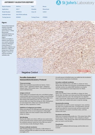
  • 1. Figure: Immunohistochemical analysis of paraffin embedded Human uterus tissue. 1: PCNA Monoclonal Antibody(12D10) was diluted at 1:200 (4 degree Celsius,overnight). 2: Sodium citrate pH 6.0 was used for antibody retrieval (>98 degree Celsius,20min). 3: Secondary antibody was diluted at 1:200 (room temperature, 30min). Negative control was used by secondary antibody only. Report Number 96933-a Host Mouse Application IHC-P Clonality Monoclonal Model Number STJ96933 Clone ID 12D10 Antibody Name Anti-PCNA antibody Testing Species HUMAN Testing Tissue UTERUS ANTIBODY VALIDATION REPORT a. (A small amount of distilled water was added into the incubation box to prevent evaporation of antibody). 128. Secondary antibody incubation a. Slides were washed 3 times, with PBS on a shaker for 5min. Shortly after the slides were dried the corresponding secondary antibody solution was added (HRP labelled), covering the tissues, and incubated at room temperature for 30min. b. 129. DAB staining a. Slides were washed 3 times, with PBS on a shaker for 5min. b. Shortly after, the slides were dried and fresh DAB staining buffer was added inside the circles. The staining time was adjusted under a microscope. Yellow-brown colour represented a positive result. Slides were washed with water to stop the staining. c. 130. Haematoxylin staining a. Haematoxylin was used to counter-staining for 1min, and then the slides were washed with water. 1% Hydrochloric acid and alcohol was added for several seconds and then washed with water. Ammonia was used to reveal blue colour, and then flushed with water. b. 131. Desolation and Clearing i. Slides were incubated sequentially into: 75% alcohol 5min, 85% alcohol 5min, Anhydrous ethanol - 5min, Anhydrous ethanol - 5min & Xylene - 5min. Shortly after slides were dried and neutral gum was used to seal the slides. ii. 132. Visualization a. Results were validated with microscope, and the slides were scanned. Paraffin-Embedded Immunohistochemistry Protocol 122. 123. Tissue processing a. Slides were incubated sequentially into Xylene; 15min – Xylene, 15min - Anhydrous ethanol, 5min - Anhydrous ethanol, 5min - 85% alcohol, 5min - 75% alcohol & 5min – wash in distilled water. b. 124. Antigen retrieval a. Tissue slides were incubated with citric acid (PH6.0) antigen retrieval buffer and microwaved for antigen retrieval (heated until boiled and then stopped heating) for 8min. Slides were then heated with medium power for 7min. During this process slides were kept from drying out. After cooling down at room temperature, slides were washed with PBS on shaker for 5min, repeated for 3 times. b. 125. Inhibition of endogenous peroxidase a. Slides were placed in 3% Hydrogen peroxide solution, and incubated for 10 min at room temperature without light exposure. Slides were then washed 3 times with PBS on a shaker for 5mins. b. 126. BSA Blocking a. Shortly after slides were dried, a PAP pen was used to draw circles around the tissue sections (and to prevent draining of the antibody solution). Inside the circles, BSA was used to cover the tissue evenly, blocking for 30min. b. 127. Primary antibody incubation After blocking solution was removed a 1:200 solution of primary antibody/PBS was added on the slide, and incubated overnight at 4°C. St John's Laboratory Ltd. www.stjohnslabs.com
  • 2. Figure: Immunohistochemical analysis of paraffin embedded Human uterus cancer tissue. 1: PCNA Monoclonal Antibody(12D10) was diluted at 1:200 (4 degree Celsius,overnight). 2: Sodium citrate pH 6.0 was used for antibody retrieval (>98 degree Celsius,20min). 3: Secondary antibody was diluted at 1:200 (room temperature, 30min). Negative control was used by secondary antibody only. Report Number 96933-b Host Mouse Application IHC-P Clonality Monoclonal Model Number STJ96933 Clone ID 12D10 Antibody Name Anti-PCNA antibody Testing Species HUMAN Testing Tissue UTERUS CANCER ANTIBODY VALIDATION REPORT a. (A small amount of distilled water was added into the incubation box to prevent evaporation of antibody). 117. Secondary antibody incubation a. Slides were washed 3 times, with PBS on a shaker for 5min. Shortly after the slides were dried the corresponding secondary antibody solution was added (HRP labelled), covering the tissues, and incubated at room temperature for 30min. b. 118. DAB staining a. Slides were washed 3 times, with PBS on a shaker for 5min. b. Shortly after, the slides were dried and fresh DAB staining buffer was added inside the circles. The staining time was adjusted under a microscope. Yellow-brown colour represented a positive result. Slides were washed with water to stop the staining. c. 119. Haematoxylin staining a. Haematoxylin was used to counter-staining for 1min, and then the slides were washed with water. 1% Hydrochloric acid and alcohol was added for several seconds and then washed with water. Ammonia was used to reveal blue colour, and then flushed with water. b. 120. Desolation and Clearing i. Slides were incubated sequentially into: 75% alcohol 5min, 85% alcohol 5min, Anhydrous ethanol - 5min, Anhydrous ethanol - 5min & Xylene - 5min. Shortly after slides were dried and neutral gum was used to seal the slides. ii. 121. Visualization a. Results were validated with microscope, and the slides were scanned. Paraffin-Embedded Immunohistochemistry Protocol 111. 112. Tissue processing a. Slides were incubated sequentially into Xylene; 15min – Xylene, 15min - Anhydrous ethanol, 5min - Anhydrous ethanol, 5min - 85% alcohol, 5min - 75% alcohol & 5min – wash in distilled water. b. 113. Antigen retrieval a. Tissue slides were incubated with citric acid (PH6.0) antigen retrieval buffer and microwaved for antigen retrieval (heated until boiled and then stopped heating) for 8min. Slides were then heated with medium power for 7min. During this process slides were kept from drying out. After cooling down at room temperature, slides were washed with PBS on shaker for 5min, repeated for 3 times. b. 114. Inhibition of endogenous peroxidase a. Slides were placed in 3% Hydrogen peroxide solution, and incubated for 10 min at room temperature without light exposure. Slides were then washed 3 times with PBS on a shaker for 5mins. b. 115. BSA Blocking a. Shortly after slides were dried, a PAP pen was used to draw circles around the tissue sections (and to prevent draining of the antibody solution). Inside the circles, BSA was used to cover the tissue evenly, blocking for 30min. b. 116. Primary antibody incubation After blocking solution was removed a 1:200 solution of primary antibody/PBS was added on the slide, and incubated overnight at 4°C. St John's Laboratory Ltd. www.stjohnslabs.com
  • 3. Figure: Immunohistochemical analysis of paraffin embedded Human Tonsil tissue. 1: PCNA Monoclonal Antibody(12D10) was diluted at 1:200 (4 degree Celsius,overnight). 2: Sodium citrate pH 6.0 was used for antibody retrieval (>98 degree Celsius,20min). 3: Secondary antibody was diluted at 1:200 (room temperature, 30min). Negative control was used by secondary antibody only. Report Number 96933-c Host Mouse Application IHC-P Clonality Monoclonal Model Number STJ96933 Clone ID 12D10 Antibody Name Anti-PCNA antibody Testing Species HUMAN Testing Tissue TONSIL ANTIBODY VALIDATION REPORT a. (A small amount of distilled water was added into the incubation box to prevent evaporation of antibody). 106. Secondary antibody incubation a. Slides were washed 3 times, with PBS on a shaker for 5min. Shortly after the slides were dried the corresponding secondary antibody solution was added (HRP labelled), covering the tissues, and incubated at room temperature for 30min. b. 107. DAB staining a. Slides were washed 3 times, with PBS on a shaker for 5min. b. Shortly after, the slides were dried and fresh DAB staining buffer was added inside the circles. The staining time was adjusted under a microscope. Yellow-brown colour represented a positive result. Slides were washed with water to stop the staining. c. 108. Haematoxylin staining a. Haematoxylin was used to counter-staining for 1min, and then the slides were washed with water. 1% Hydrochloric acid and alcohol was added for several seconds and then washed with water. Ammonia was used to reveal blue colour, and then flushed with water. b. 109. Desolation and Clearing i. Slides were incubated sequentially into: 75% alcohol 5min, 85% alcohol 5min, Anhydrous ethanol - 5min, Anhydrous ethanol - 5min & Xylene - 5min. Shortly after slides were dried and neutral gum was used to seal the slides. ii. 110. Visualization a. Results were validated with microscope, and the slides were scanned. Paraffin-Embedded Immunohistochemistry Protocol 100. 101. Tissue processing a. Slides were incubated sequentially into Xylene; 15min – Xylene, 15min - Anhydrous ethanol, 5min - Anhydrous ethanol, 5min - 85% alcohol, 5min - 75% alcohol & 5min – wash in distilled water. b. 102. Antigen retrieval a. Tissue slides were incubated with citric acid (PH6.0) antigen retrieval buffer and microwaved for antigen retrieval (heated until boiled and then stopped heating) for 8min. Slides were then heated with medium power for 7min. During this process slides were kept from drying out. After cooling down at room temperature, slides were washed with PBS on shaker for 5min, repeated for 3 times. b. 103. Inhibition of endogenous peroxidase a. Slides were placed in 3% Hydrogen peroxide solution, and incubated for 10 min at room temperature without light exposure. Slides were then washed 3 times with PBS on a shaker for 5mins. b. 104. BSA Blocking a. Shortly after slides were dried, a PAP pen was used to draw circles around the tissue sections (and to prevent draining of the antibody solution). Inside the circles, BSA was used to cover the tissue evenly, blocking for 30min. b. 105. Primary antibody incubation After blocking solution was removed a 1:200 solution of primary antibody/PBS was added on the slide, and incubated overnight at 4°C. St John's Laboratory Ltd. www.stjohnslabs.com
  • 4. Figure: Immunohistochemical analysis of paraffin embedded Human colon tissue. 1: PCNA Monoclonal Antibody(12D10) was diluted at 1:200 (4 degree Celsius,overnight). 2: Sodium citrate pH 6.0 was used for antibody retrieval (>98 degree Celsius,20min). 3: Secondary antibody was diluted at 1:200 (room temperature, 30min). Negative control was used by secondary antibody only. Report Number 96933-d Host Mouse Application IHC-P Clonality Monoclonal Model Number STJ96933 Clone ID 12D10 Antibody Name Anti-PCNA antibody Testing Species HUMAN Testing Tissue COLON ANTIBODY VALIDATION REPORT a. (A small amount of distilled water was added into the incubation box to prevent evaporation of antibody). 95. Secondary antibody incubation a. Slides were washed 3 times, with PBS on a shaker for 5min. Shortly after the slides were dried the corresponding secondary antibody solution was added (HRP labelled), covering the tissues, and incubated at room temperature for 30min. b. 96. DAB staining a. Slides were washed 3 times, with PBS on a shaker for 5min. b. Shortly after, the slides were dried and fresh DAB staining buffer was added inside the circles. The staining time was adjusted under a microscope. Yellow-brown colour represented a positive result. Slides were washed with water to stop the staining. c. 97. Haematoxylin staining a. Haematoxylin was used to counter-staining for 1min, and then the slides were washed with water. 1% Hydrochloric acid and alcohol was added for several seconds and then washed with water. Ammonia was used to reveal blue colour, and then flushed with water. b. 98. Desolation and Clearing i. Slides were incubated sequentially into: 75% alcohol 5min, 85% alcohol 5min, Anhydrous ethanol - 5min, Anhydrous ethanol - 5min & Xylene - 5min. Shortly after slides were dried and neutral gum was used to seal the slides. ii. 99. Visualization a. Results were validated with microscope, and the slides were scanned. Paraffin-Embedded Immunohistochemistry Protocol 89. 90. Tissue processing a. Slides were incubated sequentially into Xylene; 15min – Xylene, 15min - Anhydrous ethanol, 5min - Anhydrous ethanol, 5min - 85% alcohol, 5min - 75% alcohol & 5min – wash in distilled water. b. 91. Antigen retrieval a. Tissue slides were incubated with citric acid (PH6.0) antigen retrieval buffer and microwaved for antigen retrieval (heated until boiled and then stopped heating) for 8min. Slides were then heated with medium power for 7min. During this process slides were kept from drying out. After cooling down at room temperature, slides were washed with PBS on shaker for 5min, repeated for 3 times. b. 92. Inhibition of endogenous peroxidase a. Slides were placed in 3% Hydrogen peroxide solution, and incubated for 10 min at room temperature without light exposure. Slides were then washed 3 times with PBS on a shaker for 5mins. b. 93. BSA Blocking a. Shortly after slides were dried, a PAP pen was used to draw circles around the tissue sections (and to prevent draining of the antibody solution). Inside the circles, BSA was used to cover the tissue evenly, blocking for 30min. b. 94. Primary antibody incubation After blocking solution was removed a 1:200 solution of primary antibody/PBS was added on the slide, and incubated overnight at 4°C. St John's Laboratory Ltd. www.stjohnslabs.com
  • 5. Figure: Immunohistochemical analysis of paraffin embedded Human liver tissue. 1: PCNA Monoclonal Antibody(12D10) was diluted at 1:200 (4 degree Celsius,overnight). 2: Sodium citrate pH 6.0 was used for antibody retrieval (>98 degree Celsius,20min). 3: Secondary antibody was diluted at 1:200 (room temperature, 30min). Negative control was used by secondary antibody only. Report Number 96933-e Host Mouse Application IHC-P Clonality Monoclonal Model Number STJ96933 Clone ID 12D10 Antibody Name Anti-PCNA antibody Testing Species HUMAN Testing Tissue LIVER ANTIBODY VALIDATION REPORT a. (A small amount of distilled water was added into the incubation box to prevent evaporation of antibody). 84. Secondary antibody incubation a. Slides were washed 3 times, with PBS on a shaker for 5min. Shortly after the slides were dried the corresponding secondary antibody solution was added (HRP labelled), covering the tissues, and incubated at room temperature for 30min. b. 85. DAB staining a. Slides were washed 3 times, with PBS on a shaker for 5min. b. Shortly after, the slides were dried and fresh DAB staining buffer was added inside the circles. The staining time was adjusted under a microscope. Yellow-brown colour represented a positive result. Slides were washed with water to stop the staining. c. 86. Haematoxylin staining a. Haematoxylin was used to counter-staining for 1min, and then the slides were washed with water. 1% Hydrochloric acid and alcohol was added for several seconds and then washed with water. Ammonia was used to reveal blue colour, and then flushed with water. b. 87. Desolation and Clearing i. Slides were incubated sequentially into: 75% alcohol 5min, 85% alcohol 5min, Anhydrous ethanol - 5min, Anhydrous ethanol - 5min & Xylene - 5min. Shortly after slides were dried and neutral gum was used to seal the slides. ii. 88. Visualization a. Results were validated with microscope, and the slides were scanned. Paraffin-Embedded Immunohistochemistry Protocol 78. 79. Tissue processing a. Slides were incubated sequentially into Xylene; 15min – Xylene, 15min - Anhydrous ethanol, 5min - Anhydrous ethanol, 5min - 85% alcohol, 5min - 75% alcohol & 5min – wash in distilled water. b. 80. Antigen retrieval a. Tissue slides were incubated with citric acid (PH6.0) antigen retrieval buffer and microwaved for antigen retrieval (heated until boiled and then stopped heating) for 8min. Slides were then heated with medium power for 7min. During this process slides were kept from drying out. After cooling down at room temperature, slides were washed with PBS on shaker for 5min, repeated for 3 times. b. 81. Inhibition of endogenous peroxidase a. Slides were placed in 3% Hydrogen peroxide solution, and incubated for 10 min at room temperature without light exposure. Slides were then washed 3 times with PBS on a shaker for 5mins. b. 82. BSA Blocking a. Shortly after slides were dried, a PAP pen was used to draw circles around the tissue sections (and to prevent draining of the antibody solution). Inside the circles, BSA was used to cover the tissue evenly, blocking for 30min. b. 83. Primary antibody incubation After blocking solution was removed a 1:200 solution of primary antibody/PBS was added on the slide, and incubated overnight at 4°C. St John's Laboratory Ltd. www.stjohnslabs.com
  • 6. Figure: Immunohistochemical analysis of paraffin embedded Human liver cancer tissue. 1: PCNA Monoclonal Antibody(12D10) was diluted at 1:200 (4 degree Celsius,overnight). 2: Sodium citrate pH 6.0 was used for antibody retrieval (>98 degree Celsius,20min). 3: Secondary antibody was diluted at 1:200 (room temperature, 30min). Negative control was used by secondary antibody only. Report Number 96933-f Host Mouse Application IHC-P Clonality Monoclonal Model Number STJ96933 Clone ID 12D10 Antibody Name Anti-PCNA antibody Testing Species HUMAN Testing Tissue LIVER CANCER ANTIBODY VALIDATION REPORT a. (A small amount of distilled water was added into the incubation box to prevent evaporation of antibody). 73. Secondary antibody incubation a. Slides were washed 3 times, with PBS on a shaker for 5min. Shortly after the slides were dried the corresponding secondary antibody solution was added (HRP labelled), covering the tissues, and incubated at room temperature for 30min. b. 74. DAB staining a. Slides were washed 3 times, with PBS on a shaker for 5min. b. Shortly after, the slides were dried and fresh DAB staining buffer was added inside the circles. The staining time was adjusted under a microscope. Yellow-brown colour represented a positive result. Slides were washed with water to stop the staining. c. 75. Haematoxylin staining a. Haematoxylin was used to counter-staining for 1min, and then the slides were washed with water. 1% Hydrochloric acid and alcohol was added for several seconds and then washed with water. Ammonia was used to reveal blue colour, and then flushed with water. b. 76. Desolation and Clearing i. Slides were incubated sequentially into: 75% alcohol 5min, 85% alcohol 5min, Anhydrous ethanol - 5min, Anhydrous ethanol - 5min & Xylene - 5min. Shortly after slides were dried and neutral gum was used to seal the slides. ii. 77. Visualization a. Results were validated with microscope, and the slides were scanned. Paraffin-Embedded Immunohistochemistry Protocol 67. 68. Tissue processing a. Slides were incubated sequentially into Xylene; 15min – Xylene, 15min - Anhydrous ethanol, 5min - Anhydrous ethanol, 5min - 85% alcohol, 5min - 75% alcohol & 5min – wash in distilled water. b. 69. Antigen retrieval a. Tissue slides were incubated with citric acid (PH6.0) antigen retrieval buffer and microwaved for antigen retrieval (heated until boiled and then stopped heating) for 8min. Slides were then heated with medium power for 7min. During this process slides were kept from drying out. After cooling down at room temperature, slides were washed with PBS on shaker for 5min, repeated for 3 times. b. 70. Inhibition of endogenous peroxidase a. Slides were placed in 3% Hydrogen peroxide solution, and incubated for 10 min at room temperature without light exposure. Slides were then washed 3 times with PBS on a shaker for 5mins. b. 71. BSA Blocking a. Shortly after slides were dried, a PAP pen was used to draw circles around the tissue sections (and to prevent draining of the antibody solution). Inside the circles, BSA was used to cover the tissue evenly, blocking for 30min. b. 72. Primary antibody incubation After blocking solution was removed a 1:200 solution of primary antibody/PBS was added on the slide, and incubated overnight at 4°C. St John's Laboratory Ltd. www.stjohnslabs.com
  • 7. Figure: Immunohistochemical analysis of paraffin embedded Human lung tissue. 1: PCNA Monoclonal Antibody(12D10) was diluted at 1:200 (4 degree Celsius,overnight). 2: Sodium citrate pH 6.0 was used for antibody retrieval (>98 degree Celsius,20min). 3: Secondary antibody was diluted at 1:200 (room temperature, 30min). Negative control was used by secondary antibody only. Report Number 96933-g Host Mouse Application IHC-P Clonality Monoclonal Model Number STJ96933 Clone ID 12D10 Antibody Name Anti-PCNA antibody Testing Species HUMAN Testing Tissue LUNG ANTIBODY VALIDATION REPORT b. (A small amount of distilled water was added into the incubation box to prevent evaporation of antibody). 62. Secondary antibody incubation a. Slides were washed 3 times, with PBS on a shaker for 5min. Shortly after the slides were dried the corresponding secondary antibody solution was added (HRP labelled), covering the tissues, and incubated at room temperature for 30min. b. 63. DAB staining a. Slides were washed 3 times, with PBS on a shaker for 5min. b. Shortly after, the slides were dried and fresh DAB staining buffer was added inside the circles. The staining time was adjusted under a microscope. Yellow-brown colour represented a positive result. Slides were washed with water to stop the staining. c. 64. Haematoxylin staining a. Haematoxylin was used to counter-staining for 1min, and then the slides were washed with water. 1% Hydrochloric acid and alcohol was added for several seconds and then washed with water. Ammonia was used to reveal blue colour, and then flushed with water. b. 65. Desolation and Clearing i. Slides were incubated sequentially into: 75% alcohol 5min, 85% alcohol 5min, Anhydrous ethanol - 5min, Anhydrous ethanol - 5min & Xylene - 5min. Shortly after slides were dried and neutral gum was used to seal the slides. ii. 66. Visualization a. Results were validated with microscope, and the slides were scanned. Paraffin-Embedded Immunohistochemistry Protocol 56. 57. Tissue processing a. Slides were incubated sequentially into Xylene; 15min – Xylene, 15min - Anhydrous ethanol, 5min - Anhydrous ethanol, 5min - 85% alcohol, 5min - 75% alcohol & 5min – wash in distilled water. b. 58. Antigen retrieval a. Tissue slides were incubated with citric acid (PH6.0) antigen retrieval buffer and microwaved for antigen retrieval (heated until boiled and then stopped heating) for 8min. Slides were then heated with medium power for 7min. During this process slides were kept from drying out. After cooling down at room temperature, slides were washed with PBS on shaker for 5min, repeated for 3 times. b. 59. Inhibition of endogenous peroxidase a. Slides were placed in 3% Hydrogen peroxide solution, and incubated for 10 min at room temperature without light exposure. Slides were then washed 3 times with PBS on a shaker for 5mins. b. 60. BSA Blocking a. Shortly after slides were dried, a PAP pen was used to draw circles around the tissue sections (and to prevent draining of the antibody solution). Inside the circles, BSA was used to cover the tissue evenly, blocking for 30min. b. 61. Primary antibody incubation After blocking solution was removed a 1:200 solution of primary antibody/PBS was added on the slide, and incubated overnight at 4°C. St John's Laboratory Ltd. www.stjohnslabs.com
  • 8. Figure: Immunohistochemical analysis of paraffin embedded Human lung cancer tissue. 1: PCNA Monoclonal Antibody(12D10) was diluted at 1:200 (4 degree Celsius,overnight). 2: Sodium citrate pH 6.0 was used for antibody retrieval (>98 degree Celsius,20min). 3: Secondary antibody was diluted at 1:200 (room temperature, 30min). Negative control was used by secondary antibody only. Report Number 96933-h Host Mouse Application IHC-P Clonality Monoclonal Model Number STJ96933 Clone ID 12D10 Antibody Name Anti-PCNA antibody Testing Species HUMAN Testing Tissue LUNG CANCER ANTIBODY VALIDATION REPORT b. (A small amount of distilled water was added into the incubation box to prevent evaporation of antibody). 51. Secondary antibody incubation a. Slides were washed 3 times, with PBS on a shaker for 5min. Shortly after the slides were dried the corresponding secondary antibody solution was added (HRP labelled), covering the tissues, and incubated at room temperature for 30min. b. 52. DAB staining a. Slides were washed 3 times, with PBS on a shaker for 5min. b. Shortly after, the slides were dried and fresh DAB staining buffer was added inside the circles. The staining time was adjusted under a microscope. Yellow-brown colour represented a positive result. Slides were washed with water to stop the staining. c. 53. Haematoxylin staining a. Haematoxylin was used to counter-staining for 1min, and then the slides were washed with water. 1% Hydrochloric acid and alcohol was added for several seconds and then washed with water. Ammonia was used to reveal blue colour, and then flushed with water. b. 54. Desolation and Clearing i. Slides were incubated sequentially into: 75% alcohol 5min, 85% alcohol 5min, Anhydrous ethanol - 5min, Anhydrous ethanol - 5min & Xylene - 5min. Shortly after slides were dried and neutral gum was used to seal the slides. ii. 55. Visualization a. Results were validated with microscope, and the slides were scanned. Paraffin-Embedded Immunohistochemistry Protocol 45. 46. Tissue processing a. Slides were incubated sequentially into Xylene; 15min – Xylene, 15min - Anhydrous ethanol, 5min - Anhydrous ethanol, 5min - 85% alcohol, 5min - 75% alcohol & 5min – wash in distilled water. b. 47. Antigen retrieval a. Tissue slides were incubated with citric acid (PH6.0) antigen retrieval buffer and microwaved for antigen retrieval (heated until boiled and then stopped heating) for 8min. Slides were then heated with medium power for 7min. During this process slides were kept from drying out. After cooling down at room temperature, slides were washed with PBS on shaker for 5min, repeated for 3 times. b. 48. Inhibition of endogenous peroxidase a. Slides were placed in 3% Hydrogen peroxide solution, and incubated for 10 min at room temperature without light exposure. Slides were then washed 3 times with PBS on a shaker for 5mins. b. 49. BSA Blocking a. Shortly after slides were dried, a PAP pen was used to draw circles around the tissue sections (and to prevent draining of the antibody solution). Inside the circles, BSA was used to cover the tissue evenly, blocking for 30min. b. 50. Primary antibody incubation After blocking solution was removed a 1:200 solution of primary antibody/PBS was added on the slide, and incubated overnight at 4°C. St John's Laboratory Ltd. www.stjohnslabs.com
  • 9. Figure: Immunohistochemical analysis of paraffin embedded Human stomach cancer tissue. 1: PCNA Monoclonal Antibody(12D10) was diluted at 1:200 (4 degree Celsius,overnight). 2: Sodium citrate pH 6.0 was used for antibody retrieval (>98 degree Celsius,20min). 3: Secondary antibody was diluted at 1:200 (room temperature, 30min). Negative control was used by secondary antibody only. Report Number 96933-i Host Mouse Application IHC-P Clonality Monoclonal Model Number STJ96933 Clone ID 12D10 Antibody Name Anti-PCNA antibody Testing Species HUMAN Testing Tissue STOMACH CANCER ANTIBODY VALIDATION REPORT b. (A small amount of distilled water was added into the incubation box to prevent evaporation of antibody). 40. Secondary antibody incubation a. Slides were washed 3 times, with PBS on a shaker for 5min. Shortly after the slides were dried the corresponding secondary antibody solution was added (HRP labelled), covering the tissues, and incubated at room temperature for 30min. b. 41. DAB staining a. Slides were washed 3 times, with PBS on a shaker for 5min. b. Shortly after, the slides were dried and fresh DAB staining buffer was added inside the circles. The staining time was adjusted under a microscope. Yellow-brown colour represented a positive result. Slides were washed with water to stop the staining. c. 42. Haematoxylin staining a. Haematoxylin was used to counter-staining for 1min, and then the slides were washed with water. 1% Hydrochloric acid and alcohol was added for several seconds and then washed with water. Ammonia was used to reveal blue colour, and then flushed with water. b. 43. Desolation and Clearing i. Slides were incubated sequentially into: 75% alcohol 5min, 85% alcohol 5min, Anhydrous ethanol - 5min, Anhydrous ethanol - 5min & Xylene - 5min. Shortly after slides were dried and neutral gum was used to seal the slides. ii. 44. Visualization a. Results were validated with microscope, and the slides were scanned. Paraffin-Embedded Immunohistochemistry Protocol 34. 35. Tissue processing a. Slides were incubated sequentially into Xylene; 15min – Xylene, 15min - Anhydrous ethanol, 5min - Anhydrous ethanol, 5min - 85% alcohol, 5min - 75% alcohol & 5min – wash in distilled water. b. 36. Antigen retrieval a. Tissue slides were incubated with citric acid (PH6.0) antigen retrieval buffer and microwaved for antigen retrieval (heated until boiled and then stopped heating) for 8min. Slides were then heated with medium power for 7min. During this process slides were kept from drying out. After cooling down at room temperature, slides were washed with PBS on shaker for 5min, repeated for 3 times. b. 37. Inhibition of endogenous peroxidase a. Slides were placed in 3% Hydrogen peroxide solution, and incubated for 10 min at room temperature without light exposure. Slides were then washed 3 times with PBS on a shaker for 5mins. b. 38. BSA Blocking a. Shortly after slides were dried, a PAP pen was used to draw circles around the tissue sections (and to prevent draining of the antibody solution). Inside the circles, BSA was used to cover the tissue evenly, blocking for 30min. b. 39. Primary antibody incubation After blocking solution was removed a 1:200 solution of primary antibody/PBS was added on the slide, and incubated overnight at 4°C. St John's Laboratory Ltd. www.stjohnslabs.com
  • 10. Figure: Immunohistochemical analysis of paraffin embedded Human appendix tissue. 1: PCNA Monoclonal Antibody(12D10) was diluted at 1:200 (4 degree Celsius,overnight). 2: Sodium citrate pH 6.0 was used for antibody retrieval (>98 degree Celsius,20min). 3: Secondary antibody was diluted at 1:200 (room temperature, 30min). Negative control was used by secondary antibody only. Report Number 96933-j Host Mouse Application IHC-P Clonality Monoclonal Model Number STJ96933 Clone ID 12D10 Antibody Name Anti-PCNA antibody Testing Species HUMAN Testing Tissue APPENDIX ANTIBODY VALIDATION REPORT b. (A small amount of distilled water was added into the incubation box to prevent evaporation of antibody). 29. Secondary antibody incubation a. Slides were washed 3 times, with PBS on a shaker for 5min. Shortly after the slides were dried the corresponding secondary antibody solution was added (HRP labelled), covering the tissues, and incubated at room temperature for 30min. b. 30. DAB staining a. Slides were washed 3 times, with PBS on a shaker for 5min. b. Shortly after, the slides were dried and fresh DAB staining buffer was added inside the circles. The staining time was adjusted under a microscope. Yellow-brown colour represented a positive result. Slides were washed with water to stop the staining. c. 31. Haematoxylin staining a. Haematoxylin was used to counter-staining for 1min, and then the slides were washed with water. 1% Hydrochloric acid and alcohol was added for several seconds and then washed with water. Ammonia was used to reveal blue colour, and then flushed with water. b. 32. Desolation and Clearing i. Slides were incubated sequentially into: 75% alcohol 5min, 85% alcohol 5min, Anhydrous ethanol - 5min, Anhydrous ethanol - 5min & Xylene - 5min. Shortly after slides were dried and neutral gum was used to seal the slides. ii. 33. Visualization a. Results were validated with microscope, and the slides were scanned. Paraffin-Embedded Immunohistochemistry Protocol 23. 24. Tissue processing a. Slides were incubated sequentially into Xylene; 15min – Xylene, 15min - Anhydrous ethanol, 5min - Anhydrous ethanol, 5min - 85% alcohol, 5min - 75% alcohol & 5min – wash in distilled water. b. 25. Antigen retrieval a. Tissue slides were incubated with citric acid (PH6.0) antigen retrieval buffer and microwaved for antigen retrieval (heated until boiled and then stopped heating) for 8min. Slides were then heated with medium power for 7min. During this process slides were kept from drying out. After cooling down at room temperature, slides were washed with PBS on shaker for 5min, repeated for 3 times. b. 26. Inhibition of endogenous peroxidase a. Slides were placed in 3% Hydrogen peroxide solution, and incubated for 10 min at room temperature without light exposure. Slides were then washed 3 times with PBS on a shaker for 5mins. b. 27. BSA Blocking a. Shortly after slides were dried, a PAP pen was used to draw circles around the tissue sections (and to prevent draining of the antibody solution). Inside the circles, BSA was used to cover the tissue evenly, blocking for 30min. b. 28. Primary antibody incubation After blocking solution was removed a 1:200 solution of primary antibody/PBS was added on the slide, and incubated overnight at 4°C. St John's Laboratory Ltd. www.stjohnslabs.com
  • 11. Figure: Immunohistochemical analysis of paraffin embedded Rat testis tissue. 1: PCNA Monoclonal Antibody(12D10) was diluted at 1:200 (4 degree Celsius,overnight). 2: Sodium citrate pH 6.0 was used for antibody retrieval (>98 degree Celsius,20min). 3: Secondary antibody was diluted at 1:200 (room temperature, 30min). Negative control was used by secondary antibody only. Report Number 96933-k Host Mouse Application IHC-P Clonality Monoclonal Model Number STJ96933 Clone ID 12D10 Antibody Name Anti-PCNA antibody Testing Species RAT Testing Tissue TESTIS ANTIBODY VALIDATION REPORT b. (A small amount of distilled water was added into the incubation box to prevent evaporation of antibody). 18. Secondary antibody incubation a. Slides were washed 3 times, with PBS on a shaker for 5min. Shortly after the slides were dried the corresponding secondary antibody solution was added (HRP labelled), covering the tissues, and incubated at room temperature for 30min. b. 19. DAB staining a. Slides were washed 3 times, with PBS on a shaker for 5min. b. Shortly after, the slides were dried and fresh DAB staining buffer was added inside the circles. The staining time was adjusted under a microscope. Yellow-brown colour represented a positive result. Slides were washed with water to stop the staining. c. 20. Haematoxylin staining a. Haematoxylin was used to counter-staining for 1min, and then the slides were washed with water. 1% Hydrochloric acid and alcohol was added for several seconds and then washed with water. Ammonia was used to reveal blue colour, and then flushed with water. b. 21. Desolation and Clearing i. Slides were incubated sequentially into: 75% alcohol 5min, 85% alcohol 5min, Anhydrous ethanol - 5min, Anhydrous ethanol - 5min & Xylene - 5min. Shortly after slides were dried and neutral gum was used to seal the slides. ii. 22. Visualization a. Results were validated with microscope, and the slides were scanned. Paraffin-Embedded Immunohistochemistry Protocol 12. 13. Tissue processing a. Slides were incubated sequentially into Xylene; 15min – Xylene, 15min - Anhydrous ethanol, 5min - Anhydrous ethanol, 5min - 85% alcohol, 5min - 75% alcohol & 5min – wash in distilled water. b. 14. Antigen retrieval a. Tissue slides were incubated with citric acid (PH6.0) antigen retrieval buffer and microwaved for antigen retrieval (heated until boiled and then stopped heating) for 8min. Slides were then heated with medium power for 7min. During this process slides were kept from drying out. After cooling down at room temperature, slides were washed with PBS on shaker for 5min, repeated for 3 times. b. 15. Inhibition of endogenous peroxidase a. Slides were placed in 3% Hydrogen peroxide solution, and incubated for 10 min at room temperature without light exposure. Slides were then washed 3 times with PBS on a shaker for 5mins. b. 16. BSA Blocking a. Shortly after slides were dried, a PAP pen was used to draw circles around the tissue sections (and to prevent draining of the antibody solution). Inside the circles, BSA was used to cover the tissue evenly, blocking for 30min. b. 17. Primary antibody incubation After blocking solution was removed a 1:200 solution of primary antibody/PBS was added on the slide, and incubated overnight at 4°C. St John's Laboratory Ltd. www.stjohnslabs.com
  • 12. Figure: Immunohistochemical analysis of paraffin embedded Rat liver tissue. 1: PCNA Monoclonal Antibody(12D10) was diluted at 1:200 (4 degree Celsius,overnight). 2: Sodium citrate pH 6.0 was used for antibody retrieval (>98 degree Celsius,20min). 3: Secondary antibody was diluted at 1:200 (room temperature, 30min). Negative control was used by secondary antibody only. Report Number 96933-l Host Mouse Application IHC-P Clonality Monoclonal Model Number STJ96933 Clone ID 12D10 Antibody Name Anti-PCNA antibody Testing Species RAT Testing Tissue LIVER ANTIBODY VALIDATION REPORT b. (A small amount of distilled water was added into the incubation box to prevent evaporation of antibody). 7. Secondary antibody incubation a. Slides were washed 3 times, with PBS on a shaker for 5min. Shortly after the slides were dried the corresponding secondary antibody solution was added (HRP labelled), covering the tissues, and incubated at room temperature for 30min. b. 8. DAB staining a. Slides were washed 3 times, with PBS on a shaker for 5min. b. Shortly after, the slides were dried and fresh DAB staining buffer was added inside the circles. The staining time was adjusted under a microscope. Yellow-brown colour represented a positive result. Slides were washed with water to stop the staining. c. 9. Haematoxylin staining a. Haematoxylin was used to counter-staining for 1min, and then the slides were washed with water. 1% Hydrochloric acid and alcohol was added for several seconds and then washed with water. Ammonia was used to reveal blue colour, and then flushed with water. b. 10. Desolation and Clearing i. Slides were incubated sequentially into: 75% alcohol 5min, 85% alcohol 5min, Anhydrous ethanol - 5min, Anhydrous ethanol - 5min & Xylene - 5min. Shortly after slides were dried and neutral gum was used to seal the slides. ii. 11. Visualization a. Results were validated with microscope, and the slides were scanned. Paraffin-Embedded Immunohistochemistry Protocol 1. 2. Tissue processing a. Slides were incubated sequentially into Xylene; 15min – Xylene, 15min - Anhydrous ethanol, 5min - Anhydrous ethanol, 5min - 85% alcohol, 5min - 75% alcohol & 5min – wash in distilled water. b. 3. Antigen retrieval a. Tissue slides were incubated with citric acid (PH6.0) antigen retrieval buffer and microwaved for antigen retrieval (heated until boiled and then stopped heating) for 8min. Slides were then heated with medium power for 7min. During this process slides were kept from drying out. After cooling down at room temperature, slides were washed with PBS on shaker for 5min, repeated for 3 times. b. 4. Inhibition of endogenous peroxidase a. Slides were placed in 3% Hydrogen peroxide solution, and incubated for 10 min at room temperature without light exposure. Slides were then washed 3 times with PBS on a shaker for 5mins. b. 5. BSA Blocking a. Shortly after slides were dried, a PAP pen was used to draw circles around the tissue sections (and to prevent draining of the antibody solution). Inside the circles, BSA was used to cover the tissue evenly, blocking for 30min. b. 6. Primary antibody incubation After blocking solution was removed a 1:200 solution of primary antibody/PBS was added on the slide, and incubated overnight at 4°C. St John's Laboratory Ltd. www.stjohnslabs.com
  • 13. Figure: Immunohistochemical analysis of paraffin embedded Rat brain tissue. 1: PCNA Monoclonal Antibody(12D10) was diluted at 1:200 (4 degree Celsius,overnight). 2: Sodium citrate pH 6.0 was used for antibody retrieval (>98 degree Celsius,20min). 3: Secondary antibody was diluted at 1:200 (room temperature, 30min). Negative control was used by secondary antibody only. Report Number 96933-m Host Mouse Application IHC-P Clonality Monoclonal Model Number STJ96933 Clone ID 12D10 Antibody Name Anti-PCNA antibody Testing Species RAT Testing Tissue BRAIN ANTIBODY VALIDATION REPORT c. (A small amount of distilled water was added into the incubation box to prevent evaporation of antibody). 133. Secondary antibody incubation a. Slides were washed 3 times, with PBS on a shaker for 5min. Shortly after the slides were dried the corresponding secondary antibody solution was added (HRP labelled), covering the tissues, and incubated at room temperature for 30min. b. 134. DAB staining a. Slides were washed 3 times, with PBS on a shaker for 5min. b. Shortly after, the slides were dried and fresh DAB staining buffer was added inside the circles. The staining time was adjusted under a microscope. Yellow-brown colour represented a positive result. Slides were washed with water to stop the staining. c. 135. Haematoxylin staining a. Haematoxylin was used to counter-staining for 1min, and then the slides were washed with water. 1% Hydrochloric acid and alcohol was added for several seconds and then washed with water. Ammonia was used to reveal blue colour, and then flushed with water. b. 136. Desolation and Clearing i. Slides were incubated sequentially into: 75% alcohol 5min, 85% alcohol 5min, Anhydrous ethanol - 5min, Anhydrous ethanol - 5min & Xylene - 5min. Shortly after slides were dried and neutral gum was used to seal the slides. ii. 137. Visualization a. Results were validated with microscope, and the slides were scanned. Paraffin-Embedded Immunohistochemistry Protocol 138. 139. Tissue processing a. Slides were incubated sequentially into Xylene; 15min – Xylene, 15min - Anhydrous ethanol, 5min - Anhydrous ethanol, 5min - 85% alcohol, 5min - 75% alcohol & 5min – wash in distilled water. b. 140. Antigen retrieval a. Tissue slides were incubated with citric acid (PH6.0) antigen retrieval buffer and microwaved for antigen retrieval (heated until boiled and then stopped heating) for 8min. Slides were then heated with medium power for 7min. During this process slides were kept from drying out. After cooling down at room temperature, slides were washed with PBS on shaker for 5min, repeated for 3 times. b. 141. Inhibition of endogenous peroxidase a. Slides were placed in 3% Hydrogen peroxide solution, and incubated for 10 min at room temperature without light exposure. Slides were then washed 3 times with PBS on a shaker for 5mins. b. 142. BSA Blocking a. Shortly after slides were dried, a PAP pen was used to draw circles around the tissue sections (and to prevent draining of the antibody solution). Inside the circles, BSA was used to cover the tissue evenly, blocking for 30min. b. 143. Primary antibody incubation After blocking solution was removed a 1:200 solution of primary antibody/PBS was added on the slide, and incubated overnight at 4°C. St John's Laboratory Ltd. www.stjohnslabs.com
  • 14. Figure: Immunohistochemical analysis of paraffin embedded Rat spleen tissue. 1: PCNA Monoclonal Antibody(12D10) was diluted at 1:200 (4 degree Celsius,overnight). 2: Sodium citrate pH 6.0 was used for antibody retrieval (>98 degree Celsius,20min). 3: Secondary antibody was diluted at 1:200 (room temperature, 30min). Negative control was used by secondary antibody only. Report Number 96933-n Host Mouse Application IHC-P Clonality Monoclonal Model Number STJ96933 Clone ID 12D10 Antibody Name Anti-PCNA antibody Testing Species RAT Testing Tissue SPLEEN ANTIBODY VALIDATION REPORT b. (A small amount of distilled water was added into the incubation box to prevent evaporation of antibody). 144. Secondary antibody incubation a. Slides were washed 3 times, with PBS on a shaker for 5min. Shortly after the slides were dried the corresponding secondary antibody solution was added (HRP labelled), covering the tissues, and incubated at room temperature for 30min. b. 145. DAB staining a. Slides were washed 3 times, with PBS on a shaker for 5min. b. Shortly after, the slides were dried and fresh DAB staining buffer was added inside the circles. The staining time was adjusted under a microscope. Yellow-brown colour represented a positive result. Slides were washed with water to stop the staining. c. 146. Haematoxylin staining a. Haematoxylin was used to counter-staining for 1min, and then the slides were washed with water. 1% Hydrochloric acid and alcohol was added for several seconds and then washed with water. Ammonia was used to reveal blue colour, and then flushed with water. b. 147. Desolation and Clearing i. Slides were incubated sequentially into: 75% alcohol 5min, 85% alcohol 5min, Anhydrous ethanol - 5min, Anhydrous ethanol - 5min & Xylene - 5min. Shortly after slides were dried and neutral gum was used to seal the slides. ii. 148. Visualization a. Results were validated with microscope, and the slides were scanned. Paraffin-Embedded Immunohistochemistry Protocol 149. 150. Tissue processing a. Slides were incubated sequentially into Xylene; 15min – Xylene, 15min - Anhydrous ethanol, 5min - Anhydrous ethanol, 5min - 85% alcohol, 5min - 75% alcohol & 5min – wash in distilled water. b. 151. Antigen retrieval a. Tissue slides were incubated with citric acid (PH6.0) antigen retrieval buffer and microwaved for antigen retrieval (heated until boiled and then stopped heating) for 8min. Slides were then heated with medium power for 7min. During this process slides were kept from drying out. After cooling down at room temperature, slides were washed with PBS on shaker for 5min, repeated for 3 times. b. 152. Inhibition of endogenous peroxidase a. Slides were placed in 3% Hydrogen peroxide solution, and incubated for 10 min at room temperature without light exposure. Slides were then washed 3 times with PBS on a shaker for 5mins. b. 153. BSA Blocking a. Shortly after slides were dried, a PAP pen was used to draw circles around the tissue sections (and to prevent draining of the antibody solution). Inside the circles, BSA was used to cover the tissue evenly, blocking for 30min. b. 154. Primary antibody incubation After blocking solution was removed a 1:200 solution of primary antibody/PBS was added on the slide, and incubated overnight at 4°C. St John's Laboratory Ltd. www.stjohnslabs.com
  • 15. Figure: Immunohistochemical analysis of paraffin embedded Mouse heart tissue. 1: PCNA Monoclonal Antibody(12D10) was diluted at 1:200 (4 degree Celsius,overnight). 2: Sodium citrate pH 6.0 was used for antibody retrieval (>98 degree Celsius,20min). 3: Secondary antibody was diluted at 1:200 (room temperature, 30min). Negative control was used by secondary antibody only. Report Number 96933-o Host Mouse Application IHC-P Clonality Monoclonal Model Number STJ96933 Clone ID 12D10 Antibody Name Anti-PCNA antibody Testing Species MOUSE Testing Tissue HEART ANTIBODY VALIDATION REPORT b. (A small amount of distilled water was added into the incubation box to prevent evaporation of antibody). 155. Secondary antibody incubation a. Slides were washed 3 times, with PBS on a shaker for 5min. Shortly after the slides were dried the corresponding secondary antibody solution was added (HRP labelled), covering the tissues, and incubated at room temperature for 30min. b. 156. DAB staining a. Slides were washed 3 times, with PBS on a shaker for 5min. b. Shortly after, the slides were dried and fresh DAB staining buffer was added inside the circles. The staining time was adjusted under a microscope. Yellow-brown colour represented a positive result. Slides were washed with water to stop the staining. c. 157. Haematoxylin staining a. Haematoxylin was used to counter-staining for 1min, and then the slides were washed with water. 1% Hydrochloric acid and alcohol was added for several seconds and then washed with water. Ammonia was used to reveal blue colour, and then flushed with water. b. 158. Desolation and Clearing i. Slides were incubated sequentially into: 75% alcohol 5min, 85% alcohol 5min, Anhydrous ethanol - 5min, Anhydrous ethanol - 5min & Xylene - 5min. Shortly after slides were dried and neutral gum was used to seal the slides. ii. 159. Visualization a. Results were validated with microscope, and the slides were scanned. Paraffin-Embedded Immunohistochemistry Protocol 160. 161. Tissue processing a. Slides were incubated sequentially into Xylene; 15min – Xylene, 15min - Anhydrous ethanol, 5min - Anhydrous ethanol, 5min - 85% alcohol, 5min - 75% alcohol & 5min – wash in distilled water. b. 162. Antigen retrieval a. Tissue slides were incubated with citric acid (PH6.0) antigen retrieval buffer and microwaved for antigen retrieval (heated until boiled and then stopped heating) for 8min. Slides were then heated with medium power for 7min. During this process slides were kept from drying out. After cooling down at room temperature, slides were washed with PBS on shaker for 5min, repeated for 3 times. b. 163. Inhibition of endogenous peroxidase a. Slides were placed in 3% Hydrogen peroxide solution, and incubated for 10 min at room temperature without light exposure. Slides were then washed 3 times with PBS on a shaker for 5mins. b. 164. BSA Blocking a. Shortly after slides were dried, a PAP pen was used to draw circles around the tissue sections (and to prevent draining of the antibody solution). Inside the circles, BSA was used to cover the tissue evenly, blocking for 30min. b. 165. Primary antibody incubation After blocking solution was removed a 1:200 solution of primary antibody/PBS was added on the slide, and incubated overnight at 4°C. St John's Laboratory Ltd. www.stjohnslabs.com
  • 16. Figure: Immunohistochemical analysis of paraffin embedded Mouse testis tissue. 1: PCNA Monoclonal Antibody(12D10) was diluted at 1:200 (4 degree Celsius,overnight). 2: Sodium citrate pH 6.0 was used for antibody retrieval (>98 degree Celsius,20min). 3: Secondary antibody was diluted at 1:200 (room temperature, 30min). Negative control was used by secondary antibody only. Report Number 96933-p Host Mouse Application IHC-P Clonality Monoclonal Model Number STJ96933 Clone ID 12D10 Antibody Name Anti-PCNA antibody Testing Species MOUSE Testing Tissue TESTIS ANTIBODY VALIDATION REPORT b. (A small amount of distilled water was added into the incubation box to prevent evaporation of antibody). 166. Secondary antibody incubation a. Slides were washed 3 times, with PBS on a shaker for 5min. Shortly after the slides were dried the corresponding secondary antibody solution was added (HRP labelled), covering the tissues, and incubated at room temperature for 30min. b. 167. DAB staining a. Slides were washed 3 times, with PBS on a shaker for 5min. b. Shortly after, the slides were dried and fresh DAB staining buffer was added inside the circles. The staining time was adjusted under a microscope. Yellow-brown colour represented a positive result. Slides were washed with water to stop the staining. c. 168. Haematoxylin staining a. Haematoxylin was used to counter-staining for 1min, and then the slides were washed with water. 1% Hydrochloric acid and alcohol was added for several seconds and then washed with water. Ammonia was used to reveal blue colour, and then flushed with water. b. 169. Desolation and Clearing i. Slides were incubated sequentially into: 75% alcohol 5min, 85% alcohol 5min, Anhydrous ethanol - 5min, Anhydrous ethanol - 5min & Xylene - 5min. Shortly after slides were dried and neutral gum was used to seal the slides. ii. 170. Visualization a. Results were validated with microscope, and the slides were scanned. Paraffin-Embedded Immunohistochemistry Protocol 171. 172. Tissue processing a. Slides were incubated sequentially into Xylene; 15min – Xylene, 15min - Anhydrous ethanol, 5min - Anhydrous ethanol, 5min - 85% alcohol, 5min - 75% alcohol & 5min – wash in distilled water. b. 173. Antigen retrieval a. Tissue slides were incubated with citric acid (PH6.0) antigen retrieval buffer and microwaved for antigen retrieval (heated until boiled and then stopped heating) for 8min. Slides were then heated with medium power for 7min. During this process slides were kept from drying out. After cooling down at room temperature, slides were washed with PBS on shaker for 5min, repeated for 3 times. b. 174. Inhibition of endogenous peroxidase a. Slides were placed in 3% Hydrogen peroxide solution, and incubated for 10 min at room temperature without light exposure. Slides were then washed 3 times with PBS on a shaker for 5mins. b. 175. BSA Blocking a. Shortly after slides were dried, a PAP pen was used to draw circles around the tissue sections (and to prevent draining of the antibody solution). Inside the circles, BSA was used to cover the tissue evenly, blocking for 30min. b. 176. Primary antibody incubation After blocking solution was removed a 1:200 solution of primary antibody/PBS was added on the slide, and incubated overnight at 4°C. St John's Laboratory Ltd. www.stjohnslabs.com
  • 17. Figure: Immunohistochemical analysis of paraffin embedded Mouse liver tissue. 1: PCNA Monoclonal Antibody(12D10) was diluted at 1:200 (4 degree Celsius,overnight). 2: Sodium citrate pH 6.0 was used for antibody retrieval (>98 degree Celsius,20min). 3: Secondary antibody was diluted at 1:200 (room temperature, 30min). Negative control was used by secondary antibody only. Report Number 96933-q Host Mouse Application IHC-P Clonality Monoclonal Model Number STJ96933 Clone ID 12D10 Antibody Name Anti-PCNA antibody Testing Species MOUSE Testing Tissue LIVER ANTIBODY VALIDATION REPORT b. (A small amount of distilled water was added into the incubation box to prevent evaporation of antibody). 177. Secondary antibody incubation a. Slides were washed 3 times, with PBS on a shaker for 5min. Shortly after the slides were dried the corresponding secondary antibody solution was added (HRP labelled), covering the tissues, and incubated at room temperature for 30min. b. 178. DAB staining a. Slides were washed 3 times, with PBS on a shaker for 5min. b. Shortly after, the slides were dried and fresh DAB staining buffer was added inside the circles. The staining time was adjusted under a microscope. Yellow-brown colour represented a positive result. Slides were washed with water to stop the staining. c. 179. Haematoxylin staining a. Haematoxylin was used to counter-staining for 1min, and then the slides were washed with water. 1% Hydrochloric acid and alcohol was added for several seconds and then washed with water. Ammonia was used to reveal blue colour, and then flushed with water. b. 180. Desolation and Clearing i. Slides were incubated sequentially into: 75% alcohol 5min, 85% alcohol 5min, Anhydrous ethanol - 5min, Anhydrous ethanol - 5min & Xylene - 5min. Shortly after slides were dried and neutral gum was used to seal the slides. ii. 181. Visualization a. Results were validated with microscope, and the slides were scanned. Paraffin-Embedded Immunohistochemistry Protocol 182. 183. Tissue processing a. Slides were incubated sequentially into Xylene; 15min – Xylene, 15min - Anhydrous ethanol, 5min - Anhydrous ethanol, 5min - 85% alcohol, 5min - 75% alcohol & 5min – wash in distilled water. b. 184. Antigen retrieval a. Tissue slides were incubated with citric acid (PH6.0) antigen retrieval buffer and microwaved for antigen retrieval (heated until boiled and then stopped heating) for 8min. Slides were then heated with medium power for 7min. During this process slides were kept from drying out. After cooling down at room temperature, slides were washed with PBS on shaker for 5min, repeated for 3 times. b. 185. Inhibition of endogenous peroxidase a. Slides were placed in 3% Hydrogen peroxide solution, and incubated for 10 min at room temperature without light exposure. Slides were then washed 3 times with PBS on a shaker for 5mins. b. 186. BSA Blocking a. Shortly after slides were dried, a PAP pen was used to draw circles around the tissue sections (and to prevent draining of the antibody solution). Inside the circles, BSA was used to cover the tissue evenly, blocking for 30min. b. 187. Primary antibody incubation After blocking solution was removed a 1:200 solution of primary antibody/PBS was added on the slide, and incubated overnight at 4°C. St John's Laboratory Ltd. www.stjohnslabs.com
  • 18. Figure: Immunohistochemical analysis of paraffin embedded Mouse lung tissue. 1: PCNA Monoclonal Antibody(12D10) was diluted at 1:200 (4 degree Celsius,overnight). 2: Sodium citrate pH 6.0 was used for antibody retrieval (>98 degree Celsius,20min). 3: Secondary antibody was diluted at 1:200 (room temperature, 30min). Negative control was used by secondary antibody only. Report Number 96933-r Host Mouse Application IHC-P Clonality Monoclonal Model Number STJ96933 Clone ID 12D10 Antibody Name Anti-PCNA antibody Testing Species MOUSE Testing Tissue LUNG ANTIBODY VALIDATION REPORT b. (A small amount of distilled water was added into the incubation box to prevent evaporation of antibody). 188. Secondary antibody incubation a. Slides were washed 3 times, with PBS on a shaker for 5min. Shortly after the slides were dried the corresponding secondary antibody solution was added (HRP labelled), covering the tissues, and incubated at room temperature for 30min. b. 189. DAB staining a. Slides were washed 3 times, with PBS on a shaker for 5min. b. Shortly after, the slides were dried and fresh DAB staining buffer was added inside the circles. The staining time was adjusted under a microscope. Yellow-brown colour represented a positive result. Slides were washed with water to stop the staining. c. 190. Haematoxylin staining a. Haematoxylin was used to counter-staining for 1min, and then the slides were washed with water. 1% Hydrochloric acid and alcohol was added for several seconds and then washed with water. Ammonia was used to reveal blue colour, and then flushed with water. b. 191. Desolation and Clearing i. Slides were incubated sequentially into: 75% alcohol 5min, 85% alcohol 5min, Anhydrous ethanol - 5min, Anhydrous ethanol - 5min & Xylene - 5min. Shortly after slides were dried and neutral gum was used to seal the slides. ii. 192. Visualization a. Results were validated with microscope, and the slides were scanned. Paraffin-Embedded Immunohistochemistry Protocol 193. 194. Tissue processing a. Slides were incubated sequentially into Xylene; 15min – Xylene, 15min - Anhydrous ethanol, 5min - Anhydrous ethanol, 5min - 85% alcohol, 5min - 75% alcohol & 5min – wash in distilled water. b. 195. Antigen retrieval a. Tissue slides were incubated with citric acid (PH6.0) antigen retrieval buffer and microwaved for antigen retrieval (heated until boiled and then stopped heating) for 8min. Slides were then heated with medium power for 7min. During this process slides were kept from drying out. After cooling down at room temperature, slides were washed with PBS on shaker for 5min, repeated for 3 times. b. 196. Inhibition of endogenous peroxidase a. Slides were placed in 3% Hydrogen peroxide solution, and incubated for 10 min at room temperature without light exposure. Slides were then washed 3 times with PBS on a shaker for 5mins. b. 197. BSA Blocking a. Shortly after slides were dried, a PAP pen was used to draw circles around the tissue sections (and to prevent draining of the antibody solution). Inside the circles, BSA was used to cover the tissue evenly, blocking for 30min. b. 198. Primary antibody incubation After blocking solution was removed a 1:200 solution of primary antibody/PBS was added on the slide, and incubated overnight at 4°C. St John's Laboratory Ltd. www.stjohnslabs.com
  • 19. Figure: Immunohistochemical analysis of paraffin embedded Mouse kidney tissue. 1: PCNA Monoclonal Antibody(12D10) was diluted at 1:200 (4 degree Celsius,overnight). 2: Sodium citrate pH 6.0 was used for antibody retrieval (>98 degree Celsius,20min). 3: Secondary antibody was diluted at 1:200 (room temperature, 30min). Negative control was used by secondary antibody only. Report Number 96933-s Host Mouse Application IHC-P Clonality Monoclonal Model Number STJ96933 Clone ID 12D10 Antibody Name Anti-PCNA antibody Testing Species MOUSE Testing Tissue KIDNEY ANTIBODY VALIDATION REPORT b. (A small amount of distilled water was added into the incubation box to prevent evaporation of antibody). 199. Secondary antibody incubation a. Slides were washed 3 times, with PBS on a shaker for 5min. Shortly after the slides were dried the corresponding secondary antibody solution was added (HRP labelled), covering the tissues, and incubated at room temperature for 30min. b. 200. DAB staining a. Slides were washed 3 times, with PBS on a shaker for 5min. b. Shortly after, the slides were dried and fresh DAB staining buffer was added inside the circles. The staining time was adjusted under a microscope. Yellow-brown colour represented a positive result. Slides were washed with water to stop the staining. c. 201. Haematoxylin staining a. Haematoxylin was used to counter-staining for 1min, and then the slides were washed with water. 1% Hydrochloric acid and alcohol was added for several seconds and then washed with water. Ammonia was used to reveal blue colour, and then flushed with water. b. 202. Desolation and Clearing i. Slides were incubated sequentially into: 75% alcohol 5min, 85% alcohol 5min, Anhydrous ethanol - 5min, Anhydrous ethanol - 5min & Xylene - 5min. Shortly after slides were dried and neutral gum was used to seal the slides. ii. 203. Visualization a. Results were validated with microscope, and the slides were scanned. Paraffin-Embedded Immunohistochemistry Protocol 204. 205. Tissue processing a. Slides were incubated sequentially into Xylene; 15min – Xylene, 15min - Anhydrous ethanol, 5min - Anhydrous ethanol, 5min - 85% alcohol, 5min - 75% alcohol & 5min – wash in distilled water. b. 206. Antigen retrieval a. Tissue slides were incubated with citric acid (PH6.0) antigen retrieval buffer and microwaved for antigen retrieval (heated until boiled and then stopped heating) for 8min. Slides were then heated with medium power for 7min. During this process slides were kept from drying out. After cooling down at room temperature, slides were washed with PBS on shaker for 5min, repeated for 3 times. b. 207. Inhibition of endogenous peroxidase a. Slides were placed in 3% Hydrogen peroxide solution, and incubated for 10 min at room temperature without light exposure. Slides were then washed 3 times with PBS on a shaker for 5mins. b. 208. BSA Blocking a. Shortly after slides were dried, a PAP pen was used to draw circles around the tissue sections (and to prevent draining of the antibody solution). Inside the circles, BSA was used to cover the tissue evenly, blocking for 30min. b. 209. Primary antibody incubation After blocking solution was removed a 1:200 solution of primary antibody/PBS was added on the slide, and incubated overnight at 4°C. St John's Laboratory Ltd. www.stjohnslabs.com