SlideShare a Scribd company logo
ANTIBODY VALIDATION REPORT
Report Number 96944-a
Application Immunofluorescence
Model Number STJ96944
Antibody Name Anti-HSP70 antibody
Host Mouse
Clonality Monoclonal
Clone ID 3G10
Species HUMAN Tissue BREAST CANCER
Image
Description
Immunofluorescence analysis of Human
breast cancer tissue. 1: HSP70
Monoclonal Antibody(3G10)(red) was
diluted at 1:200 (4 degree
Celsius,overnight). 2: Cy3 labled
Secondary antibody was diluted at
1:300 (room temperature, 50min).3:
Picture B: DAPI(blue) 10min. Picture
A:Target. Picture B: DAPI. Picture C:
merge of A+B.
Primary Antibody Incubation
After blocking solution was removed a 1:200 primary antibody/PBS
solution was added on the slide, and incubated overnight at 4°C (a
small amount of distilled water was added into the incubation box to
prevent evaporation of antibody).
Secondary Antibody Incubation
slides were washed with PBS on a shaker for 5min, and repeated 3
times. Shortly after the slides were dried and corresponding
secondary antibody solution was added (HRP labelled), covering the
tissues, and incubated at room temperature for 50min.
DAPI Counter-Staining
slides were washed with PBS on a shaker for 5min, repeated 3 times
and then dried. DAPI staining solution was added inside the PAP
circles and incubated for 10 min at room temperature without light
exposure.
Mounting
Slides were washed with PBS on a shaker for 5min, and repeated 3
times. Shortly after slides were dried, anti-quench mountings were
used to mount slides.
Visualization
The slides were observed and placed under a NIKON inverted
fluorescence microscope (Ultra violet excitation 330-380nm,
emission 420nm; FITC green excitation 465-495nm, emission 515-
555 nm; CY3 red excitation 510-560nm, emission 590nm)
Immunofluorescence Protocol
Tissue Processing
Slides were incubated sequentially into: Xylene - 15min, Anhydrous
ethanol – 15 min, Anhydrous ethanol – 5 min, 85% alcohol – 5 min,
75% alcohol – 5 min & washed with distilled water – 5 min.
Antigen Retrieval
Tissue slides were incubated with citric acid (PH6.0) antigen
retrieval buffer, and microwaved for antigen retrieval (heated until
boiled and then stop heating) for 8min. Slides were then heated with
medium power for 7min. During this process slides are kept from
drying out. After cooling down at room temperature, slides were
washed with PBS on a shaker for 5min, and repeated 3 times.
Anti-Quench
shortly after slides were dried, a PAP pen was used to draw circles
around the tissues (to prevent draining of the antibody). Inside the
circles, anti-quench mountings were added and incubated for 5 min,
and then flushed with water for 10min.
BSA Blocking
Inside the circles, BSA was used to cover the tissue evenly, blocking
for 30min.
St John's Laboratory Ltd.
www.stjohnslabs.com
ANTIBODY VALIDATION REPORT
Report Number 96944-b
Application Immunofluorescence
Model Number STJ96944
Antibody Name Anti-HSP70 antibody
Host Mouse
Clonality Monoclonal
Clone ID 3G10
Species HUMAN Tissue BREAST CANCER
Image
Description
Immunofluorescence analysis of Human
breast cancer tissue. 1: HSP70
Monoclonal Antibody(3G10)(red) was
diluted at 1:200 (4 degree
Celsius,overnight). 2: Cy3 labled
Secondary antibody was diluted at
1:300 (room temperature, 50min).3:
Picture B: DAPI(blue) 10min. Picture
A:Target. Picture B: DAPI. Picture C:
merge of A+B.
Primary Antibody Incubation
After blocking solution was removed a 1:200 primary antibody/PBS
solution was added on the slide, and incubated overnight at 4°C (a
small amount of distilled water was added into the incubation box to
prevent evaporation of antibody).
Secondary Antibody Incubation
slides were washed with PBS on a shaker for 5min, and repeated 3
times. Shortly after the slides were dried and corresponding
secondary antibody solution was added (HRP labelled), covering the
tissues, and incubated at room temperature for 50min.
DAPI Counter-Staining
slides were washed with PBS on a shaker for 5min, repeated 3 times
and then dried. DAPI staining solution was added inside the PAP
circles and incubated for 10 min at room temperature without light
exposure.
Mounting
Slides were washed with PBS on a shaker for 5min, and repeated 3
times. Shortly after slides were dried, anti-quench mountings were
used to mount slides.
Visualization
The slides were observed and placed under a NIKON inverted
fluorescence microscope (Ultra violet excitation 330-380nm,
emission 420nm; FITC green excitation 465-495nm, emission 515-
555 nm; CY3 red excitation 510-560nm, emission 590nm)
Immunofluorescence Protocol
Tissue Processing
Slides were incubated sequentially into: Xylene - 15min, Anhydrous
ethanol – 15 min, Anhydrous ethanol – 5 min, 85% alcohol – 5 min,
75% alcohol – 5 min & washed with distilled water – 5 min.
Antigen Retrieval
Tissue slides were incubated with citric acid (PH6.0) antigen
retrieval buffer, and microwaved for antigen retrieval (heated until
boiled and then stop heating) for 8min. Slides were then heated with
medium power for 7min. During this process slides are kept from
drying out. After cooling down at room temperature, slides were
washed with PBS on a shaker for 5min, and repeated 3 times.
Anti-Quench
shortly after slides were dried, a PAP pen was used to draw circles
around the tissues (to prevent draining of the antibody). Inside the
circles, anti-quench mountings were added and incubated for 5 min,
and then flushed with water for 10min.
BSA Blocking
Inside the circles, BSA was used to cover the tissue evenly, blocking
for 30min.
St John's Laboratory Ltd.
www.stjohnslabs.com
ANTIBODY VALIDATION REPORT
Report Number 96944-c
Application Immunofluorescence
Model Number STJ96944
Antibody Name Anti-HSP70 antibody
Host Mouse
Clonality Monoclonal
Clone ID 3G10
Species HUMAN Tissue BREAST CANCER
Image
Description
Immunofluorescence analysis of Human
breast cancer tissue. 1: HSP70
Monoclonal Antibody(3G10)(red) was
diluted at 1:200 (4 degree
Celsius,overnight). 2: Cy3 labled
Secondary antibody was diluted at
1:300 (room temperature, 50min).3:
Picture B: DAPI(blue) 10min. Picture
A:Target. Picture B: DAPI. Picture C:
merge of A+B.
Primary Antibody Incubation
After blocking solution was removed a 1:200 primary antibody/PBS
solution was added on the slide, and incubated overnight at 4°C (a
small amount of distilled water was added into the incubation box to
prevent evaporation of antibody).
Secondary Antibody Incubation
slides were washed with PBS on a shaker for 5min, and repeated 3
times. Shortly after the slides were dried and corresponding
secondary antibody solution was added (HRP labelled), covering the
tissues, and incubated at room temperature for 50min.
DAPI Counter-Staining
slides were washed with PBS on a shaker for 5min, repeated 3 times
and then dried. DAPI staining solution was added inside the PAP
circles and incubated for 10 min at room temperature without light
exposure.
Mounting
Slides were washed with PBS on a shaker for 5min, and repeated 3
times. Shortly after slides were dried, anti-quench mountings were
used to mount slides.
Visualization
The slides were observed and placed under a NIKON inverted
fluorescence microscope (Ultra violet excitation 330-380nm,
emission 420nm; FITC green excitation 465-495nm, emission 515-
555 nm; CY3 red excitation 510-560nm, emission 590nm)
Immunofluorescence Protocol
Tissue Processing
Slides were incubated sequentially into: Xylene - 15min, Anhydrous
ethanol – 15 min, Anhydrous ethanol – 5 min, 85% alcohol – 5 min,
75% alcohol – 5 min & washed with distilled water – 5 min.
Antigen Retrieval
Tissue slides were incubated with citric acid (PH6.0) antigen
retrieval buffer, and microwaved for antigen retrieval (heated until
boiled and then stop heating) for 8min. Slides were then heated with
medium power for 7min. During this process slides are kept from
drying out. After cooling down at room temperature, slides were
washed with PBS on a shaker for 5min, and repeated 3 times.
Anti-Quench
shortly after slides were dried, a PAP pen was used to draw circles
around the tissues (to prevent draining of the antibody). Inside the
circles, anti-quench mountings were added and incubated for 5 min,
and then flushed with water for 10min.
BSA Blocking
Inside the circles, BSA was used to cover the tissue evenly, blocking
for 30min.
St John's Laboratory Ltd.
www.stjohnslabs.com
ANTIBODY VALIDATION REPORT
Report Number 96944-d
Application Immunofluorescence
Model Number STJ96944
Antibody Name Anti-HSP70 antibody
Host Mouse
Clonality Monoclonal
Clone ID 3G10
Species MOUSE Tissue LUNG
Image
Description
Immunofluorescence analysis of Mouse
lung tissue. 1: HSP70 Monoclonal
Antibody(3G10)(red) was diluted at
1:200 (4 degree Celsius,overnight). 2:
Cy3 labled Secondary antibody was
diluted at 1:300 (room temperature,
50min).3: Picture B: DAPI(blue) 10min.
Picture A:Target. Picture B: DAPI.
Picture C: merge of A+B.
Primary Antibody Incubation
After blocking solution was removed a 1:200 primary antibody/PBS
solution was added on the slide, and incubated overnight at 4°C (a
small amount of distilled water was added into the incubation box to
prevent evaporation of antibody).
Secondary Antibody Incubation
slides were washed with PBS on a shaker for 5min, and repeated 3
times. Shortly after the slides were dried and corresponding
secondary antibody solution was added (HRP labelled), covering the
tissues, and incubated at room temperature for 50min.
DAPI Counter-Staining
slides were washed with PBS on a shaker for 5min, repeated 3 times
and then dried. DAPI staining solution was added inside the PAP
circles and incubated for 10 min at room temperature without light
exposure.
Mounting
Slides were washed with PBS on a shaker for 5min, and repeated 3
times. Shortly after slides were dried, anti-quench mountings were
used to mount slides.
Visualization
The slides were observed and placed under a NIKON inverted
fluorescence microscope (Ultra violet excitation 330-380nm,
emission 420nm; FITC green excitation 465-495nm, emission 515-
555 nm; CY3 red excitation 510-560nm, emission 590nm)
Immunofluorescence Protocol
Tissue Processing
Slides were incubated sequentially into: Xylene - 15min, Anhydrous
ethanol – 15 min, Anhydrous ethanol – 5 min, 85% alcohol – 5 min,
75% alcohol – 5 min & washed with distilled water – 5 min.
Antigen Retrieval
Tissue slides were incubated with citric acid (PH6.0) antigen
retrieval buffer, and microwaved for antigen retrieval (heated until
boiled and then stop heating) for 8min. Slides were then heated with
medium power for 7min. During this process slides are kept from
drying out. After cooling down at room temperature, slides were
washed with PBS on a shaker for 5min, and repeated 3 times.
Anti-Quench
shortly after slides were dried, a PAP pen was used to draw circles
around the tissues (to prevent draining of the antibody). Inside the
circles, anti-quench mountings were added and incubated for 5 min,
and then flushed with water for 10min.
BSA Blocking
Inside the circles, BSA was used to cover the tissue evenly, blocking
for 30min.
St John's Laboratory Ltd.
www.stjohnslabs.com
ANTIBODY VALIDATION REPORT
Report Number 96944-e
Application Immunofluorescence
Model Number STJ96944
Antibody Name Anti-HSP70 antibody
Host Mouse
Clonality Monoclonal
Clone ID 3G10
Species MOUSE Tissue LUNG
Image
Description
Immunofluorescence analysis of Mouse
lung tissue. 1: HSP70 Monoclonal
Antibody(3G10)(red) was diluted at
1:200 (4 degree Celsius,overnight). 2:
Cy3 labled Secondary antibody was
diluted at 1:300 (room temperature,
50min).3: Picture B: DAPI(blue) 10min.
Picture A:Target. Picture B: DAPI.
Picture C: merge of A+B.
Primary Antibody Incubation
After blocking solution was removed a 1:200 primary antibody/PBS
solution was added on the slide, and incubated overnight at 4°C (a
small amount of distilled water was added into the incubation box to
prevent evaporation of antibody).
Secondary Antibody Incubation
slides were washed with PBS on a shaker for 5min, and repeated 3
times. Shortly after the slides were dried and corresponding
secondary antibody solution was added (HRP labelled), covering the
tissues, and incubated at room temperature for 50min.
DAPI Counter-Staining
slides were washed with PBS on a shaker for 5min, repeated 3 times
and then dried. DAPI staining solution was added inside the PAP
circles and incubated for 10 min at room temperature without light
exposure.
Mounting
Slides were washed with PBS on a shaker for 5min, and repeated 3
times. Shortly after slides were dried, anti-quench mountings were
used to mount slides.
Visualization
The slides were observed and placed under a NIKON inverted
fluorescence microscope (Ultra violet excitation 330-380nm,
emission 420nm; FITC green excitation 465-495nm, emission 515-
555 nm; CY3 red excitation 510-560nm, emission 590nm)
Immunofluorescence Protocol
Tissue Processing
Slides were incubated sequentially into: Xylene - 15min, Anhydrous
ethanol – 15 min, Anhydrous ethanol – 5 min, 85% alcohol – 5 min,
75% alcohol – 5 min & washed with distilled water – 5 min.
Antigen Retrieval
Tissue slides were incubated with citric acid (PH6.0) antigen
retrieval buffer, and microwaved for antigen retrieval (heated until
boiled and then stop heating) for 8min. Slides were then heated with
medium power for 7min. During this process slides are kept from
drying out. After cooling down at room temperature, slides were
washed with PBS on a shaker for 5min, and repeated 3 times.
Anti-Quench
shortly after slides were dried, a PAP pen was used to draw circles
around the tissues (to prevent draining of the antibody). Inside the
circles, anti-quench mountings were added and incubated for 5 min,
and then flushed with water for 10min.
BSA Blocking
Inside the circles, BSA was used to cover the tissue evenly, blocking
for 30min.
St John's Laboratory Ltd.
www.stjohnslabs.com
ANTIBODY VALIDATION REPORT
Report Number 96944-f
Application Immunofluorescence
Model Number STJ96944
Antibody Name Anti-HSP70 antibody
Host Mouse
Clonality Monoclonal
Clone ID 3G10
Species MOUSE Tissue LUNG
Image
Description
Immunofluorescence analysis of Mouse
lung tissue. 1: HSP70 Monoclonal
Antibody(3G10)(red) was diluted at
1:200 (4 degree Celsius,overnight). 2:
Cy3 labled Secondary antibody was
diluted at 1:300 (room temperature,
50min).3: Picture B: DAPI(blue) 10min.
Picture A:Target. Picture B: DAPI.
Picture C: merge of A+B.
Primary Antibody Incubation
After blocking solution was removed a 1:200 primary antibody/PBS
solution was added on the slide, and incubated overnight at 4°C (a
small amount of distilled water was added into the incubation box to
prevent evaporation of antibody).
Secondary Antibody Incubation
slides were washed with PBS on a shaker for 5min, and repeated 3
times. Shortly after the slides were dried and corresponding
secondary antibody solution was added (HRP labelled), covering the
tissues, and incubated at room temperature for 50min.
DAPI Counter-Staining
slides were washed with PBS on a shaker for 5min, repeated 3 times
and then dried. DAPI staining solution was added inside the PAP
circles and incubated for 10 min at room temperature without light
exposure.
Mounting
Slides were washed with PBS on a shaker for 5min, and repeated 3
times. Shortly after slides were dried, anti-quench mountings were
used to mount slides.
Visualization
The slides were observed and placed under a NIKON inverted
fluorescence microscope (Ultra violet excitation 330-380nm,
emission 420nm; FITC green excitation 465-495nm, emission 515-
555 nm; CY3 red excitation 510-560nm, emission 590nm)
Immunofluorescence Protocol
Tissue Processing
Slides were incubated sequentially into: Xylene - 15min, Anhydrous
ethanol – 15 min, Anhydrous ethanol – 5 min, 85% alcohol – 5 min,
75% alcohol – 5 min & washed with distilled water – 5 min.
Antigen Retrieval
Tissue slides were incubated with citric acid (PH6.0) antigen
retrieval buffer, and microwaved for antigen retrieval (heated until
boiled and then stop heating) for 8min. Slides were then heated with
medium power for 7min. During this process slides are kept from
drying out. After cooling down at room temperature, slides were
washed with PBS on a shaker for 5min, and repeated 3 times.
Anti-Quench
shortly after slides were dried, a PAP pen was used to draw circles
around the tissues (to prevent draining of the antibody). Inside the
circles, anti-quench mountings were added and incubated for 5 min,
and then flushed with water for 10min.
BSA Blocking
Inside the circles, BSA was used to cover the tissue evenly, blocking
for 30min.
St John's Laboratory Ltd.
www.stjohnslabs.com
ANTIBODY VALIDATION REPORT
Report Number 96944-g
Application Immunofluorescence
Model Number STJ96944
Antibody Name Anti-HSP70 antibody
Host Mouse
Clonality Monoclonal
Clone ID 3G10
Species RAT Tissue TESTIS
Image
Description
Immunofluorescence analysis of Rat
testis tissue. 1: HSP70 Monoclonal
Antibody(3G10)(red) was diluted at
1:200 (4 degree Celsius,overnight). 2:
Cy3 labled Secondary antibody was
diluted at 1:300 (room temperature,
50min).3: Picture B: DAPI(blue) 10min.
Picture A:Target. Picture B: DAPI.
Picture C: merge of A+B.
Primary Antibody Incubation
After blocking solution was removed a 1:200 primary antibody/PBS
solution was added on the slide, and incubated overnight at 4°C (a
small amount of distilled water was added into the incubation box to
prevent evaporation of antibody).
Secondary Antibody Incubation
slides were washed with PBS on a shaker for 5min, and repeated 3
times. Shortly after the slides were dried and corresponding
secondary antibody solution was added (HRP labelled), covering the
tissues, and incubated at room temperature for 50min.
DAPI Counter-Staining
slides were washed with PBS on a shaker for 5min, repeated 3 times
and then dried. DAPI staining solution was added inside the PAP
circles and incubated for 10 min at room temperature without light
exposure.
Mounting
Slides were washed with PBS on a shaker for 5min, and repeated 3
times. Shortly after slides were dried, anti-quench mountings were
used to mount slides.
Visualization
The slides were observed and placed under a NIKON inverted
fluorescence microscope (Ultra violet excitation 330-380nm,
emission 420nm; FITC green excitation 465-495nm, emission 515-
555 nm; CY3 red excitation 510-560nm, emission 590nm)
Immunofluorescence Protocol
Tissue Processing
Slides were incubated sequentially into: Xylene - 15min, Anhydrous
ethanol – 15 min, Anhydrous ethanol – 5 min, 85% alcohol – 5 min,
75% alcohol – 5 min & washed with distilled water – 5 min.
Antigen Retrieval
Tissue slides were incubated with citric acid (PH6.0) antigen
retrieval buffer, and microwaved for antigen retrieval (heated until
boiled and then stop heating) for 8min. Slides were then heated with
medium power for 7min. During this process slides are kept from
drying out. After cooling down at room temperature, slides were
washed with PBS on a shaker for 5min, and repeated 3 times.
Anti-Quench
shortly after slides were dried, a PAP pen was used to draw circles
around the tissues (to prevent draining of the antibody). Inside the
circles, anti-quench mountings were added and incubated for 5 min,
and then flushed with water for 10min.
BSA Blocking
Inside the circles, BSA was used to cover the tissue evenly, blocking
for 30min.
St John's Laboratory Ltd.
www.stjohnslabs.com
ANTIBODY VALIDATION REPORT
Report Number 96944-h
Application Immunofluorescence
Model Number STJ96944
Antibody Name Anti-HSP70 antibody
Host Mouse
Clonality Monoclonal
Clone ID 3G10
Species RAT Tissue TESTIS
Image
Description
Immunofluorescence analysis of Rat
testis tissue. 1: HSP70 Monoclonal
Antibody(3G10)(red) was diluted at
1:200 (4 degree Celsius,overnight). 2:
Cy3 labled Secondary antibody was
diluted at 1:300 (room temperature,
50min).3: Picture B: DAPI(blue) 10min.
Picture A:Target. Picture B: DAPI.
Picture C: merge of A+B.
Primary Antibody Incubation
After blocking solution was removed a 1:200 primary antibody/PBS
solution was added on the slide, and incubated overnight at 4°C (a
small amount of distilled water was added into the incubation box to
prevent evaporation of antibody).
Secondary Antibody Incubation
slides were washed with PBS on a shaker for 5min, and repeated 3
times. Shortly after the slides were dried and corresponding
secondary antibody solution was added (HRP labelled), covering the
tissues, and incubated at room temperature for 50min.
DAPI Counter-Staining
slides were washed with PBS on a shaker for 5min, repeated 3 times
and then dried. DAPI staining solution was added inside the PAP
circles and incubated for 10 min at room temperature without light
exposure.
Mounting
Slides were washed with PBS on a shaker for 5min, and repeated 3
times. Shortly after slides were dried, anti-quench mountings were
used to mount slides.
Visualization
The slides were observed and placed under a NIKON inverted
fluorescence microscope (Ultra violet excitation 330-380nm,
emission 420nm; FITC green excitation 465-495nm, emission 515-
555 nm; CY3 red excitation 510-560nm, emission 590nm)
Immunofluorescence Protocol
Tissue Processing
Slides were incubated sequentially into: Xylene - 15min, Anhydrous
ethanol – 15 min, Anhydrous ethanol – 5 min, 85% alcohol – 5 min,
75% alcohol – 5 min & washed with distilled water – 5 min.
Antigen Retrieval
Tissue slides were incubated with citric acid (PH6.0) antigen
retrieval buffer, and microwaved for antigen retrieval (heated until
boiled and then stop heating) for 8min. Slides were then heated with
medium power for 7min. During this process slides are kept from
drying out. After cooling down at room temperature, slides were
washed with PBS on a shaker for 5min, and repeated 3 times.
Anti-Quench
shortly after slides were dried, a PAP pen was used to draw circles
around the tissues (to prevent draining of the antibody). Inside the
circles, anti-quench mountings were added and incubated for 5 min,
and then flushed with water for 10min.
BSA Blocking
Inside the circles, BSA was used to cover the tissue evenly, blocking
for 30min.
St John's Laboratory Ltd.
www.stjohnslabs.com
ANTIBODY VALIDATION REPORT
Report Number 96944-i
Application Immunofluorescence
Model Number STJ96944
Antibody Name Anti-HSP70 antibody
Host Mouse
Clonality Monoclonal
Clone ID 3G10
Species RAT Tissue TESTIS
Image
Description
Immunofluorescence analysis of Rat
testis tissue. 1: HSP70 Monoclonal
Antibody(3G10)(red) was diluted at
1:200 (4 degree Celsius,overnight). 2:
Cy3 labled Secondary antibody was
diluted at 1:300 (room temperature,
50min).3: Picture B: DAPI(blue) 10min.
Picture A:Target. Picture B: DAPI.
Picture C: merge of A+B.
Primary Antibody Incubation
After blocking solution was removed a 1:200 primary antibody/PBS
solution was added on the slide, and incubated overnight at 4°C (a
small amount of distilled water was added into the incubation box to
prevent evaporation of antibody).
Secondary Antibody Incubation
slides were washed with PBS on a shaker for 5min, and repeated 3
times. Shortly after the slides were dried and corresponding
secondary antibody solution was added (HRP labelled), covering the
tissues, and incubated at room temperature for 50min.
DAPI Counter-Staining
slides were washed with PBS on a shaker for 5min, repeated 3 times
and then dried. DAPI staining solution was added inside the PAP
circles and incubated for 10 min at room temperature without light
exposure.
Mounting
Slides were washed with PBS on a shaker for 5min, and repeated 3
times. Shortly after slides were dried, anti-quench mountings were
used to mount slides.
Visualization
The slides were observed and placed under a NIKON inverted
fluorescence microscope (Ultra violet excitation 330-380nm,
emission 420nm; FITC green excitation 465-495nm, emission 515-
555 nm; CY3 red excitation 510-560nm, emission 590nm)
Immunofluorescence Protocol
Tissue Processing
Slides were incubated sequentially into: Xylene - 15min, Anhydrous
ethanol – 15 min, Anhydrous ethanol – 5 min, 85% alcohol – 5 min,
75% alcohol – 5 min & washed with distilled water – 5 min.
Antigen Retrieval
Tissue slides were incubated with citric acid (PH6.0) antigen
retrieval buffer, and microwaved for antigen retrieval (heated until
boiled and then stop heating) for 8min. Slides were then heated with
medium power for 7min. During this process slides are kept from
drying out. After cooling down at room temperature, slides were
washed with PBS on a shaker for 5min, and repeated 3 times.
Anti-Quench
shortly after slides were dried, a PAP pen was used to draw circles
around the tissues (to prevent draining of the antibody). Inside the
circles, anti-quench mountings were added and incubated for 5 min,
and then flushed with water for 10min.
BSA Blocking
Inside the circles, BSA was used to cover the tissue evenly, blocking
for 30min.
St John's Laboratory Ltd.
www.stjohnslabs.com

More Related Content

What's hot

Immunofluorescence Antibody Validation Report for Anti-Cleaved-PARP-1 (D214) ...
Immunofluorescence Antibody Validation Report for Anti-Cleaved-PARP-1 (D214) ...Immunofluorescence Antibody Validation Report for Anti-Cleaved-PARP-1 (D214) ...
Immunofluorescence Antibody Validation Report for Anti-Cleaved-PARP-1 (D214) ...
St John's Laboratory Ltd
 
Immunofluorescence Antibody Validation Report for Anti-TIMP-1 Antibody (STJ96...
Immunofluorescence Antibody Validation Report for Anti-TIMP-1 Antibody (STJ96...Immunofluorescence Antibody Validation Report for Anti-TIMP-1 Antibody (STJ96...
Immunofluorescence Antibody Validation Report for Anti-TIMP-1 Antibody (STJ96...
St John's Laboratory Ltd
 
Immunohistochemistry Antibody Validation Report for Anti-VE-Cadherin Antibody...
Immunohistochemistry Antibody Validation Report for Anti-VE-Cadherin Antibody...Immunohistochemistry Antibody Validation Report for Anti-VE-Cadherin Antibody...
Immunohistochemistry Antibody Validation Report for Anti-VE-Cadherin Antibody...
St John's Laboratory Ltd
 
Immunohistochemistry Antibody Validation Report for Anti-α-tubulin Antibody (...
Immunohistochemistry Antibody Validation Report for Anti-α-tubulin Antibody (...Immunohistochemistry Antibody Validation Report for Anti-α-tubulin Antibody (...
Immunohistochemistry Antibody Validation Report for Anti-α-tubulin Antibody (...
St John's Laboratory Ltd
 
Immunofluorescence Antibody Validation Report for Anti-E-cadherin Antibody (S...
Immunofluorescence Antibody Validation Report for Anti-E-cadherin Antibody (S...Immunofluorescence Antibody Validation Report for Anti-E-cadherin Antibody (S...
Immunofluorescence Antibody Validation Report for Anti-E-cadherin Antibody (S...
St John's Laboratory Ltd
 
Immunofluorescence Antibody Validation Report for Anti-Active Caspase-3 Antib...
Immunofluorescence Antibody Validation Report for Anti-Active Caspase-3 Antib...Immunofluorescence Antibody Validation Report for Anti-Active Caspase-3 Antib...
Immunofluorescence Antibody Validation Report for Anti-Active Caspase-3 Antib...
St John's Laboratory Ltd
 
Immunofluorescence Antibody Validation Report for Anti-SIRT1 Antibody (STJ95667)
Immunofluorescence Antibody Validation Report for Anti-SIRT1 Antibody (STJ95667)Immunofluorescence Antibody Validation Report for Anti-SIRT1 Antibody (STJ95667)
Immunofluorescence Antibody Validation Report for Anti-SIRT1 Antibody (STJ95667)
St John's Laboratory Ltd
 
Immunofluorescence Antibody Validation Report for Anti-Cleaved-Notch 2 (D1733...
Immunofluorescence Antibody Validation Report for Anti-Cleaved-Notch 2 (D1733...Immunofluorescence Antibody Validation Report for Anti-Cleaved-Notch 2 (D1733...
Immunofluorescence Antibody Validation Report for Anti-Cleaved-Notch 2 (D1733...
St John's Laboratory Ltd
 
Immunofluorescence Antibody Validation Report for Anti-Lamin B1 Antibody (STJ...
Immunofluorescence Antibody Validation Report for Anti-Lamin B1 Antibody (STJ...Immunofluorescence Antibody Validation Report for Anti-Lamin B1 Antibody (STJ...
Immunofluorescence Antibody Validation Report for Anti-Lamin B1 Antibody (STJ...
St John's Laboratory Ltd
 
Immunohistochemistry Antibody Validation Report for Anti-Cleaved-PARP-1 (D214...
Immunohistochemistry Antibody Validation Report for Anti-Cleaved-PARP-1 (D214...Immunohistochemistry Antibody Validation Report for Anti-Cleaved-PARP-1 (D214...
Immunohistochemistry Antibody Validation Report for Anti-Cleaved-PARP-1 (D214...
St John's Laboratory Ltd
 
Immunohistochemistry Antibody Validation Report for Anti-GAPDH Antibody (STJ9...
Immunohistochemistry Antibody Validation Report for Anti-GAPDH Antibody (STJ9...Immunohistochemistry Antibody Validation Report for Anti-GAPDH Antibody (STJ9...
Immunohistochemistry Antibody Validation Report for Anti-GAPDH Antibody (STJ9...
St John's Laboratory Ltd
 
Immunohistochemistry Antibody Validation Report for Anti-Active Caspase-3 Ant...
Immunohistochemistry Antibody Validation Report for Anti-Active Caspase-3 Ant...Immunohistochemistry Antibody Validation Report for Anti-Active Caspase-3 Ant...
Immunohistochemistry Antibody Validation Report for Anti-Active Caspase-3 Ant...
St John's Laboratory Ltd
 
Immunohistochemistry Antibody Validation Report for Anti-E-cadherin Antibody ...
Immunohistochemistry Antibody Validation Report for Anti-E-cadherin Antibody ...Immunohistochemistry Antibody Validation Report for Anti-E-cadherin Antibody ...
Immunohistochemistry Antibody Validation Report for Anti-E-cadherin Antibody ...
St John's Laboratory Ltd
 
Immunohistochemistry Antibody Validation Report for Anti-SIRT1 Antibody (STJ9...
Immunohistochemistry Antibody Validation Report for Anti-SIRT1 Antibody (STJ9...Immunohistochemistry Antibody Validation Report for Anti-SIRT1 Antibody (STJ9...
Immunohistochemistry Antibody Validation Report for Anti-SIRT1 Antibody (STJ9...
St John's Laboratory Ltd
 
Immunofluorescence Antibody Validation Report for Anti-Amyloid-β Antibody (ST...
Immunofluorescence Antibody Validation Report for Anti-Amyloid-β Antibody (ST...Immunofluorescence Antibody Validation Report for Anti-Amyloid-β Antibody (ST...
Immunofluorescence Antibody Validation Report for Anti-Amyloid-β Antibody (ST...
St John's Laboratory Ltd
 
Immunohistochemistry Antibody Validation Report for Anti-TIMP-1 Antibody (STJ...
Immunohistochemistry Antibody Validation Report for Anti-TIMP-1 Antibody (STJ...Immunohistochemistry Antibody Validation Report for Anti-TIMP-1 Antibody (STJ...
Immunohistochemistry Antibody Validation Report for Anti-TIMP-1 Antibody (STJ...
St John's Laboratory Ltd
 
Immunohistochemistry Antibody Validation Report for Anti-Cleaved-Notch 2 (D17...
Immunohistochemistry Antibody Validation Report for Anti-Cleaved-Notch 2 (D17...Immunohistochemistry Antibody Validation Report for Anti-Cleaved-Notch 2 (D17...
Immunohistochemistry Antibody Validation Report for Anti-Cleaved-Notch 2 (D17...
St John's Laboratory Ltd
 
Immunofluorescence Antibody Validation Report for Anti-ERK 1/2 Antibody (STJ9...
Immunofluorescence Antibody Validation Report for Anti-ERK 1/2 Antibody (STJ9...Immunofluorescence Antibody Validation Report for Anti-ERK 1/2 Antibody (STJ9...
Immunofluorescence Antibody Validation Report for Anti-ERK 1/2 Antibody (STJ9...
St John's Laboratory Ltd
 
Immunofluorescence Antibody Validation Report for Anti-Lamin B1 Antibody (STJ...
Immunofluorescence Antibody Validation Report for Anti-Lamin B1 Antibody (STJ...Immunofluorescence Antibody Validation Report for Anti-Lamin B1 Antibody (STJ...
Immunofluorescence Antibody Validation Report for Anti-Lamin B1 Antibody (STJ...
St John's Laboratory Ltd
 
Immunohistochemistry Antibody Validation Report for Anti-Lamin B1 Antibody (S...
Immunohistochemistry Antibody Validation Report for Anti-Lamin B1 Antibody (S...Immunohistochemistry Antibody Validation Report for Anti-Lamin B1 Antibody (S...
Immunohistochemistry Antibody Validation Report for Anti-Lamin B1 Antibody (S...
St John's Laboratory Ltd
 

What's hot (20)

Immunofluorescence Antibody Validation Report for Anti-Cleaved-PARP-1 (D214) ...
Immunofluorescence Antibody Validation Report for Anti-Cleaved-PARP-1 (D214) ...Immunofluorescence Antibody Validation Report for Anti-Cleaved-PARP-1 (D214) ...
Immunofluorescence Antibody Validation Report for Anti-Cleaved-PARP-1 (D214) ...
 
Immunofluorescence Antibody Validation Report for Anti-TIMP-1 Antibody (STJ96...
Immunofluorescence Antibody Validation Report for Anti-TIMP-1 Antibody (STJ96...Immunofluorescence Antibody Validation Report for Anti-TIMP-1 Antibody (STJ96...
Immunofluorescence Antibody Validation Report for Anti-TIMP-1 Antibody (STJ96...
 
Immunohistochemistry Antibody Validation Report for Anti-VE-Cadherin Antibody...
Immunohistochemistry Antibody Validation Report for Anti-VE-Cadherin Antibody...Immunohistochemistry Antibody Validation Report for Anti-VE-Cadherin Antibody...
Immunohistochemistry Antibody Validation Report for Anti-VE-Cadherin Antibody...
 
Immunohistochemistry Antibody Validation Report for Anti-α-tubulin Antibody (...
Immunohistochemistry Antibody Validation Report for Anti-α-tubulin Antibody (...Immunohistochemistry Antibody Validation Report for Anti-α-tubulin Antibody (...
Immunohistochemistry Antibody Validation Report for Anti-α-tubulin Antibody (...
 
Immunofluorescence Antibody Validation Report for Anti-E-cadherin Antibody (S...
Immunofluorescence Antibody Validation Report for Anti-E-cadherin Antibody (S...Immunofluorescence Antibody Validation Report for Anti-E-cadherin Antibody (S...
Immunofluorescence Antibody Validation Report for Anti-E-cadherin Antibody (S...
 
Immunofluorescence Antibody Validation Report for Anti-Active Caspase-3 Antib...
Immunofluorescence Antibody Validation Report for Anti-Active Caspase-3 Antib...Immunofluorescence Antibody Validation Report for Anti-Active Caspase-3 Antib...
Immunofluorescence Antibody Validation Report for Anti-Active Caspase-3 Antib...
 
Immunofluorescence Antibody Validation Report for Anti-SIRT1 Antibody (STJ95667)
Immunofluorescence Antibody Validation Report for Anti-SIRT1 Antibody (STJ95667)Immunofluorescence Antibody Validation Report for Anti-SIRT1 Antibody (STJ95667)
Immunofluorescence Antibody Validation Report for Anti-SIRT1 Antibody (STJ95667)
 
Immunofluorescence Antibody Validation Report for Anti-Cleaved-Notch 2 (D1733...
Immunofluorescence Antibody Validation Report for Anti-Cleaved-Notch 2 (D1733...Immunofluorescence Antibody Validation Report for Anti-Cleaved-Notch 2 (D1733...
Immunofluorescence Antibody Validation Report for Anti-Cleaved-Notch 2 (D1733...
 
Immunofluorescence Antibody Validation Report for Anti-Lamin B1 Antibody (STJ...
Immunofluorescence Antibody Validation Report for Anti-Lamin B1 Antibody (STJ...Immunofluorescence Antibody Validation Report for Anti-Lamin B1 Antibody (STJ...
Immunofluorescence Antibody Validation Report for Anti-Lamin B1 Antibody (STJ...
 
Immunohistochemistry Antibody Validation Report for Anti-Cleaved-PARP-1 (D214...
Immunohistochemistry Antibody Validation Report for Anti-Cleaved-PARP-1 (D214...Immunohistochemistry Antibody Validation Report for Anti-Cleaved-PARP-1 (D214...
Immunohistochemistry Antibody Validation Report for Anti-Cleaved-PARP-1 (D214...
 
Immunohistochemistry Antibody Validation Report for Anti-GAPDH Antibody (STJ9...
Immunohistochemistry Antibody Validation Report for Anti-GAPDH Antibody (STJ9...Immunohistochemistry Antibody Validation Report for Anti-GAPDH Antibody (STJ9...
Immunohistochemistry Antibody Validation Report for Anti-GAPDH Antibody (STJ9...
 
Immunohistochemistry Antibody Validation Report for Anti-Active Caspase-3 Ant...
Immunohistochemistry Antibody Validation Report for Anti-Active Caspase-3 Ant...Immunohistochemistry Antibody Validation Report for Anti-Active Caspase-3 Ant...
Immunohistochemistry Antibody Validation Report for Anti-Active Caspase-3 Ant...
 
Immunohistochemistry Antibody Validation Report for Anti-E-cadherin Antibody ...
Immunohistochemistry Antibody Validation Report for Anti-E-cadherin Antibody ...Immunohistochemistry Antibody Validation Report for Anti-E-cadherin Antibody ...
Immunohistochemistry Antibody Validation Report for Anti-E-cadherin Antibody ...
 
Immunohistochemistry Antibody Validation Report for Anti-SIRT1 Antibody (STJ9...
Immunohistochemistry Antibody Validation Report for Anti-SIRT1 Antibody (STJ9...Immunohistochemistry Antibody Validation Report for Anti-SIRT1 Antibody (STJ9...
Immunohistochemistry Antibody Validation Report for Anti-SIRT1 Antibody (STJ9...
 
Immunofluorescence Antibody Validation Report for Anti-Amyloid-β Antibody (ST...
Immunofluorescence Antibody Validation Report for Anti-Amyloid-β Antibody (ST...Immunofluorescence Antibody Validation Report for Anti-Amyloid-β Antibody (ST...
Immunofluorescence Antibody Validation Report for Anti-Amyloid-β Antibody (ST...
 
Immunohistochemistry Antibody Validation Report for Anti-TIMP-1 Antibody (STJ...
Immunohistochemistry Antibody Validation Report for Anti-TIMP-1 Antibody (STJ...Immunohistochemistry Antibody Validation Report for Anti-TIMP-1 Antibody (STJ...
Immunohistochemistry Antibody Validation Report for Anti-TIMP-1 Antibody (STJ...
 
Immunohistochemistry Antibody Validation Report for Anti-Cleaved-Notch 2 (D17...
Immunohistochemistry Antibody Validation Report for Anti-Cleaved-Notch 2 (D17...Immunohistochemistry Antibody Validation Report for Anti-Cleaved-Notch 2 (D17...
Immunohistochemistry Antibody Validation Report for Anti-Cleaved-Notch 2 (D17...
 
Immunofluorescence Antibody Validation Report for Anti-ERK 1/2 Antibody (STJ9...
Immunofluorescence Antibody Validation Report for Anti-ERK 1/2 Antibody (STJ9...Immunofluorescence Antibody Validation Report for Anti-ERK 1/2 Antibody (STJ9...
Immunofluorescence Antibody Validation Report for Anti-ERK 1/2 Antibody (STJ9...
 
Immunofluorescence Antibody Validation Report for Anti-Lamin B1 Antibody (STJ...
Immunofluorescence Antibody Validation Report for Anti-Lamin B1 Antibody (STJ...Immunofluorescence Antibody Validation Report for Anti-Lamin B1 Antibody (STJ...
Immunofluorescence Antibody Validation Report for Anti-Lamin B1 Antibody (STJ...
 
Immunohistochemistry Antibody Validation Report for Anti-Lamin B1 Antibody (S...
Immunohistochemistry Antibody Validation Report for Anti-Lamin B1 Antibody (S...Immunohistochemistry Antibody Validation Report for Anti-Lamin B1 Antibody (S...
Immunohistochemistry Antibody Validation Report for Anti-Lamin B1 Antibody (S...
 

Viewers also liked

Immunohistochemistry Antibody Validation Report for Anti-CD4 Antibody (STJ96972)
Immunohistochemistry Antibody Validation Report for Anti-CD4 Antibody (STJ96972)Immunohistochemistry Antibody Validation Report for Anti-CD4 Antibody (STJ96972)
Immunohistochemistry Antibody Validation Report for Anti-CD4 Antibody (STJ96972)
St John's Laboratory Ltd
 
Immunofluorescence Antibody Validation Report for Anti-Aquaporin 4 Antibody (...
Immunofluorescence Antibody Validation Report for Anti-Aquaporin 4 Antibody (...Immunofluorescence Antibody Validation Report for Anti-Aquaporin 4 Antibody (...
Immunofluorescence Antibody Validation Report for Anti-Aquaporin 4 Antibody (...
St John's Laboratory Ltd
 
Immunofluorescence Antibody Validation Report for Anti-CD4 Antibody (STJ96972)
Immunofluorescence Antibody Validation Report for Anti-CD4 Antibody (STJ96972)Immunofluorescence Antibody Validation Report for Anti-CD4 Antibody (STJ96972)
Immunofluorescence Antibody Validation Report for Anti-CD4 Antibody (STJ96972)
St John's Laboratory Ltd
 
Immunofluorescence Antibody Validation Report for Anti-Histone H3 Antibody (S...
Immunofluorescence Antibody Validation Report for Anti-Histone H3 Antibody (S...Immunofluorescence Antibody Validation Report for Anti-Histone H3 Antibody (S...
Immunofluorescence Antibody Validation Report for Anti-Histone H3 Antibody (S...
St John's Laboratory Ltd
 
Immunohistochemistry Antibody Validation Report for Anti-Aquaporin 4 Antibody...
Immunohistochemistry Antibody Validation Report for Anti-Aquaporin 4 Antibody...Immunohistochemistry Antibody Validation Report for Anti-Aquaporin 4 Antibody...
Immunohistochemistry Antibody Validation Report for Anti-Aquaporin 4 Antibody...
St John's Laboratory Ltd
 
Immunohistochemistry Antibody Validation Report for Anti-Histone H3 Antibody ...
Immunohistochemistry Antibody Validation Report for Anti-Histone H3 Antibody ...Immunohistochemistry Antibody Validation Report for Anti-Histone H3 Antibody ...
Immunohistochemistry Antibody Validation Report for Anti-Histone H3 Antibody ...
St John's Laboratory Ltd
 
Western Blot Customer Review Anti-β-tubulin Antibody (STJ97037)
Western Blot Customer Review Anti-β-tubulin Antibody (STJ97037)Western Blot Customer Review Anti-β-tubulin Antibody (STJ97037)
Western Blot Customer Review Anti-β-tubulin Antibody (STJ97037)
St John's Laboratory Ltd
 
Western Blot Customer Review Anti-Endophilin I Antibody (STJ92924)
Western Blot Customer Review Anti-Endophilin I Antibody (STJ92924)Western Blot Customer Review Anti-Endophilin I Antibody (STJ92924)
Western Blot Customer Review Anti-Endophilin I Antibody (STJ92924)
St John's Laboratory Ltd
 
Immunofluorescence Antibody Validation Report for Anti-Galectin-3 Antibody (S...
Immunofluorescence Antibody Validation Report for Anti-Galectin-3 Antibody (S...Immunofluorescence Antibody Validation Report for Anti-Galectin-3 Antibody (S...
Immunofluorescence Antibody Validation Report for Anti-Galectin-3 Antibody (S...
St John's Laboratory Ltd
 
Immunohistochemistry Antibody Validation Report for Anti-Cleaved-Caspase-1 (D...
Immunohistochemistry Antibody Validation Report for Anti-Cleaved-Caspase-1 (D...Immunohistochemistry Antibody Validation Report for Anti-Cleaved-Caspase-1 (D...
Immunohistochemistry Antibody Validation Report for Anti-Cleaved-Caspase-1 (D...
St John's Laboratory Ltd
 
Independent Antibody Validation For mCherry Tag Monoclonal Antibody (STJ34373)
Independent Antibody Validation For mCherry Tag Monoclonal Antibody (STJ34373)Independent Antibody Validation For mCherry Tag Monoclonal Antibody (STJ34373)
Independent Antibody Validation For mCherry Tag Monoclonal Antibody (STJ34373)
St John's Laboratory Ltd
 
Immunofluorescence Antibody Validation Report for Anti-p21 Antibody (STJ94862)
Immunofluorescence Antibody Validation Report for Anti-p21 Antibody (STJ94862)Immunofluorescence Antibody Validation Report for Anti-p21 Antibody (STJ94862)
Immunofluorescence Antibody Validation Report for Anti-p21 Antibody (STJ94862)
St John's Laboratory Ltd
 
Immunofluorescence Antibody Validation Report for Anti-EFHD1 Antibody (STJ96987)
Immunofluorescence Antibody Validation Report for Anti-EFHD1 Antibody (STJ96987)Immunofluorescence Antibody Validation Report for Anti-EFHD1 Antibody (STJ96987)
Immunofluorescence Antibody Validation Report for Anti-EFHD1 Antibody (STJ96987)
St John's Laboratory Ltd
 

Viewers also liked (13)

Immunohistochemistry Antibody Validation Report for Anti-CD4 Antibody (STJ96972)
Immunohistochemistry Antibody Validation Report for Anti-CD4 Antibody (STJ96972)Immunohistochemistry Antibody Validation Report for Anti-CD4 Antibody (STJ96972)
Immunohistochemistry Antibody Validation Report for Anti-CD4 Antibody (STJ96972)
 
Immunofluorescence Antibody Validation Report for Anti-Aquaporin 4 Antibody (...
Immunofluorescence Antibody Validation Report for Anti-Aquaporin 4 Antibody (...Immunofluorescence Antibody Validation Report for Anti-Aquaporin 4 Antibody (...
Immunofluorescence Antibody Validation Report for Anti-Aquaporin 4 Antibody (...
 
Immunofluorescence Antibody Validation Report for Anti-CD4 Antibody (STJ96972)
Immunofluorescence Antibody Validation Report for Anti-CD4 Antibody (STJ96972)Immunofluorescence Antibody Validation Report for Anti-CD4 Antibody (STJ96972)
Immunofluorescence Antibody Validation Report for Anti-CD4 Antibody (STJ96972)
 
Immunofluorescence Antibody Validation Report for Anti-Histone H3 Antibody (S...
Immunofluorescence Antibody Validation Report for Anti-Histone H3 Antibody (S...Immunofluorescence Antibody Validation Report for Anti-Histone H3 Antibody (S...
Immunofluorescence Antibody Validation Report for Anti-Histone H3 Antibody (S...
 
Immunohistochemistry Antibody Validation Report for Anti-Aquaporin 4 Antibody...
Immunohistochemistry Antibody Validation Report for Anti-Aquaporin 4 Antibody...Immunohistochemistry Antibody Validation Report for Anti-Aquaporin 4 Antibody...
Immunohistochemistry Antibody Validation Report for Anti-Aquaporin 4 Antibody...
 
Immunohistochemistry Antibody Validation Report for Anti-Histone H3 Antibody ...
Immunohistochemistry Antibody Validation Report for Anti-Histone H3 Antibody ...Immunohistochemistry Antibody Validation Report for Anti-Histone H3 Antibody ...
Immunohistochemistry Antibody Validation Report for Anti-Histone H3 Antibody ...
 
Western Blot Customer Review Anti-β-tubulin Antibody (STJ97037)
Western Blot Customer Review Anti-β-tubulin Antibody (STJ97037)Western Blot Customer Review Anti-β-tubulin Antibody (STJ97037)
Western Blot Customer Review Anti-β-tubulin Antibody (STJ97037)
 
Western Blot Customer Review Anti-Endophilin I Antibody (STJ92924)
Western Blot Customer Review Anti-Endophilin I Antibody (STJ92924)Western Blot Customer Review Anti-Endophilin I Antibody (STJ92924)
Western Blot Customer Review Anti-Endophilin I Antibody (STJ92924)
 
Immunofluorescence Antibody Validation Report for Anti-Galectin-3 Antibody (S...
Immunofluorescence Antibody Validation Report for Anti-Galectin-3 Antibody (S...Immunofluorescence Antibody Validation Report for Anti-Galectin-3 Antibody (S...
Immunofluorescence Antibody Validation Report for Anti-Galectin-3 Antibody (S...
 
Immunohistochemistry Antibody Validation Report for Anti-Cleaved-Caspase-1 (D...
Immunohistochemistry Antibody Validation Report for Anti-Cleaved-Caspase-1 (D...Immunohistochemistry Antibody Validation Report for Anti-Cleaved-Caspase-1 (D...
Immunohistochemistry Antibody Validation Report for Anti-Cleaved-Caspase-1 (D...
 
Independent Antibody Validation For mCherry Tag Monoclonal Antibody (STJ34373)
Independent Antibody Validation For mCherry Tag Monoclonal Antibody (STJ34373)Independent Antibody Validation For mCherry Tag Monoclonal Antibody (STJ34373)
Independent Antibody Validation For mCherry Tag Monoclonal Antibody (STJ34373)
 
Immunofluorescence Antibody Validation Report for Anti-p21 Antibody (STJ94862)
Immunofluorescence Antibody Validation Report for Anti-p21 Antibody (STJ94862)Immunofluorescence Antibody Validation Report for Anti-p21 Antibody (STJ94862)
Immunofluorescence Antibody Validation Report for Anti-p21 Antibody (STJ94862)
 
Immunofluorescence Antibody Validation Report for Anti-EFHD1 Antibody (STJ96987)
Immunofluorescence Antibody Validation Report for Anti-EFHD1 Antibody (STJ96987)Immunofluorescence Antibody Validation Report for Anti-EFHD1 Antibody (STJ96987)
Immunofluorescence Antibody Validation Report for Anti-EFHD1 Antibody (STJ96987)
 

Similar to Immunofluorescence Antibody Validation Report for Anti-HSP70 Antibody (STJ96944)

Immunofluorescence Antibody Validation Report for Anti-CD10 Antibody (STJ96974)
Immunofluorescence Antibody Validation Report for Anti-CD10 Antibody (STJ96974)Immunofluorescence Antibody Validation Report for Anti-CD10 Antibody (STJ96974)
Immunofluorescence Antibody Validation Report for Anti-CD10 Antibody (STJ96974)
St John's Laboratory Ltd
 
Immunofluorescence Antibody Validation Report for Anti-Luciferase Antibody (S...
Immunofluorescence Antibody Validation Report for Anti-Luciferase Antibody (S...Immunofluorescence Antibody Validation Report for Anti-Luciferase Antibody (S...
Immunofluorescence Antibody Validation Report for Anti-Luciferase Antibody (S...
St John's Laboratory Ltd
 
Immunofluorescence Antibody Validation Report for Anti-HMG-1 Antibody (STJ96867)
Immunofluorescence Antibody Validation Report for Anti-HMG-1 Antibody (STJ96867)Immunofluorescence Antibody Validation Report for Anti-HMG-1 Antibody (STJ96867)
Immunofluorescence Antibody Validation Report for Anti-HMG-1 Antibody (STJ96867)
St John's Laboratory Ltd
 
Immunofluorescence Antibody Validation Report for Anti-β-tubulin (HRP)Antibo...
Immunofluorescence Antibody Validation Report for Anti-β-tubulin  (HRP)Antibo...Immunofluorescence Antibody Validation Report for Anti-β-tubulin  (HRP)Antibo...
Immunofluorescence Antibody Validation Report for Anti-β-tubulin (HRP)Antibo...
St John's Laboratory Ltd
 
Immunofluorescence Antibody Validation Report for Anti-HSP90β Antibody (STJ97...
Immunofluorescence Antibody Validation Report for Anti-HSP90β Antibody (STJ97...Immunofluorescence Antibody Validation Report for Anti-HSP90β Antibody (STJ97...
Immunofluorescence Antibody Validation Report for Anti-HSP90β Antibody (STJ97...
St John's Laboratory Ltd
 
Immunofluorescence Antibody Validation Report for Anti-CD63 Antibody (STJ96890)
Immunofluorescence Antibody Validation Report for Anti-CD63 Antibody (STJ96890)Immunofluorescence Antibody Validation Report for Anti-CD63 Antibody (STJ96890)
Immunofluorescence Antibody Validation Report for Anti-CD63 Antibody (STJ96890)
St John's Laboratory Ltd
 
Immunofluorescence Antibody Validation Report for Anti-Ub Antibody (STJ97236)
Immunofluorescence Antibody Validation Report for Anti-Ub Antibody (STJ97236)Immunofluorescence Antibody Validation Report for Anti-Ub Antibody (STJ97236)
Immunofluorescence Antibody Validation Report for Anti-Ub Antibody (STJ97236)
St John's Laboratory Ltd
 
Immunofluorescence Antibody Validation Report for Anti-HAO1 Antibody (STJ97395)
Immunofluorescence Antibody Validation Report for Anti-HAO1 Antibody (STJ97395)Immunofluorescence Antibody Validation Report for Anti-HAO1 Antibody (STJ97395)
Immunofluorescence Antibody Validation Report for Anti-HAO1 Antibody (STJ97395)
St John's Laboratory Ltd
 
Immunofluorescence Antibody Validation Report for Anti-β-actin Antibody (STJ9...
Immunofluorescence Antibody Validation Report for Anti-β-actin Antibody (STJ9...Immunofluorescence Antibody Validation Report for Anti-β-actin Antibody (STJ9...
Immunofluorescence Antibody Validation Report for Anti-β-actin Antibody (STJ9...
St John's Laboratory Ltd
 
Immunofluorescence Antibody Validation Report for Anti-Transferrin Antibody (...
Immunofluorescence Antibody Validation Report for Anti-Transferrin Antibody (...Immunofluorescence Antibody Validation Report for Anti-Transferrin Antibody (...
Immunofluorescence Antibody Validation Report for Anti-Transferrin Antibody (...
St John's Laboratory Ltd
 
Immunofluorescence Antibody Validation Report for Anti-Histone H2B Antibody (...
Immunofluorescence Antibody Validation Report for Anti-Histone H2B Antibody (...Immunofluorescence Antibody Validation Report for Anti-Histone H2B Antibody (...
Immunofluorescence Antibody Validation Report for Anti-Histone H2B Antibody (...
St John's Laboratory Ltd
 
Immunofluorescence Antibody Validation Report for Anti-IDE Antibody (STJ96985)
Immunofluorescence Antibody Validation Report for Anti-IDE Antibody (STJ96985)Immunofluorescence Antibody Validation Report for Anti-IDE Antibody (STJ96985)
Immunofluorescence Antibody Validation Report for Anti-IDE Antibody (STJ96985)
St John's Laboratory Ltd
 
Immunofluorescence Antibody Validation Report for Anti-LC3A Antibody (STJ97755)
Immunofluorescence Antibody Validation Report for Anti-LC3A Antibody (STJ97755)Immunofluorescence Antibody Validation Report for Anti-LC3A Antibody (STJ97755)
Immunofluorescence Antibody Validation Report for Anti-LC3A Antibody (STJ97755)
St John's Laboratory Ltd
 
Immunofluorescence Antibody Validation Report for Anti-OCT1 Antibody (STJ96991)
Immunofluorescence Antibody Validation Report for Anti-OCT1 Antibody (STJ96991)Immunofluorescence Antibody Validation Report for Anti-OCT1 Antibody (STJ96991)
Immunofluorescence Antibody Validation Report for Anti-OCT1 Antibody (STJ96991)
St John's Laboratory Ltd
 
Immunofluorescence Antibody Validation Report for Anti-ABCG2 Antibody (STJ97253)
Immunofluorescence Antibody Validation Report for Anti-ABCG2 Antibody (STJ97253)Immunofluorescence Antibody Validation Report for Anti-ABCG2 Antibody (STJ97253)
Immunofluorescence Antibody Validation Report for Anti-ABCG2 Antibody (STJ97253)
St John's Laboratory Ltd
 
Immunofluorescence Antibody Validation Report for Anti-CD68 Antibody (STJ96952)
Immunofluorescence Antibody Validation Report for Anti-CD68 Antibody (STJ96952)Immunofluorescence Antibody Validation Report for Anti-CD68 Antibody (STJ96952)
Immunofluorescence Antibody Validation Report for Anti-CD68 Antibody (STJ96952)
St John's Laboratory Ltd
 
Immunofluorescence Antibody Validation Report for Anti-β I tubulin Antibody (...
Immunofluorescence Antibody Validation Report for Anti-β I tubulin Antibody (...Immunofluorescence Antibody Validation Report for Anti-β I tubulin Antibody (...
Immunofluorescence Antibody Validation Report for Anti-β I tubulin Antibody (...
St John's Laboratory Ltd
 
Immunofluorescence Antibody Validation Report for Anti-CD1 Antibody (STJ96970)
Immunofluorescence Antibody Validation Report for Anti-CD1 Antibody (STJ96970)Immunofluorescence Antibody Validation Report for Anti-CD1 Antibody (STJ96970)
Immunofluorescence Antibody Validation Report for Anti-CD1 Antibody (STJ96970)
St John's Laboratory Ltd
 
Immunofluorescence Antibody Validation Report for Anti-Cleaved PARP Antibody ...
Immunofluorescence Antibody Validation Report for Anti-Cleaved PARP Antibody ...Immunofluorescence Antibody Validation Report for Anti-Cleaved PARP Antibody ...
Immunofluorescence Antibody Validation Report for Anti-Cleaved PARP Antibody ...
St John's Laboratory Ltd
 
Immunofluorescence Antibody Validation Report for Anti-XRCC4 Antibody (STJ96988)
Immunofluorescence Antibody Validation Report for Anti-XRCC4 Antibody (STJ96988)Immunofluorescence Antibody Validation Report for Anti-XRCC4 Antibody (STJ96988)
Immunofluorescence Antibody Validation Report for Anti-XRCC4 Antibody (STJ96988)
St John's Laboratory Ltd
 

Similar to Immunofluorescence Antibody Validation Report for Anti-HSP70 Antibody (STJ96944) (20)

Immunofluorescence Antibody Validation Report for Anti-CD10 Antibody (STJ96974)
Immunofluorescence Antibody Validation Report for Anti-CD10 Antibody (STJ96974)Immunofluorescence Antibody Validation Report for Anti-CD10 Antibody (STJ96974)
Immunofluorescence Antibody Validation Report for Anti-CD10 Antibody (STJ96974)
 
Immunofluorescence Antibody Validation Report for Anti-Luciferase Antibody (S...
Immunofluorescence Antibody Validation Report for Anti-Luciferase Antibody (S...Immunofluorescence Antibody Validation Report for Anti-Luciferase Antibody (S...
Immunofluorescence Antibody Validation Report for Anti-Luciferase Antibody (S...
 
Immunofluorescence Antibody Validation Report for Anti-HMG-1 Antibody (STJ96867)
Immunofluorescence Antibody Validation Report for Anti-HMG-1 Antibody (STJ96867)Immunofluorescence Antibody Validation Report for Anti-HMG-1 Antibody (STJ96867)
Immunofluorescence Antibody Validation Report for Anti-HMG-1 Antibody (STJ96867)
 
Immunofluorescence Antibody Validation Report for Anti-β-tubulin (HRP)Antibo...
Immunofluorescence Antibody Validation Report for Anti-β-tubulin  (HRP)Antibo...Immunofluorescence Antibody Validation Report for Anti-β-tubulin  (HRP)Antibo...
Immunofluorescence Antibody Validation Report for Anti-β-tubulin (HRP)Antibo...
 
Immunofluorescence Antibody Validation Report for Anti-HSP90β Antibody (STJ97...
Immunofluorescence Antibody Validation Report for Anti-HSP90β Antibody (STJ97...Immunofluorescence Antibody Validation Report for Anti-HSP90β Antibody (STJ97...
Immunofluorescence Antibody Validation Report for Anti-HSP90β Antibody (STJ97...
 
Immunofluorescence Antibody Validation Report for Anti-CD63 Antibody (STJ96890)
Immunofluorescence Antibody Validation Report for Anti-CD63 Antibody (STJ96890)Immunofluorescence Antibody Validation Report for Anti-CD63 Antibody (STJ96890)
Immunofluorescence Antibody Validation Report for Anti-CD63 Antibody (STJ96890)
 
Immunofluorescence Antibody Validation Report for Anti-Ub Antibody (STJ97236)
Immunofluorescence Antibody Validation Report for Anti-Ub Antibody (STJ97236)Immunofluorescence Antibody Validation Report for Anti-Ub Antibody (STJ97236)
Immunofluorescence Antibody Validation Report for Anti-Ub Antibody (STJ97236)
 
Immunofluorescence Antibody Validation Report for Anti-HAO1 Antibody (STJ97395)
Immunofluorescence Antibody Validation Report for Anti-HAO1 Antibody (STJ97395)Immunofluorescence Antibody Validation Report for Anti-HAO1 Antibody (STJ97395)
Immunofluorescence Antibody Validation Report for Anti-HAO1 Antibody (STJ97395)
 
Immunofluorescence Antibody Validation Report for Anti-β-actin Antibody (STJ9...
Immunofluorescence Antibody Validation Report for Anti-β-actin Antibody (STJ9...Immunofluorescence Antibody Validation Report for Anti-β-actin Antibody (STJ9...
Immunofluorescence Antibody Validation Report for Anti-β-actin Antibody (STJ9...
 
Immunofluorescence Antibody Validation Report for Anti-Transferrin Antibody (...
Immunofluorescence Antibody Validation Report for Anti-Transferrin Antibody (...Immunofluorescence Antibody Validation Report for Anti-Transferrin Antibody (...
Immunofluorescence Antibody Validation Report for Anti-Transferrin Antibody (...
 
Immunofluorescence Antibody Validation Report for Anti-Histone H2B Antibody (...
Immunofluorescence Antibody Validation Report for Anti-Histone H2B Antibody (...Immunofluorescence Antibody Validation Report for Anti-Histone H2B Antibody (...
Immunofluorescence Antibody Validation Report for Anti-Histone H2B Antibody (...
 
Immunofluorescence Antibody Validation Report for Anti-IDE Antibody (STJ96985)
Immunofluorescence Antibody Validation Report for Anti-IDE Antibody (STJ96985)Immunofluorescence Antibody Validation Report for Anti-IDE Antibody (STJ96985)
Immunofluorescence Antibody Validation Report for Anti-IDE Antibody (STJ96985)
 
Immunofluorescence Antibody Validation Report for Anti-LC3A Antibody (STJ97755)
Immunofluorescence Antibody Validation Report for Anti-LC3A Antibody (STJ97755)Immunofluorescence Antibody Validation Report for Anti-LC3A Antibody (STJ97755)
Immunofluorescence Antibody Validation Report for Anti-LC3A Antibody (STJ97755)
 
Immunofluorescence Antibody Validation Report for Anti-OCT1 Antibody (STJ96991)
Immunofluorescence Antibody Validation Report for Anti-OCT1 Antibody (STJ96991)Immunofluorescence Antibody Validation Report for Anti-OCT1 Antibody (STJ96991)
Immunofluorescence Antibody Validation Report for Anti-OCT1 Antibody (STJ96991)
 
Immunofluorescence Antibody Validation Report for Anti-ABCG2 Antibody (STJ97253)
Immunofluorescence Antibody Validation Report for Anti-ABCG2 Antibody (STJ97253)Immunofluorescence Antibody Validation Report for Anti-ABCG2 Antibody (STJ97253)
Immunofluorescence Antibody Validation Report for Anti-ABCG2 Antibody (STJ97253)
 
Immunofluorescence Antibody Validation Report for Anti-CD68 Antibody (STJ96952)
Immunofluorescence Antibody Validation Report for Anti-CD68 Antibody (STJ96952)Immunofluorescence Antibody Validation Report for Anti-CD68 Antibody (STJ96952)
Immunofluorescence Antibody Validation Report for Anti-CD68 Antibody (STJ96952)
 
Immunofluorescence Antibody Validation Report for Anti-β I tubulin Antibody (...
Immunofluorescence Antibody Validation Report for Anti-β I tubulin Antibody (...Immunofluorescence Antibody Validation Report for Anti-β I tubulin Antibody (...
Immunofluorescence Antibody Validation Report for Anti-β I tubulin Antibody (...
 
Immunofluorescence Antibody Validation Report for Anti-CD1 Antibody (STJ96970)
Immunofluorescence Antibody Validation Report for Anti-CD1 Antibody (STJ96970)Immunofluorescence Antibody Validation Report for Anti-CD1 Antibody (STJ96970)
Immunofluorescence Antibody Validation Report for Anti-CD1 Antibody (STJ96970)
 
Immunofluorescence Antibody Validation Report for Anti-Cleaved PARP Antibody ...
Immunofluorescence Antibody Validation Report for Anti-Cleaved PARP Antibody ...Immunofluorescence Antibody Validation Report for Anti-Cleaved PARP Antibody ...
Immunofluorescence Antibody Validation Report for Anti-Cleaved PARP Antibody ...
 
Immunofluorescence Antibody Validation Report for Anti-XRCC4 Antibody (STJ96988)
Immunofluorescence Antibody Validation Report for Anti-XRCC4 Antibody (STJ96988)Immunofluorescence Antibody Validation Report for Anti-XRCC4 Antibody (STJ96988)
Immunofluorescence Antibody Validation Report for Anti-XRCC4 Antibody (STJ96988)
 

More from St John's Laboratory Ltd

Immunohistochemistry Customer Review Anti-Smo antibody (STJ95710)
Immunohistochemistry Customer Review Anti-Smo antibody (STJ95710)Immunohistochemistry Customer Review Anti-Smo antibody (STJ95710)
Immunohistochemistry Customer Review Anti-Smo antibody (STJ95710)
St John's Laboratory Ltd
 
Western Blot Customer Review Anti Glucocorticoid Receptor antibody (STJ97101)
Western Blot Customer Review Anti Glucocorticoid Receptor antibody (STJ97101)Western Blot Customer Review Anti Glucocorticoid Receptor antibody (STJ97101)
Western Blot Customer Review Anti Glucocorticoid Receptor antibody (STJ97101)
St John's Laboratory Ltd
 
Western Blot Customer Review Anti-Phospho-Cofilin (S3) Antibody (STJ90230)
Western Blot Customer Review Anti-Phospho-Cofilin (S3) Antibody (STJ90230)Western Blot Customer Review Anti-Phospho-Cofilin (S3) Antibody (STJ90230)
Western Blot Customer Review Anti-Phospho-Cofilin (S3) Antibody (STJ90230)
St John's Laboratory Ltd
 
Scientist of the Month - May Byron Baron
Scientist of the Month - May Byron BaronScientist of the Month - May Byron Baron
Scientist of the Month - May Byron Baron
St John's Laboratory Ltd
 
Western Blot Customer Review Anti-WAVE2 Antibody (STJ96263)
Western Blot Customer Review Anti-WAVE2 Antibody (STJ96263)Western Blot Customer Review Anti-WAVE2 Antibody (STJ96263)
Western Blot Customer Review Anti-WAVE2 Antibody (STJ96263)
St John's Laboratory Ltd
 
Immunohistochemistry Antibody Validation Report for Anti-Caspase-8 Antibody (...
Immunohistochemistry Antibody Validation Report for Anti-Caspase-8 Antibody (...Immunohistochemistry Antibody Validation Report for Anti-Caspase-8 Antibody (...
Immunohistochemistry Antibody Validation Report for Anti-Caspase-8 Antibody (...
St John's Laboratory Ltd
 
Immunohistochemistry Antibody Validation Report for Anti-Gamma Tubulin Antibo...
Immunohistochemistry Antibody Validation Report for Anti-Gamma Tubulin Antibo...Immunohistochemistry Antibody Validation Report for Anti-Gamma Tubulin Antibo...
Immunohistochemistry Antibody Validation Report for Anti-Gamma Tubulin Antibo...
St John's Laboratory Ltd
 
Immunohistochemistry Antibody Validation Report for Anti-Epsilon Tubulin Anti...
Immunohistochemistry Antibody Validation Report for Anti-Epsilon Tubulin Anti...Immunohistochemistry Antibody Validation Report for Anti-Epsilon Tubulin Anti...
Immunohistochemistry Antibody Validation Report for Anti-Epsilon Tubulin Anti...
St John's Laboratory Ltd
 
Immunohistochemistry Antibody Validation Report for Anti-Luciferase Antibody ...
Immunohistochemistry Antibody Validation Report for Anti-Luciferase Antibody ...Immunohistochemistry Antibody Validation Report for Anti-Luciferase Antibody ...
Immunohistochemistry Antibody Validation Report for Anti-Luciferase Antibody ...
St John's Laboratory Ltd
 
Immunohistochemistry Antibody Validation Report for Anti-LC3A Antibody (STJ97...
Immunohistochemistry Antibody Validation Report for Anti-LC3A Antibody (STJ97...Immunohistochemistry Antibody Validation Report for Anti-LC3A Antibody (STJ97...
Immunohistochemistry Antibody Validation Report for Anti-LC3A Antibody (STJ97...
St John's Laboratory Ltd
 
Immunohistochemistry Antibody Validation Report for Anti-CHOP Antibody (STJ97...
Immunohistochemistry Antibody Validation Report for Anti-CHOP Antibody (STJ97...Immunohistochemistry Antibody Validation Report for Anti-CHOP Antibody (STJ97...
Immunohistochemistry Antibody Validation Report for Anti-CHOP Antibody (STJ97...
St John's Laboratory Ltd
 
Immunohistochemistry Antibody Validation Report for Anti-phospho-MLKL (S358) ...
Immunohistochemistry Antibody Validation Report for Anti-phospho-MLKL (S358) ...Immunohistochemistry Antibody Validation Report for Anti-phospho-MLKL (S358) ...
Immunohistochemistry Antibody Validation Report for Anti-phospho-MLKL (S358) ...
St John's Laboratory Ltd
 
Immunohistochemistry Antibody Validation Report for Anti-ERK1 Antibody (STJ97...
Immunohistochemistry Antibody Validation Report for Anti-ERK1 Antibody (STJ97...Immunohistochemistry Antibody Validation Report for Anti-ERK1 Antibody (STJ97...
Immunohistochemistry Antibody Validation Report for Anti-ERK1 Antibody (STJ97...
St John's Laboratory Ltd
 
Immunohistochemistry Antibody Validation Report for Anti-PDGFRα Antibody (STJ...
Immunohistochemistry Antibody Validation Report for Anti-PDGFRα Antibody (STJ...Immunohistochemistry Antibody Validation Report for Anti-PDGFRα Antibody (STJ...
Immunohistochemistry Antibody Validation Report for Anti-PDGFRα Antibody (STJ...
St John's Laboratory Ltd
 
Immunohistochemistry Antibody Validation Report for Anti-Caspase-1 Antibody (...
Immunohistochemistry Antibody Validation Report for Anti-Caspase-1 Antibody (...Immunohistochemistry Antibody Validation Report for Anti-Caspase-1 Antibody (...
Immunohistochemistry Antibody Validation Report for Anti-Caspase-1 Antibody (...
St John's Laboratory Ltd
 
Immunohistochemistry Antibody Validation Report for Anti-Amyloid-β Antibody (...
Immunohistochemistry Antibody Validation Report for Anti-Amyloid-β Antibody (...Immunohistochemistry Antibody Validation Report for Anti-Amyloid-β Antibody (...
Immunohistochemistry Antibody Validation Report for Anti-Amyloid-β Antibody (...
St John's Laboratory Ltd
 
Immunohistochemistry Antibody Validation Report for Anti-Bcl-2 Antibody (STJ9...
Immunohistochemistry Antibody Validation Report for Anti-Bcl-2 Antibody (STJ9...Immunohistochemistry Antibody Validation Report for Anti-Bcl-2 Antibody (STJ9...
Immunohistochemistry Antibody Validation Report for Anti-Bcl-2 Antibody (STJ9...
St John's Laboratory Ltd
 
Immunohistochemistry Antibody Validation Report for Anti-α-SMA Antibody (STJ9...
Immunohistochemistry Antibody Validation Report for Anti-α-SMA Antibody (STJ9...Immunohistochemistry Antibody Validation Report for Anti-α-SMA Antibody (STJ9...
Immunohistochemistry Antibody Validation Report for Anti-α-SMA Antibody (STJ9...
St John's Laboratory Ltd
 
Immunohistochemistry Antibody Validation Report for Anti-E2F-1 Antibody (STJ9...
Immunohistochemistry Antibody Validation Report for Anti-E2F-1 Antibody (STJ9...Immunohistochemistry Antibody Validation Report for Anti-E2F-1 Antibody (STJ9...
Immunohistochemistry Antibody Validation Report for Anti-E2F-1 Antibody (STJ9...
St John's Laboratory Ltd
 
Immunohistochemistry Antibody Validation Report for Anti-Annexin I Antibody (...
Immunohistochemistry Antibody Validation Report for Anti-Annexin I Antibody (...Immunohistochemistry Antibody Validation Report for Anti-Annexin I Antibody (...
Immunohistochemistry Antibody Validation Report for Anti-Annexin I Antibody (...
St John's Laboratory Ltd
 

More from St John's Laboratory Ltd (20)

Immunohistochemistry Customer Review Anti-Smo antibody (STJ95710)
Immunohistochemistry Customer Review Anti-Smo antibody (STJ95710)Immunohistochemistry Customer Review Anti-Smo antibody (STJ95710)
Immunohistochemistry Customer Review Anti-Smo antibody (STJ95710)
 
Western Blot Customer Review Anti Glucocorticoid Receptor antibody (STJ97101)
Western Blot Customer Review Anti Glucocorticoid Receptor antibody (STJ97101)Western Blot Customer Review Anti Glucocorticoid Receptor antibody (STJ97101)
Western Blot Customer Review Anti Glucocorticoid Receptor antibody (STJ97101)
 
Western Blot Customer Review Anti-Phospho-Cofilin (S3) Antibody (STJ90230)
Western Blot Customer Review Anti-Phospho-Cofilin (S3) Antibody (STJ90230)Western Blot Customer Review Anti-Phospho-Cofilin (S3) Antibody (STJ90230)
Western Blot Customer Review Anti-Phospho-Cofilin (S3) Antibody (STJ90230)
 
Scientist of the Month - May Byron Baron
Scientist of the Month - May Byron BaronScientist of the Month - May Byron Baron
Scientist of the Month - May Byron Baron
 
Western Blot Customer Review Anti-WAVE2 Antibody (STJ96263)
Western Blot Customer Review Anti-WAVE2 Antibody (STJ96263)Western Blot Customer Review Anti-WAVE2 Antibody (STJ96263)
Western Blot Customer Review Anti-WAVE2 Antibody (STJ96263)
 
Immunohistochemistry Antibody Validation Report for Anti-Caspase-8 Antibody (...
Immunohistochemistry Antibody Validation Report for Anti-Caspase-8 Antibody (...Immunohistochemistry Antibody Validation Report for Anti-Caspase-8 Antibody (...
Immunohistochemistry Antibody Validation Report for Anti-Caspase-8 Antibody (...
 
Immunohistochemistry Antibody Validation Report for Anti-Gamma Tubulin Antibo...
Immunohistochemistry Antibody Validation Report for Anti-Gamma Tubulin Antibo...Immunohistochemistry Antibody Validation Report for Anti-Gamma Tubulin Antibo...
Immunohistochemistry Antibody Validation Report for Anti-Gamma Tubulin Antibo...
 
Immunohistochemistry Antibody Validation Report for Anti-Epsilon Tubulin Anti...
Immunohistochemistry Antibody Validation Report for Anti-Epsilon Tubulin Anti...Immunohistochemistry Antibody Validation Report for Anti-Epsilon Tubulin Anti...
Immunohistochemistry Antibody Validation Report for Anti-Epsilon Tubulin Anti...
 
Immunohistochemistry Antibody Validation Report for Anti-Luciferase Antibody ...
Immunohistochemistry Antibody Validation Report for Anti-Luciferase Antibody ...Immunohistochemistry Antibody Validation Report for Anti-Luciferase Antibody ...
Immunohistochemistry Antibody Validation Report for Anti-Luciferase Antibody ...
 
Immunohistochemistry Antibody Validation Report for Anti-LC3A Antibody (STJ97...
Immunohistochemistry Antibody Validation Report for Anti-LC3A Antibody (STJ97...Immunohistochemistry Antibody Validation Report for Anti-LC3A Antibody (STJ97...
Immunohistochemistry Antibody Validation Report for Anti-LC3A Antibody (STJ97...
 
Immunohistochemistry Antibody Validation Report for Anti-CHOP Antibody (STJ97...
Immunohistochemistry Antibody Validation Report for Anti-CHOP Antibody (STJ97...Immunohistochemistry Antibody Validation Report for Anti-CHOP Antibody (STJ97...
Immunohistochemistry Antibody Validation Report for Anti-CHOP Antibody (STJ97...
 
Immunohistochemistry Antibody Validation Report for Anti-phospho-MLKL (S358) ...
Immunohistochemistry Antibody Validation Report for Anti-phospho-MLKL (S358) ...Immunohistochemistry Antibody Validation Report for Anti-phospho-MLKL (S358) ...
Immunohistochemistry Antibody Validation Report for Anti-phospho-MLKL (S358) ...
 
Immunohistochemistry Antibody Validation Report for Anti-ERK1 Antibody (STJ97...
Immunohistochemistry Antibody Validation Report for Anti-ERK1 Antibody (STJ97...Immunohistochemistry Antibody Validation Report for Anti-ERK1 Antibody (STJ97...
Immunohistochemistry Antibody Validation Report for Anti-ERK1 Antibody (STJ97...
 
Immunohistochemistry Antibody Validation Report for Anti-PDGFRα Antibody (STJ...
Immunohistochemistry Antibody Validation Report for Anti-PDGFRα Antibody (STJ...Immunohistochemistry Antibody Validation Report for Anti-PDGFRα Antibody (STJ...
Immunohistochemistry Antibody Validation Report for Anti-PDGFRα Antibody (STJ...
 
Immunohistochemistry Antibody Validation Report for Anti-Caspase-1 Antibody (...
Immunohistochemistry Antibody Validation Report for Anti-Caspase-1 Antibody (...Immunohistochemistry Antibody Validation Report for Anti-Caspase-1 Antibody (...
Immunohistochemistry Antibody Validation Report for Anti-Caspase-1 Antibody (...
 
Immunohistochemistry Antibody Validation Report for Anti-Amyloid-β Antibody (...
Immunohistochemistry Antibody Validation Report for Anti-Amyloid-β Antibody (...Immunohistochemistry Antibody Validation Report for Anti-Amyloid-β Antibody (...
Immunohistochemistry Antibody Validation Report for Anti-Amyloid-β Antibody (...
 
Immunohistochemistry Antibody Validation Report for Anti-Bcl-2 Antibody (STJ9...
Immunohistochemistry Antibody Validation Report for Anti-Bcl-2 Antibody (STJ9...Immunohistochemistry Antibody Validation Report for Anti-Bcl-2 Antibody (STJ9...
Immunohistochemistry Antibody Validation Report for Anti-Bcl-2 Antibody (STJ9...
 
Immunohistochemistry Antibody Validation Report for Anti-α-SMA Antibody (STJ9...
Immunohistochemistry Antibody Validation Report for Anti-α-SMA Antibody (STJ9...Immunohistochemistry Antibody Validation Report for Anti-α-SMA Antibody (STJ9...
Immunohistochemistry Antibody Validation Report for Anti-α-SMA Antibody (STJ9...
 
Immunohistochemistry Antibody Validation Report for Anti-E2F-1 Antibody (STJ9...
Immunohistochemistry Antibody Validation Report for Anti-E2F-1 Antibody (STJ9...Immunohistochemistry Antibody Validation Report for Anti-E2F-1 Antibody (STJ9...
Immunohistochemistry Antibody Validation Report for Anti-E2F-1 Antibody (STJ9...
 
Immunohistochemistry Antibody Validation Report for Anti-Annexin I Antibody (...
Immunohistochemistry Antibody Validation Report for Anti-Annexin I Antibody (...Immunohistochemistry Antibody Validation Report for Anti-Annexin I Antibody (...
Immunohistochemistry Antibody Validation Report for Anti-Annexin I Antibody (...
 

Recently uploaded

Anemia_ different types_causes_ conditions
Anemia_ different types_causes_ conditionsAnemia_ different types_causes_ conditions
Anemia_ different types_causes_ conditions
muralinath2
 
filosofia boliviana introducción jsjdjd.pptx
filosofia boliviana introducción jsjdjd.pptxfilosofia boliviana introducción jsjdjd.pptx
filosofia boliviana introducción jsjdjd.pptx
IvanMallco1
 
EY - Supply Chain Services 2018_template.pptx
EY - Supply Chain Services 2018_template.pptxEY - Supply Chain Services 2018_template.pptx
EY - Supply Chain Services 2018_template.pptx
AlguinaldoKong
 
erythropoiesis-I_mechanism& clinical significance.pptx
erythropoiesis-I_mechanism& clinical significance.pptxerythropoiesis-I_mechanism& clinical significance.pptx
erythropoiesis-I_mechanism& clinical significance.pptx
muralinath2
 
platelets- lifespan -Clot retraction-disorders.pptx
platelets- lifespan -Clot retraction-disorders.pptxplatelets- lifespan -Clot retraction-disorders.pptx
platelets- lifespan -Clot retraction-disorders.pptx
muralinath2
 
general properties of oerganologametal.ppt
general properties of oerganologametal.pptgeneral properties of oerganologametal.ppt
general properties of oerganologametal.ppt
IqrimaNabilatulhusni
 
ESR_factors_affect-clinic significance-Pathysiology.pptx
ESR_factors_affect-clinic significance-Pathysiology.pptxESR_factors_affect-clinic significance-Pathysiology.pptx
ESR_factors_affect-clinic significance-Pathysiology.pptx
muralinath2
 
SCHIZOPHRENIA Disorder/ Brain Disorder.pdf
SCHIZOPHRENIA Disorder/ Brain Disorder.pdfSCHIZOPHRENIA Disorder/ Brain Disorder.pdf
SCHIZOPHRENIA Disorder/ Brain Disorder.pdf
SELF-EXPLANATORY
 
insect taxonomy importance systematics and classification
insect taxonomy importance systematics and classificationinsect taxonomy importance systematics and classification
insect taxonomy importance systematics and classification
anitaento25
 
Hemoglobin metabolism_pathophysiology.pptx
Hemoglobin metabolism_pathophysiology.pptxHemoglobin metabolism_pathophysiology.pptx
Hemoglobin metabolism_pathophysiology.pptx
muralinath2
 
Structures and textures of metamorphic rocks
Structures and textures of metamorphic rocksStructures and textures of metamorphic rocks
Structures and textures of metamorphic rocks
kumarmathi863
 
Hemostasis_importance& clinical significance.pptx
Hemostasis_importance& clinical significance.pptxHemostasis_importance& clinical significance.pptx
Hemostasis_importance& clinical significance.pptx
muralinath2
 
Lab report on liquid viscosity of glycerin
Lab report on liquid viscosity of glycerinLab report on liquid viscosity of glycerin
Lab report on liquid viscosity of glycerin
ossaicprecious19
 
Richard's entangled aventures in wonderland
Richard's entangled aventures in wonderlandRichard's entangled aventures in wonderland
Richard's entangled aventures in wonderland
Richard Gill
 
THE IMPORTANCE OF MARTIAN ATMOSPHERE SAMPLE RETURN.
THE IMPORTANCE OF MARTIAN ATMOSPHERE SAMPLE RETURN.THE IMPORTANCE OF MARTIAN ATMOSPHERE SAMPLE RETURN.
THE IMPORTANCE OF MARTIAN ATMOSPHERE SAMPLE RETURN.
Sérgio Sacani
 
PRESENTATION ABOUT PRINCIPLE OF COSMATIC EVALUATION
PRESENTATION ABOUT PRINCIPLE OF COSMATIC EVALUATIONPRESENTATION ABOUT PRINCIPLE OF COSMATIC EVALUATION
PRESENTATION ABOUT PRINCIPLE OF COSMATIC EVALUATION
ChetanK57
 
FAIR & AI Ready KGs for Explainable Predictions
FAIR & AI Ready KGs for Explainable PredictionsFAIR & AI Ready KGs for Explainable Predictions
FAIR & AI Ready KGs for Explainable Predictions
Michel Dumontier
 
Predicting property prices with machine learning algorithms.pdf
Predicting property prices with machine learning algorithms.pdfPredicting property prices with machine learning algorithms.pdf
Predicting property prices with machine learning algorithms.pdf
binhminhvu04
 
Circulatory system_ Laplace law. Ohms law.reynaults law,baro-chemo-receptors-...
Circulatory system_ Laplace law. Ohms law.reynaults law,baro-chemo-receptors-...Circulatory system_ Laplace law. Ohms law.reynaults law,baro-chemo-receptors-...
Circulatory system_ Laplace law. Ohms law.reynaults law,baro-chemo-receptors-...
muralinath2
 
Astronomy Update- Curiosity’s exploration of Mars _ Local Briefs _ leadertele...
Astronomy Update- Curiosity’s exploration of Mars _ Local Briefs _ leadertele...Astronomy Update- Curiosity’s exploration of Mars _ Local Briefs _ leadertele...
Astronomy Update- Curiosity’s exploration of Mars _ Local Briefs _ leadertele...
NathanBaughman3
 

Recently uploaded (20)

Anemia_ different types_causes_ conditions
Anemia_ different types_causes_ conditionsAnemia_ different types_causes_ conditions
Anemia_ different types_causes_ conditions
 
filosofia boliviana introducción jsjdjd.pptx
filosofia boliviana introducción jsjdjd.pptxfilosofia boliviana introducción jsjdjd.pptx
filosofia boliviana introducción jsjdjd.pptx
 
EY - Supply Chain Services 2018_template.pptx
EY - Supply Chain Services 2018_template.pptxEY - Supply Chain Services 2018_template.pptx
EY - Supply Chain Services 2018_template.pptx
 
erythropoiesis-I_mechanism& clinical significance.pptx
erythropoiesis-I_mechanism& clinical significance.pptxerythropoiesis-I_mechanism& clinical significance.pptx
erythropoiesis-I_mechanism& clinical significance.pptx
 
platelets- lifespan -Clot retraction-disorders.pptx
platelets- lifespan -Clot retraction-disorders.pptxplatelets- lifespan -Clot retraction-disorders.pptx
platelets- lifespan -Clot retraction-disorders.pptx
 
general properties of oerganologametal.ppt
general properties of oerganologametal.pptgeneral properties of oerganologametal.ppt
general properties of oerganologametal.ppt
 
ESR_factors_affect-clinic significance-Pathysiology.pptx
ESR_factors_affect-clinic significance-Pathysiology.pptxESR_factors_affect-clinic significance-Pathysiology.pptx
ESR_factors_affect-clinic significance-Pathysiology.pptx
 
SCHIZOPHRENIA Disorder/ Brain Disorder.pdf
SCHIZOPHRENIA Disorder/ Brain Disorder.pdfSCHIZOPHRENIA Disorder/ Brain Disorder.pdf
SCHIZOPHRENIA Disorder/ Brain Disorder.pdf
 
insect taxonomy importance systematics and classification
insect taxonomy importance systematics and classificationinsect taxonomy importance systematics and classification
insect taxonomy importance systematics and classification
 
Hemoglobin metabolism_pathophysiology.pptx
Hemoglobin metabolism_pathophysiology.pptxHemoglobin metabolism_pathophysiology.pptx
Hemoglobin metabolism_pathophysiology.pptx
 
Structures and textures of metamorphic rocks
Structures and textures of metamorphic rocksStructures and textures of metamorphic rocks
Structures and textures of metamorphic rocks
 
Hemostasis_importance& clinical significance.pptx
Hemostasis_importance& clinical significance.pptxHemostasis_importance& clinical significance.pptx
Hemostasis_importance& clinical significance.pptx
 
Lab report on liquid viscosity of glycerin
Lab report on liquid viscosity of glycerinLab report on liquid viscosity of glycerin
Lab report on liquid viscosity of glycerin
 
Richard's entangled aventures in wonderland
Richard's entangled aventures in wonderlandRichard's entangled aventures in wonderland
Richard's entangled aventures in wonderland
 
THE IMPORTANCE OF MARTIAN ATMOSPHERE SAMPLE RETURN.
THE IMPORTANCE OF MARTIAN ATMOSPHERE SAMPLE RETURN.THE IMPORTANCE OF MARTIAN ATMOSPHERE SAMPLE RETURN.
THE IMPORTANCE OF MARTIAN ATMOSPHERE SAMPLE RETURN.
 
PRESENTATION ABOUT PRINCIPLE OF COSMATIC EVALUATION
PRESENTATION ABOUT PRINCIPLE OF COSMATIC EVALUATIONPRESENTATION ABOUT PRINCIPLE OF COSMATIC EVALUATION
PRESENTATION ABOUT PRINCIPLE OF COSMATIC EVALUATION
 
FAIR & AI Ready KGs for Explainable Predictions
FAIR & AI Ready KGs for Explainable PredictionsFAIR & AI Ready KGs for Explainable Predictions
FAIR & AI Ready KGs for Explainable Predictions
 
Predicting property prices with machine learning algorithms.pdf
Predicting property prices with machine learning algorithms.pdfPredicting property prices with machine learning algorithms.pdf
Predicting property prices with machine learning algorithms.pdf
 
Circulatory system_ Laplace law. Ohms law.reynaults law,baro-chemo-receptors-...
Circulatory system_ Laplace law. Ohms law.reynaults law,baro-chemo-receptors-...Circulatory system_ Laplace law. Ohms law.reynaults law,baro-chemo-receptors-...
Circulatory system_ Laplace law. Ohms law.reynaults law,baro-chemo-receptors-...
 
Astronomy Update- Curiosity’s exploration of Mars _ Local Briefs _ leadertele...
Astronomy Update- Curiosity’s exploration of Mars _ Local Briefs _ leadertele...Astronomy Update- Curiosity’s exploration of Mars _ Local Briefs _ leadertele...
Astronomy Update- Curiosity’s exploration of Mars _ Local Briefs _ leadertele...
 

Immunofluorescence Antibody Validation Report for Anti-HSP70 Antibody (STJ96944)

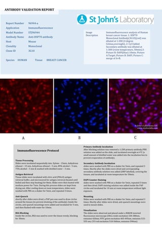
  • 1. ANTIBODY VALIDATION REPORT Report Number 96944-a Application Immunofluorescence Model Number STJ96944 Antibody Name Anti-HSP70 antibody Host Mouse Clonality Monoclonal Clone ID 3G10 Species HUMAN Tissue BREAST CANCER Image Description Immunofluorescence analysis of Human breast cancer tissue. 1: HSP70 Monoclonal Antibody(3G10)(red) was diluted at 1:200 (4 degree Celsius,overnight). 2: Cy3 labled Secondary antibody was diluted at 1:300 (room temperature, 50min).3: Picture B: DAPI(blue) 10min. Picture A:Target. Picture B: DAPI. Picture C: merge of A+B. Primary Antibody Incubation After blocking solution was removed a 1:200 primary antibody/PBS solution was added on the slide, and incubated overnight at 4°C (a small amount of distilled water was added into the incubation box to prevent evaporation of antibody). Secondary Antibody Incubation slides were washed with PBS on a shaker for 5min, and repeated 3 times. Shortly after the slides were dried and corresponding secondary antibody solution was added (HRP labelled), covering the tissues, and incubated at room temperature for 50min. DAPI Counter-Staining slides were washed with PBS on a shaker for 5min, repeated 3 times and then dried. DAPI staining solution was added inside the PAP circles and incubated for 10 min at room temperature without light exposure. Mounting Slides were washed with PBS on a shaker for 5min, and repeated 3 times. Shortly after slides were dried, anti-quench mountings were used to mount slides. Visualization The slides were observed and placed under a NIKON inverted fluorescence microscope (Ultra violet excitation 330-380nm, emission 420nm; FITC green excitation 465-495nm, emission 515- 555 nm; CY3 red excitation 510-560nm, emission 590nm) Immunofluorescence Protocol Tissue Processing Slides were incubated sequentially into: Xylene - 15min, Anhydrous ethanol – 15 min, Anhydrous ethanol – 5 min, 85% alcohol – 5 min, 75% alcohol – 5 min & washed with distilled water – 5 min. Antigen Retrieval Tissue slides were incubated with citric acid (PH6.0) antigen retrieval buffer, and microwaved for antigen retrieval (heated until boiled and then stop heating) for 8min. Slides were then heated with medium power for 7min. During this process slides are kept from drying out. After cooling down at room temperature, slides were washed with PBS on a shaker for 5min, and repeated 3 times. Anti-Quench shortly after slides were dried, a PAP pen was used to draw circles around the tissues (to prevent draining of the antibody). Inside the circles, anti-quench mountings were added and incubated for 5 min, and then flushed with water for 10min. BSA Blocking Inside the circles, BSA was used to cover the tissue evenly, blocking for 30min. St John's Laboratory Ltd. www.stjohnslabs.com
  • 2. ANTIBODY VALIDATION REPORT Report Number 96944-b Application Immunofluorescence Model Number STJ96944 Antibody Name Anti-HSP70 antibody Host Mouse Clonality Monoclonal Clone ID 3G10 Species HUMAN Tissue BREAST CANCER Image Description Immunofluorescence analysis of Human breast cancer tissue. 1: HSP70 Monoclonal Antibody(3G10)(red) was diluted at 1:200 (4 degree Celsius,overnight). 2: Cy3 labled Secondary antibody was diluted at 1:300 (room temperature, 50min).3: Picture B: DAPI(blue) 10min. Picture A:Target. Picture B: DAPI. Picture C: merge of A+B. Primary Antibody Incubation After blocking solution was removed a 1:200 primary antibody/PBS solution was added on the slide, and incubated overnight at 4°C (a small amount of distilled water was added into the incubation box to prevent evaporation of antibody). Secondary Antibody Incubation slides were washed with PBS on a shaker for 5min, and repeated 3 times. Shortly after the slides were dried and corresponding secondary antibody solution was added (HRP labelled), covering the tissues, and incubated at room temperature for 50min. DAPI Counter-Staining slides were washed with PBS on a shaker for 5min, repeated 3 times and then dried. DAPI staining solution was added inside the PAP circles and incubated for 10 min at room temperature without light exposure. Mounting Slides were washed with PBS on a shaker for 5min, and repeated 3 times. Shortly after slides were dried, anti-quench mountings were used to mount slides. Visualization The slides were observed and placed under a NIKON inverted fluorescence microscope (Ultra violet excitation 330-380nm, emission 420nm; FITC green excitation 465-495nm, emission 515- 555 nm; CY3 red excitation 510-560nm, emission 590nm) Immunofluorescence Protocol Tissue Processing Slides were incubated sequentially into: Xylene - 15min, Anhydrous ethanol – 15 min, Anhydrous ethanol – 5 min, 85% alcohol – 5 min, 75% alcohol – 5 min & washed with distilled water – 5 min. Antigen Retrieval Tissue slides were incubated with citric acid (PH6.0) antigen retrieval buffer, and microwaved for antigen retrieval (heated until boiled and then stop heating) for 8min. Slides were then heated with medium power for 7min. During this process slides are kept from drying out. After cooling down at room temperature, slides were washed with PBS on a shaker for 5min, and repeated 3 times. Anti-Quench shortly after slides were dried, a PAP pen was used to draw circles around the tissues (to prevent draining of the antibody). Inside the circles, anti-quench mountings were added and incubated for 5 min, and then flushed with water for 10min. BSA Blocking Inside the circles, BSA was used to cover the tissue evenly, blocking for 30min. St John's Laboratory Ltd. www.stjohnslabs.com
  • 3. ANTIBODY VALIDATION REPORT Report Number 96944-c Application Immunofluorescence Model Number STJ96944 Antibody Name Anti-HSP70 antibody Host Mouse Clonality Monoclonal Clone ID 3G10 Species HUMAN Tissue BREAST CANCER Image Description Immunofluorescence analysis of Human breast cancer tissue. 1: HSP70 Monoclonal Antibody(3G10)(red) was diluted at 1:200 (4 degree Celsius,overnight). 2: Cy3 labled Secondary antibody was diluted at 1:300 (room temperature, 50min).3: Picture B: DAPI(blue) 10min. Picture A:Target. Picture B: DAPI. Picture C: merge of A+B. Primary Antibody Incubation After blocking solution was removed a 1:200 primary antibody/PBS solution was added on the slide, and incubated overnight at 4°C (a small amount of distilled water was added into the incubation box to prevent evaporation of antibody). Secondary Antibody Incubation slides were washed with PBS on a shaker for 5min, and repeated 3 times. Shortly after the slides were dried and corresponding secondary antibody solution was added (HRP labelled), covering the tissues, and incubated at room temperature for 50min. DAPI Counter-Staining slides were washed with PBS on a shaker for 5min, repeated 3 times and then dried. DAPI staining solution was added inside the PAP circles and incubated for 10 min at room temperature without light exposure. Mounting Slides were washed with PBS on a shaker for 5min, and repeated 3 times. Shortly after slides were dried, anti-quench mountings were used to mount slides. Visualization The slides were observed and placed under a NIKON inverted fluorescence microscope (Ultra violet excitation 330-380nm, emission 420nm; FITC green excitation 465-495nm, emission 515- 555 nm; CY3 red excitation 510-560nm, emission 590nm) Immunofluorescence Protocol Tissue Processing Slides were incubated sequentially into: Xylene - 15min, Anhydrous ethanol – 15 min, Anhydrous ethanol – 5 min, 85% alcohol – 5 min, 75% alcohol – 5 min & washed with distilled water – 5 min. Antigen Retrieval Tissue slides were incubated with citric acid (PH6.0) antigen retrieval buffer, and microwaved for antigen retrieval (heated until boiled and then stop heating) for 8min. Slides were then heated with medium power for 7min. During this process slides are kept from drying out. After cooling down at room temperature, slides were washed with PBS on a shaker for 5min, and repeated 3 times. Anti-Quench shortly after slides were dried, a PAP pen was used to draw circles around the tissues (to prevent draining of the antibody). Inside the circles, anti-quench mountings were added and incubated for 5 min, and then flushed with water for 10min. BSA Blocking Inside the circles, BSA was used to cover the tissue evenly, blocking for 30min. St John's Laboratory Ltd. www.stjohnslabs.com
  • 4. ANTIBODY VALIDATION REPORT Report Number 96944-d Application Immunofluorescence Model Number STJ96944 Antibody Name Anti-HSP70 antibody Host Mouse Clonality Monoclonal Clone ID 3G10 Species MOUSE Tissue LUNG Image Description Immunofluorescence analysis of Mouse lung tissue. 1: HSP70 Monoclonal Antibody(3G10)(red) was diluted at 1:200 (4 degree Celsius,overnight). 2: Cy3 labled Secondary antibody was diluted at 1:300 (room temperature, 50min).3: Picture B: DAPI(blue) 10min. Picture A:Target. Picture B: DAPI. Picture C: merge of A+B. Primary Antibody Incubation After blocking solution was removed a 1:200 primary antibody/PBS solution was added on the slide, and incubated overnight at 4°C (a small amount of distilled water was added into the incubation box to prevent evaporation of antibody). Secondary Antibody Incubation slides were washed with PBS on a shaker for 5min, and repeated 3 times. Shortly after the slides were dried and corresponding secondary antibody solution was added (HRP labelled), covering the tissues, and incubated at room temperature for 50min. DAPI Counter-Staining slides were washed with PBS on a shaker for 5min, repeated 3 times and then dried. DAPI staining solution was added inside the PAP circles and incubated for 10 min at room temperature without light exposure. Mounting Slides were washed with PBS on a shaker for 5min, and repeated 3 times. Shortly after slides were dried, anti-quench mountings were used to mount slides. Visualization The slides were observed and placed under a NIKON inverted fluorescence microscope (Ultra violet excitation 330-380nm, emission 420nm; FITC green excitation 465-495nm, emission 515- 555 nm; CY3 red excitation 510-560nm, emission 590nm) Immunofluorescence Protocol Tissue Processing Slides were incubated sequentially into: Xylene - 15min, Anhydrous ethanol – 15 min, Anhydrous ethanol – 5 min, 85% alcohol – 5 min, 75% alcohol – 5 min & washed with distilled water – 5 min. Antigen Retrieval Tissue slides were incubated with citric acid (PH6.0) antigen retrieval buffer, and microwaved for antigen retrieval (heated until boiled and then stop heating) for 8min. Slides were then heated with medium power for 7min. During this process slides are kept from drying out. After cooling down at room temperature, slides were washed with PBS on a shaker for 5min, and repeated 3 times. Anti-Quench shortly after slides were dried, a PAP pen was used to draw circles around the tissues (to prevent draining of the antibody). Inside the circles, anti-quench mountings were added and incubated for 5 min, and then flushed with water for 10min. BSA Blocking Inside the circles, BSA was used to cover the tissue evenly, blocking for 30min. St John's Laboratory Ltd. www.stjohnslabs.com
  • 5. ANTIBODY VALIDATION REPORT Report Number 96944-e Application Immunofluorescence Model Number STJ96944 Antibody Name Anti-HSP70 antibody Host Mouse Clonality Monoclonal Clone ID 3G10 Species MOUSE Tissue LUNG Image Description Immunofluorescence analysis of Mouse lung tissue. 1: HSP70 Monoclonal Antibody(3G10)(red) was diluted at 1:200 (4 degree Celsius,overnight). 2: Cy3 labled Secondary antibody was diluted at 1:300 (room temperature, 50min).3: Picture B: DAPI(blue) 10min. Picture A:Target. Picture B: DAPI. Picture C: merge of A+B. Primary Antibody Incubation After blocking solution was removed a 1:200 primary antibody/PBS solution was added on the slide, and incubated overnight at 4°C (a small amount of distilled water was added into the incubation box to prevent evaporation of antibody). Secondary Antibody Incubation slides were washed with PBS on a shaker for 5min, and repeated 3 times. Shortly after the slides were dried and corresponding secondary antibody solution was added (HRP labelled), covering the tissues, and incubated at room temperature for 50min. DAPI Counter-Staining slides were washed with PBS on a shaker for 5min, repeated 3 times and then dried. DAPI staining solution was added inside the PAP circles and incubated for 10 min at room temperature without light exposure. Mounting Slides were washed with PBS on a shaker for 5min, and repeated 3 times. Shortly after slides were dried, anti-quench mountings were used to mount slides. Visualization The slides were observed and placed under a NIKON inverted fluorescence microscope (Ultra violet excitation 330-380nm, emission 420nm; FITC green excitation 465-495nm, emission 515- 555 nm; CY3 red excitation 510-560nm, emission 590nm) Immunofluorescence Protocol Tissue Processing Slides were incubated sequentially into: Xylene - 15min, Anhydrous ethanol – 15 min, Anhydrous ethanol – 5 min, 85% alcohol – 5 min, 75% alcohol – 5 min & washed with distilled water – 5 min. Antigen Retrieval Tissue slides were incubated with citric acid (PH6.0) antigen retrieval buffer, and microwaved for antigen retrieval (heated until boiled and then stop heating) for 8min. Slides were then heated with medium power for 7min. During this process slides are kept from drying out. After cooling down at room temperature, slides were washed with PBS on a shaker for 5min, and repeated 3 times. Anti-Quench shortly after slides were dried, a PAP pen was used to draw circles around the tissues (to prevent draining of the antibody). Inside the circles, anti-quench mountings were added and incubated for 5 min, and then flushed with water for 10min. BSA Blocking Inside the circles, BSA was used to cover the tissue evenly, blocking for 30min. St John's Laboratory Ltd. www.stjohnslabs.com
  • 6. ANTIBODY VALIDATION REPORT Report Number 96944-f Application Immunofluorescence Model Number STJ96944 Antibody Name Anti-HSP70 antibody Host Mouse Clonality Monoclonal Clone ID 3G10 Species MOUSE Tissue LUNG Image Description Immunofluorescence analysis of Mouse lung tissue. 1: HSP70 Monoclonal Antibody(3G10)(red) was diluted at 1:200 (4 degree Celsius,overnight). 2: Cy3 labled Secondary antibody was diluted at 1:300 (room temperature, 50min).3: Picture B: DAPI(blue) 10min. Picture A:Target. Picture B: DAPI. Picture C: merge of A+B. Primary Antibody Incubation After blocking solution was removed a 1:200 primary antibody/PBS solution was added on the slide, and incubated overnight at 4°C (a small amount of distilled water was added into the incubation box to prevent evaporation of antibody). Secondary Antibody Incubation slides were washed with PBS on a shaker for 5min, and repeated 3 times. Shortly after the slides were dried and corresponding secondary antibody solution was added (HRP labelled), covering the tissues, and incubated at room temperature for 50min. DAPI Counter-Staining slides were washed with PBS on a shaker for 5min, repeated 3 times and then dried. DAPI staining solution was added inside the PAP circles and incubated for 10 min at room temperature without light exposure. Mounting Slides were washed with PBS on a shaker for 5min, and repeated 3 times. Shortly after slides were dried, anti-quench mountings were used to mount slides. Visualization The slides were observed and placed under a NIKON inverted fluorescence microscope (Ultra violet excitation 330-380nm, emission 420nm; FITC green excitation 465-495nm, emission 515- 555 nm; CY3 red excitation 510-560nm, emission 590nm) Immunofluorescence Protocol Tissue Processing Slides were incubated sequentially into: Xylene - 15min, Anhydrous ethanol – 15 min, Anhydrous ethanol – 5 min, 85% alcohol – 5 min, 75% alcohol – 5 min & washed with distilled water – 5 min. Antigen Retrieval Tissue slides were incubated with citric acid (PH6.0) antigen retrieval buffer, and microwaved for antigen retrieval (heated until boiled and then stop heating) for 8min. Slides were then heated with medium power for 7min. During this process slides are kept from drying out. After cooling down at room temperature, slides were washed with PBS on a shaker for 5min, and repeated 3 times. Anti-Quench shortly after slides were dried, a PAP pen was used to draw circles around the tissues (to prevent draining of the antibody). Inside the circles, anti-quench mountings were added and incubated for 5 min, and then flushed with water for 10min. BSA Blocking Inside the circles, BSA was used to cover the tissue evenly, blocking for 30min. St John's Laboratory Ltd. www.stjohnslabs.com
  • 7. ANTIBODY VALIDATION REPORT Report Number 96944-g Application Immunofluorescence Model Number STJ96944 Antibody Name Anti-HSP70 antibody Host Mouse Clonality Monoclonal Clone ID 3G10 Species RAT Tissue TESTIS Image Description Immunofluorescence analysis of Rat testis tissue. 1: HSP70 Monoclonal Antibody(3G10)(red) was diluted at 1:200 (4 degree Celsius,overnight). 2: Cy3 labled Secondary antibody was diluted at 1:300 (room temperature, 50min).3: Picture B: DAPI(blue) 10min. Picture A:Target. Picture B: DAPI. Picture C: merge of A+B. Primary Antibody Incubation After blocking solution was removed a 1:200 primary antibody/PBS solution was added on the slide, and incubated overnight at 4°C (a small amount of distilled water was added into the incubation box to prevent evaporation of antibody). Secondary Antibody Incubation slides were washed with PBS on a shaker for 5min, and repeated 3 times. Shortly after the slides were dried and corresponding secondary antibody solution was added (HRP labelled), covering the tissues, and incubated at room temperature for 50min. DAPI Counter-Staining slides were washed with PBS on a shaker for 5min, repeated 3 times and then dried. DAPI staining solution was added inside the PAP circles and incubated for 10 min at room temperature without light exposure. Mounting Slides were washed with PBS on a shaker for 5min, and repeated 3 times. Shortly after slides were dried, anti-quench mountings were used to mount slides. Visualization The slides were observed and placed under a NIKON inverted fluorescence microscope (Ultra violet excitation 330-380nm, emission 420nm; FITC green excitation 465-495nm, emission 515- 555 nm; CY3 red excitation 510-560nm, emission 590nm) Immunofluorescence Protocol Tissue Processing Slides were incubated sequentially into: Xylene - 15min, Anhydrous ethanol – 15 min, Anhydrous ethanol – 5 min, 85% alcohol – 5 min, 75% alcohol – 5 min & washed with distilled water – 5 min. Antigen Retrieval Tissue slides were incubated with citric acid (PH6.0) antigen retrieval buffer, and microwaved for antigen retrieval (heated until boiled and then stop heating) for 8min. Slides were then heated with medium power for 7min. During this process slides are kept from drying out. After cooling down at room temperature, slides were washed with PBS on a shaker for 5min, and repeated 3 times. Anti-Quench shortly after slides were dried, a PAP pen was used to draw circles around the tissues (to prevent draining of the antibody). Inside the circles, anti-quench mountings were added and incubated for 5 min, and then flushed with water for 10min. BSA Blocking Inside the circles, BSA was used to cover the tissue evenly, blocking for 30min. St John's Laboratory Ltd. www.stjohnslabs.com
  • 8. ANTIBODY VALIDATION REPORT Report Number 96944-h Application Immunofluorescence Model Number STJ96944 Antibody Name Anti-HSP70 antibody Host Mouse Clonality Monoclonal Clone ID 3G10 Species RAT Tissue TESTIS Image Description Immunofluorescence analysis of Rat testis tissue. 1: HSP70 Monoclonal Antibody(3G10)(red) was diluted at 1:200 (4 degree Celsius,overnight). 2: Cy3 labled Secondary antibody was diluted at 1:300 (room temperature, 50min).3: Picture B: DAPI(blue) 10min. Picture A:Target. Picture B: DAPI. Picture C: merge of A+B. Primary Antibody Incubation After blocking solution was removed a 1:200 primary antibody/PBS solution was added on the slide, and incubated overnight at 4°C (a small amount of distilled water was added into the incubation box to prevent evaporation of antibody). Secondary Antibody Incubation slides were washed with PBS on a shaker for 5min, and repeated 3 times. Shortly after the slides were dried and corresponding secondary antibody solution was added (HRP labelled), covering the tissues, and incubated at room temperature for 50min. DAPI Counter-Staining slides were washed with PBS on a shaker for 5min, repeated 3 times and then dried. DAPI staining solution was added inside the PAP circles and incubated for 10 min at room temperature without light exposure. Mounting Slides were washed with PBS on a shaker for 5min, and repeated 3 times. Shortly after slides were dried, anti-quench mountings were used to mount slides. Visualization The slides were observed and placed under a NIKON inverted fluorescence microscope (Ultra violet excitation 330-380nm, emission 420nm; FITC green excitation 465-495nm, emission 515- 555 nm; CY3 red excitation 510-560nm, emission 590nm) Immunofluorescence Protocol Tissue Processing Slides were incubated sequentially into: Xylene - 15min, Anhydrous ethanol – 15 min, Anhydrous ethanol – 5 min, 85% alcohol – 5 min, 75% alcohol – 5 min & washed with distilled water – 5 min. Antigen Retrieval Tissue slides were incubated with citric acid (PH6.0) antigen retrieval buffer, and microwaved for antigen retrieval (heated until boiled and then stop heating) for 8min. Slides were then heated with medium power for 7min. During this process slides are kept from drying out. After cooling down at room temperature, slides were washed with PBS on a shaker for 5min, and repeated 3 times. Anti-Quench shortly after slides were dried, a PAP pen was used to draw circles around the tissues (to prevent draining of the antibody). Inside the circles, anti-quench mountings were added and incubated for 5 min, and then flushed with water for 10min. BSA Blocking Inside the circles, BSA was used to cover the tissue evenly, blocking for 30min. St John's Laboratory Ltd. www.stjohnslabs.com
  • 9. ANTIBODY VALIDATION REPORT Report Number 96944-i Application Immunofluorescence Model Number STJ96944 Antibody Name Anti-HSP70 antibody Host Mouse Clonality Monoclonal Clone ID 3G10 Species RAT Tissue TESTIS Image Description Immunofluorescence analysis of Rat testis tissue. 1: HSP70 Monoclonal Antibody(3G10)(red) was diluted at 1:200 (4 degree Celsius,overnight). 2: Cy3 labled Secondary antibody was diluted at 1:300 (room temperature, 50min).3: Picture B: DAPI(blue) 10min. Picture A:Target. Picture B: DAPI. Picture C: merge of A+B. Primary Antibody Incubation After blocking solution was removed a 1:200 primary antibody/PBS solution was added on the slide, and incubated overnight at 4°C (a small amount of distilled water was added into the incubation box to prevent evaporation of antibody). Secondary Antibody Incubation slides were washed with PBS on a shaker for 5min, and repeated 3 times. Shortly after the slides were dried and corresponding secondary antibody solution was added (HRP labelled), covering the tissues, and incubated at room temperature for 50min. DAPI Counter-Staining slides were washed with PBS on a shaker for 5min, repeated 3 times and then dried. DAPI staining solution was added inside the PAP circles and incubated for 10 min at room temperature without light exposure. Mounting Slides were washed with PBS on a shaker for 5min, and repeated 3 times. Shortly after slides were dried, anti-quench mountings were used to mount slides. Visualization The slides were observed and placed under a NIKON inverted fluorescence microscope (Ultra violet excitation 330-380nm, emission 420nm; FITC green excitation 465-495nm, emission 515- 555 nm; CY3 red excitation 510-560nm, emission 590nm) Immunofluorescence Protocol Tissue Processing Slides were incubated sequentially into: Xylene - 15min, Anhydrous ethanol – 15 min, Anhydrous ethanol – 5 min, 85% alcohol – 5 min, 75% alcohol – 5 min & washed with distilled water – 5 min. Antigen Retrieval Tissue slides were incubated with citric acid (PH6.0) antigen retrieval buffer, and microwaved for antigen retrieval (heated until boiled and then stop heating) for 8min. Slides were then heated with medium power for 7min. During this process slides are kept from drying out. After cooling down at room temperature, slides were washed with PBS on a shaker for 5min, and repeated 3 times. Anti-Quench shortly after slides were dried, a PAP pen was used to draw circles around the tissues (to prevent draining of the antibody). Inside the circles, anti-quench mountings were added and incubated for 5 min, and then flushed with water for 10min. BSA Blocking Inside the circles, BSA was used to cover the tissue evenly, blocking for 30min. St John's Laboratory Ltd. www.stjohnslabs.com