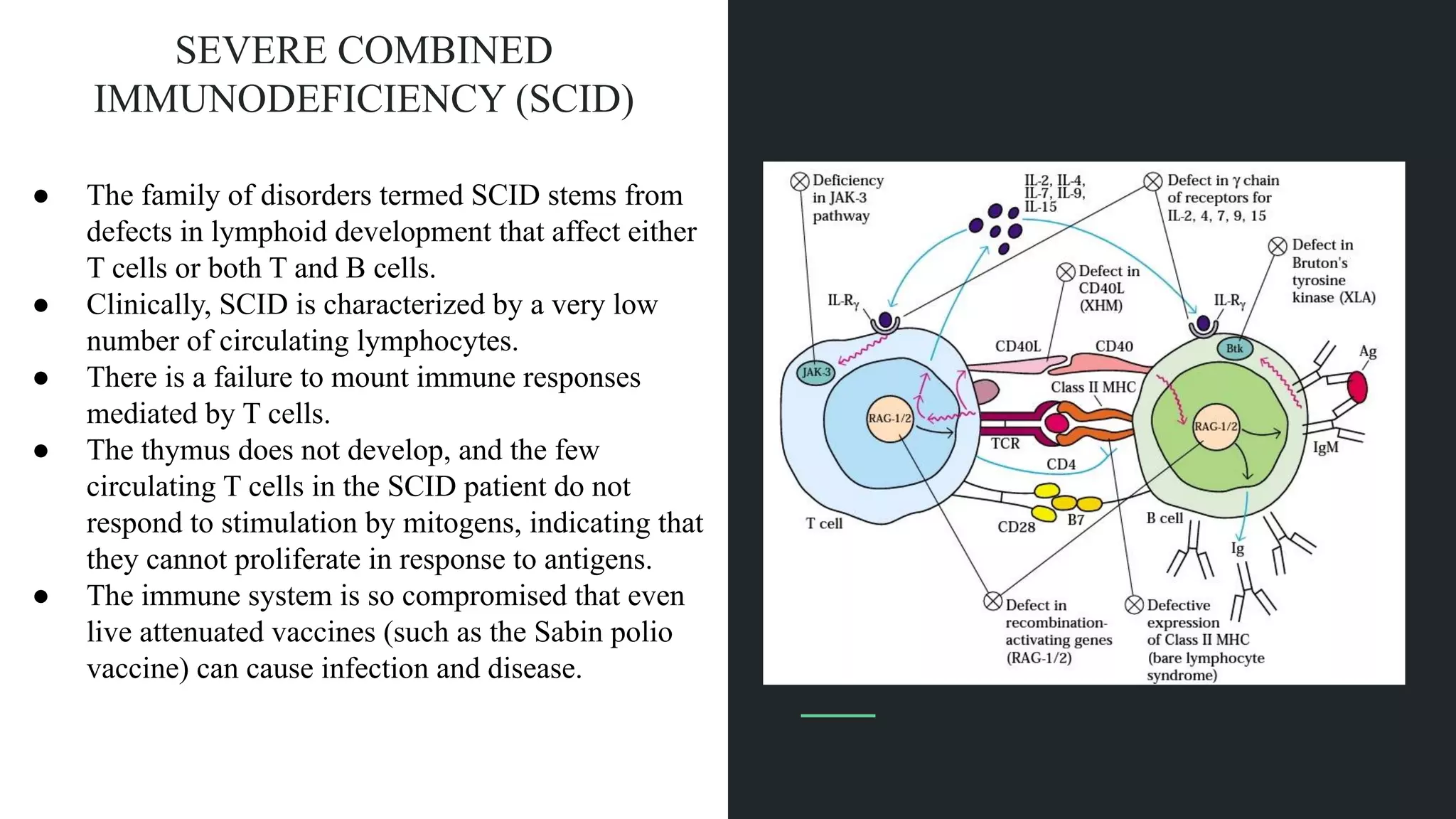
IMMUNODEFICIENCY
DISEASES
By
Sreehari A P
Primary Immunodeficiencies
● A primary immunodeficiency may affect either adaptive or innate immune
functions.
● Deficiencies involving components ofadaptive immunity,such as T or B cells,are
thus differentiated from immunodeficiencies in which the nonspecific mediators
ofinnate immunity,such as phagocytes or complement,are impaired
● the two main cell lineages important to immune function are lymphoid and
myeloid.Most defects that lead to immunodeficiencies affect either one or the
other.
● The lymphoid cell disorders may affect T cells,B cells, or,in combined
immunodeficiencies,both B and T cells.The myeloid cell disorders affect phagocytic
function.Most ofthe primary immunodeficiencies are inherited,
SEVERE COMBINED
IMMUNODEFICIENCY (SCID)
● The family of disorders termed SCID stems from
defects in lymphoid development that affect either
T cells or both T and B cells.
● Clinically, SCID is characterized by a very low
number of circulating lymphocytes.
● There is a failure to mount immune responses
mediated by T cells.
● The thymus does not develop, and the few
circulating T cells in the SCID patient do not
respond to stimulation by mitogens, indicating that
they cannot proliferate in response to antigens.
● The immune system is so compromised that even
live attenuated vaccines (such as the Sabin polio
vaccine) can cause infection and disease.
Different causes for SCID
● The search for defects that underlie SCID has revealed several
● The most common cause (64 cases) was deficiency of the common gamma chain
of the IL-2 receptor (IL-2R;see Figure 12-7). Defects in this chain impede
signaling through receptors for IL-4,-7,-9, and -15 as well as the IL-2 receptor,
because the chain is present in receptors for all of these cytokines
● Deficiency in the kinase JAK-3, which has a similar phenotype because the IL
receptors signal through this molecule
● Adenosine deaminase or ADA deficiency .
○ Adenosine deaminase catalyzes conversion ofadenosine to inosine,and its
deficiency results in accumulation ofadenosine,which interferes with purine
metabolism and DNA synthesis.
● Depletion of CD8+T
○ CD8+ T cells that involves the tyrosine kinase ZAP-70,an important element in
T-cell signal transduction.
○ Infants with defects in ZAP-70 may have normal levels of immunoglobulin and
CD4+lymphocytes,but their CD4+T cells are nonfunctional.
○ A deficiency in the enzyme purine nucleoside phosphorylase (PNP) causes
immunodeficiency by a mechanism similar to the ADA defect
● A defect in the genes that encode RAG-1 and RAG-2
○ A defect in the genes that encode mediators of the rearrangement processes
(recombination-activating proteins RAG-1 and RAG-2) precludes development of B and T
cells with functional receptors and leads to SCID.
● Failure to transcribe the genes that encode class II MHC molecules
○ A defect leading to general failure ofimmunity similar to SCID is failure to transcribe the
genes that encode class II MHC molecules.Without these molecules,the patient’s
lymphocytes cannot participate in cellular interactions with T helper cells.This type
ofimmunodeficiency is also called the bare-lymphocyte syndrome
● Mutation in the TAP genes
○ This rare variant ofimmunodeficiency was ascribed to mutation in the TAP genes that are
vital to antigen processing by class I MHC molecules (see Clinical Focus Chapter 8).This
defect causes a deficit in CD8-mediated immunity,characterized by susceptibility to viral
infection
WISKOTT-ALDRICH SYNDROME (WAS)
● The severity of this X-linked disorder increases with age and usually results in fatal
infection or lymphoid malignancy. Initially, T and B lymphocytes are present in normal
numbers.
● WAS first manifests itself by defective responses to bacterial polysaccharides and by
lower-than-average IgM levels.
● As the WAS sufferer ages, there are recurrent bacterial infections and a gradual loss of
humoral and cellular responses.
● The syndrome includes thrombocytopenia which may lead to fatal bleeding. Eczema
(skin rashes) in varying degrees of severity may also occur, usually beginning around one
year of age
● The defect in WAS has been mapped to the short arm of the X chromosome and
involves a cytoskeletal glycoprotein present in lymphoid cells called sialophorin (CD43).
● The WAS protein is required for assembly of actin filaments required for the formation of
microvesicles
INTERFERON-GAMMA–RECEPTOR DEFECT
● Involves a defect in the receptor for interferon gamma.
● Most of those carrying this autosomal recessive trait are from families with a history of
inbreeding.
● This immunodeficiency points to a specific role for IFN-and its receptor in protection from
infection with mycobacteria.
X-LINKED AGAMMAGLOBULINEMIA
● A B-cell defect called X-linked agammaglobulinemia (XLA) or Bruton’s
hypogammaglobulinemia is characterized by extremely low IgG levels and by the
absence of other immunoglobulin classes
● Individuals with XLA have no peripheral B cells and suffer from recurrent bacterial
infections, beginning at about nine months of age,
● There is a defect in B-cell signal transduction in this disorder, due to a defect in a
transduction molecule called Bruton’s tyrosine kinase (Btk), after the investigator who
described the syndrome.
X-LINKED HYPER-IgM SYNDROME
● X-linked hyperIgM (XHM) syndrome is characterized by a deficiency of IgG,
IgA, and IgE, and elevated levels of IgM, sometimes as high as 10 mg/ml
(normal IgM concentration is 1.5 mg/ml)
● X-linked recessive disorder
● XHM patients often have high levels of autoantibodies to neutrophils, platelets,
and red blood cells
● The defect in XHM is in the gene encoding the CD40 ligand (CD40L), which
maps to the X chromosome
COMMON VARIABLE IMMUNODEFICIENCY (CVI)
● CVI is characterized by a profound decrease in numbers of antibody-producing
plasma cells, low levels of most immunoglobulin isotypes
(hypogammaglobulinemia), and recurrent infections.
● B cells fail to mature into plasma cells; however in vitro studies show that CVI B
cells are capable of maturing in response to appropriate differentiation signals.
● The underlying defect in CVI is not known,but must involve either an in vivo
blockage of the maturation of B cells to the plasma-cell stage or their inability
to produce the secreted form of immunoglobulins.
HYPER-IgE SYNDROME (JOB SYNDROME)
● A primary immunodeficiency characterized by skin abcesses, recurrent
pneumonia, eczema, and elevated levels of IgE accompanies facial
abnormalities and bone fragility.
● This multi-system disorder is autosomal dominant and has variable
expressivity. The gene for hyper IgE syndrome, or HIES, maps to chromosome
4.
● HIES immunologic signs include recurrent infection and eosinophilia in addition
to elevated IgE levels.
ATAXIA TELANGIECTASIA
● Ataxia telangiectasia is a disease syndrome that includes deficiency of IgA and
sometimes of IgE.
● The syndrome is characterized by difficulty in maintaining balance (ataxia) and
by the appearance of broken capillaries (telangiectasia)
● The primary defect appears to be in a kinase involved in regulation of the cell
cycle. The relationship between the immune deficiency and the other defects
in ataxia telangiectasia remains obscure.
Immune Disorders Involving
The Thymus
DIGEORGE SYNDROME
● DiGeorge syndrome, or congenital thymic aplasia, in its most severe form is the complete absence of a
thymus.
● This developmental defect, which is associated with the deletion in the embryo of a region on chromosome
22, causes immunodeficiency along with characteristic facial abnormalities, hypoparathyroidism, and
congenital heart disease
● The stage at which the causative developmental defect occurs has been determined, and the syndrome is
sometimes called the third and fourth pharyngeal pouch syndrome to reflect its precise embryonic origin.
● The immune defect includes a profound depression of T-cell numbers and absence of T-cell responses
● Thymic transplantation is of some value for correcting the T-cell defects, but many DiGeorge patients have
such severe heart disease that their chances for long-term survival are poor, even if the immune defects are
corrected.
● DiGeorge syndrome results from an intrauterine or developmental anomaly, thymic hypoplasia, or the Nezelof
syndrome, is an inherited disorder.
● Individuals suffer from chronic diarrhea, viral and fungal infections, and a general failure to thrive.
Immunodeficiencies Of The
Myeloid Lineage Affect
Innate Immunity
CHRONIC GRANULOMATOUS DISEASE (CGD)
● This disease is rooted in a defect in the oxidative pathway by which phagocytes
generate hydrogen peroxide and the resulting reactive products, such as hypochlorous
acid, that kill phagocytosed bacteria.
● CGD sufferers undergo excessive inflammatory reactions that result in gingivitis, swollen
lymph nodes, and nonmalignant granulomas (lumpy subcutaneous cell masses); they are
also susceptible to bacterial and fungal infection
● In addition to the general defect in the killer function of phagocytes, there is also a
decrease in the ability of mononuclear cells to serve as APC.
● Both processing and presentation of antigen are impaired. Increased amounts of antigen
are required to trigger T-cell help when mononuclear cells from CGD patients are used
as APCs.
CHEDIAK-HIGASHI SYNDROME
● This autosomal recessive disease is characterized by recurrent bacterial
infections,partial oculo-cutaneous albinism (lack of skin and eye pigment),and
aggressive but nonmalignant infiltration oforgans by lymphoid cells.
● Phagocytes from patients with this immune defect contain giant granules but
do not have the ability to kill bacteria.
● The molecular basis ofthe defect is a mutation in a protein (LYST) involved in
the regulation ofintracellular trafficking.The mutation impairs the targeting
ofproteins to secretory lysosomes,which makes them unable to lyse bacteria.
LEUKOCYTE ADHESION DEFICIENCY (LAD)
● Cell-surface molecules belonging to the integrin family ofproteins function as adhesion molecules
and are required to facilitate cellular interaction.Three of these,LFA-1,Mac-1,and gp150/95
(CD11a,b,and c,respectively) have a common chain (CD18) and are variably present on different
monocytic cells;CD11a is also expressed on B cells
● An immunodeficiency related to dysfunction ofthe adhesion molecules is rooted in a defect localized
to the common chain and affects expression ofall three ofthe molecules that use this chain.
● This defect,called leukocyte adhesion deficiency (LAD),causes susceptibility to infection with both
gram-positive and gram-negative bacteria as well as various fungi
● Impairment ofadhesion ofleukocytes to vascular endothelium limits recruitment ofcells to sites
ofinflammation.Viral immunity is somewhat impaired, as would be predicted from the defective T-B
cell cooperation arising from the adhesion defect.
● LAD varies in its severity; some affected individuals die within a few years,others survive into their
forties.
Complement Defects Result
in Immunodeficiency or
Immune-Complex Disease
Complement Defects Result in Immunodeficiency or Immune-Complex
Disease
● Many complement deficiencies are associated with increased susceptibility to
bacterial infections and/or immune-complex diseases.
● One ofthese complement disorders,a deficiency in properdin,which stabilizes
the C3 convertase in the alternative complement pathway,is caused by a
defect in a gene located on the X chromosome
● Homozygous deficiencies in any of the early components of the classical
pathway (C1q, C1r, C1s, C4, and C2) exhibit similar symptoms, notably a marked
increase in immune-complex diseases such as systemic lupus erythematosus,
glomerulonephritis, and vasculitis.
●
● Homozygous deficiencies in any of the early components of the classical pathway (C1q,
C1r, C1s, C4, and C2) exhibit similar symptoms, notably a marked increase in
immune-complex diseases such as systemic lupus erythematosus, glomerulonephritis,
and vasculitis.
● These deficiencies highlight the importance of the early complement reactions in
generating C3b, and the critical role of C3b in solubilization and clearance of immune
complexes.
● Individuals with such complement deficiencies may suffer from recurrent infections by
pyogenic (pusforming) bacteria such as streptococci and staphylococci.
● These organisms are gram-positive and therefore resistant to the lytic effects of the
MAC. Nevertheless, the early complement components ordinarily prevent recurrent
infection by mediating a localized inflammatory response and opsonizing the bacteria.
● Deficiencies in factor D and properdin—early components of the alternative
pathway—appear to be associated with Neisseria infections but not with
immune-complex disease.
C3 deficiency
● Patients with C3 deficiencies have the most severe clinical manifestations,
reflecting the central role of C3 in activation of C5 and formation of the MAC.
● The first patient identified with a C3 deficiency was a child suffering from
frequent severe bacterial infections and initially diagnosed as having
agammaglobulinemia. After tests revealed normal immunoglobulin levels, a
deficiency in C3 was discovered.
● This case highlights the critical function of the complement system in
converting a humoral antibody response into an effective defense
mechanism.The majority of patients with C3 deficiency have recurrent bacterial
infections and may have immunecomplex diseases.
Homozygous Deficiencies In The Components
● Individuals with homozygous deficiencies in the components involved in the
MAC develop recurrent meningococcal and gonococcal infections caused by
Neisseria species.
● In normal individuals, these gram-negative bacteria are generally susceptible
to complement-mediated lysis or are cleared by the opsonizing activity of C3b.
● MAC-deficient individuals rarely have immune-complex disease, which
suggests that they produce enough C3b to clear immune
complexes.Interestingly, a deficiency in C9 results in no clinical symptoms,
suggesting that the entire MAC is not always necessary for
complement-mediated lysis.
● Congenital deficiencies of complement regulatory proteins have also been
reported.
● The C1 inhibitor (C1Inh) regulates activation of the classical pathway by
preventing excessive C4 and C2 activation by C1. Deficiency of C1Inh is an
autosomal dominant condition with a frequency of 1 in 1000.
● The deficiency gives rise to a condition called hereditary angioedema, which
manifests clinically as localized edema of the tissue, often following trauma,
but sometimes with no known cause.
● The edema can be in subcutaneous tissues or within the bowel, where it
causes abdominal pain, or in the upper respiratory tract, where it causes
obstruction of the airway.
AIDS and Other Acquired or
Secondary
Immunodeficiencies
● All other forms of immunodeficiency have been overshadowed by an epidemic of severe
immunodeficiency caused by the infectious agent called human immunodeficiency virus 1, or HIV-1.
● The disease that HIV-1 causes, acquired immunodeficiency syndrome (AIDS) was first reported in the
United States in 1981 in Los Angeles, New York, and San Francisco.
● A group of patients displayed unusual infections, including the opportunistic fungal pathogen
Pneumocystis carinii, which causes a pneumonia called PCP (P. carinii pneumonia) in persons with
immunodeficiency. In addition to PCP, some patients had Kaposi’s sarcoma, an extremely rare skin
tumor, as well as other, rarely encountered opportunistic infections.
● More complete evaluation of the patients showed that they had in common a marked deficiency in
cellular immune responses and a significant decrease in the subpopulation of T cells that carry the
CD4 marker (T helper cells.)
HIV-1 Spreads by Sexual Contact, Infected Blood, and
from Mother to Infant
● Although the precise mechanism by which HIV-1 infects an individual is not
known, epidemiological data indicate that common means of transmission
include homosexual and heterosexual intercourse, receipt of infected blood or
blood products, and passage from mothers to infants.
● Before tests for HIV in the blood supply were routinely used, patients who
received blood transfusions and hemophiliacs who received blood products
were at risk for HIV-1 infection.Exposure to infected blood accounts for the high
incidence of AIDS among intravenous drug users who normally share
hypodermic needles.
A Retrovirus, HIV-1, Is the Causative Agent of AIDS
● This immunodeficiency syndrome was novel at the time in that the type of virus
causing it was a retrovirus.
● Retroviruses carry their genetic information in the form of RNA.When the virus
enters a cell, the RNA is reverse transcribed to DNA by a virally encoded
enzyme, reverse transcriptase (RT)
● As the name implies, RT reverses the normal transcription process and makes
a DNA copy of the viral RNA genome. This copy, which is called a provirus, is
integrated into the cell genome and is replicated along with the cell DNA.
● Cross-sectional schematic diagram of HIV virion.
Each virion expresses 72 glycoprotein projections
composed of gp120 and gp41.
● The gp41 molecule is a transmembrane molecule
that crosses the lipid bilayer of the viral envelope.
● Gp120 is associated with gp41 and serves as the
viral receptor for CD4 on host cells.
● The viral envelope derives from the host cell and
contains some host-cell membrane proteins,
including class I and class II MHC molecules.
● Within the envelope is the viral core, or
nucleocapsid, which includes a layer of a protein
called p17 and an inner layer of a protein called
p24.
● The HIV genome consists of two copies of
single-stranded RNA, which are associated with
two molecules of reverse transcriptase (p64) and
nucleoid proteins p10, a protease, and p32, an
integrase.
STRUCTURE OF HIV
INFECTION OF HIV VIRUS
THE END

Immunodeficiency diseases.pdf

  • 1.
  • 2.
    Primary Immunodeficiencies ● Aprimary immunodeficiency may affect either adaptive or innate immune functions. ● Deficiencies involving components ofadaptive immunity,such as T or B cells,are thus differentiated from immunodeficiencies in which the nonspecific mediators ofinnate immunity,such as phagocytes or complement,are impaired ● the two main cell lineages important to immune function are lymphoid and myeloid.Most defects that lead to immunodeficiencies affect either one or the other.
  • 3.
    ● The lymphoidcell disorders may affect T cells,B cells, or,in combined immunodeficiencies,both B and T cells.The myeloid cell disorders affect phagocytic function.Most ofthe primary immunodeficiencies are inherited,
  • 6.
    SEVERE COMBINED IMMUNODEFICIENCY (SCID) ●The family of disorders termed SCID stems from defects in lymphoid development that affect either T cells or both T and B cells. ● Clinically, SCID is characterized by a very low number of circulating lymphocytes. ● There is a failure to mount immune responses mediated by T cells. ● The thymus does not develop, and the few circulating T cells in the SCID patient do not respond to stimulation by mitogens, indicating that they cannot proliferate in response to antigens. ● The immune system is so compromised that even live attenuated vaccines (such as the Sabin polio vaccine) can cause infection and disease.
  • 7.
    Different causes forSCID ● The search for defects that underlie SCID has revealed several ● The most common cause (64 cases) was deficiency of the common gamma chain of the IL-2 receptor (IL-2R;see Figure 12-7). Defects in this chain impede signaling through receptors for IL-4,-7,-9, and -15 as well as the IL-2 receptor, because the chain is present in receptors for all of these cytokines ● Deficiency in the kinase JAK-3, which has a similar phenotype because the IL receptors signal through this molecule
  • 8.
    ● Adenosine deaminaseor ADA deficiency . ○ Adenosine deaminase catalyzes conversion ofadenosine to inosine,and its deficiency results in accumulation ofadenosine,which interferes with purine metabolism and DNA synthesis. ● Depletion of CD8+T ○ CD8+ T cells that involves the tyrosine kinase ZAP-70,an important element in T-cell signal transduction. ○ Infants with defects in ZAP-70 may have normal levels of immunoglobulin and CD4+lymphocytes,but their CD4+T cells are nonfunctional. ○ A deficiency in the enzyme purine nucleoside phosphorylase (PNP) causes immunodeficiency by a mechanism similar to the ADA defect
  • 9.
    ● A defectin the genes that encode RAG-1 and RAG-2 ○ A defect in the genes that encode mediators of the rearrangement processes (recombination-activating proteins RAG-1 and RAG-2) precludes development of B and T cells with functional receptors and leads to SCID. ● Failure to transcribe the genes that encode class II MHC molecules ○ A defect leading to general failure ofimmunity similar to SCID is failure to transcribe the genes that encode class II MHC molecules.Without these molecules,the patient’s lymphocytes cannot participate in cellular interactions with T helper cells.This type ofimmunodeficiency is also called the bare-lymphocyte syndrome ● Mutation in the TAP genes ○ This rare variant ofimmunodeficiency was ascribed to mutation in the TAP genes that are vital to antigen processing by class I MHC molecules (see Clinical Focus Chapter 8).This defect causes a deficit in CD8-mediated immunity,characterized by susceptibility to viral infection
  • 10.
    WISKOTT-ALDRICH SYNDROME (WAS) ●The severity of this X-linked disorder increases with age and usually results in fatal infection or lymphoid malignancy. Initially, T and B lymphocytes are present in normal numbers. ● WAS first manifests itself by defective responses to bacterial polysaccharides and by lower-than-average IgM levels. ● As the WAS sufferer ages, there are recurrent bacterial infections and a gradual loss of humoral and cellular responses. ● The syndrome includes thrombocytopenia which may lead to fatal bleeding. Eczema (skin rashes) in varying degrees of severity may also occur, usually beginning around one year of age ● The defect in WAS has been mapped to the short arm of the X chromosome and involves a cytoskeletal glycoprotein present in lymphoid cells called sialophorin (CD43). ● The WAS protein is required for assembly of actin filaments required for the formation of microvesicles
  • 11.
    INTERFERON-GAMMA–RECEPTOR DEFECT ● Involvesa defect in the receptor for interferon gamma. ● Most of those carrying this autosomal recessive trait are from families with a history of inbreeding. ● This immunodeficiency points to a specific role for IFN-and its receptor in protection from infection with mycobacteria. X-LINKED AGAMMAGLOBULINEMIA ● A B-cell defect called X-linked agammaglobulinemia (XLA) or Bruton’s hypogammaglobulinemia is characterized by extremely low IgG levels and by the absence of other immunoglobulin classes ● Individuals with XLA have no peripheral B cells and suffer from recurrent bacterial infections, beginning at about nine months of age, ● There is a defect in B-cell signal transduction in this disorder, due to a defect in a transduction molecule called Bruton’s tyrosine kinase (Btk), after the investigator who described the syndrome.
  • 12.
    X-LINKED HYPER-IgM SYNDROME ●X-linked hyperIgM (XHM) syndrome is characterized by a deficiency of IgG, IgA, and IgE, and elevated levels of IgM, sometimes as high as 10 mg/ml (normal IgM concentration is 1.5 mg/ml) ● X-linked recessive disorder ● XHM patients often have high levels of autoantibodies to neutrophils, platelets, and red blood cells ● The defect in XHM is in the gene encoding the CD40 ligand (CD40L), which maps to the X chromosome
  • 13.
    COMMON VARIABLE IMMUNODEFICIENCY(CVI) ● CVI is characterized by a profound decrease in numbers of antibody-producing plasma cells, low levels of most immunoglobulin isotypes (hypogammaglobulinemia), and recurrent infections. ● B cells fail to mature into plasma cells; however in vitro studies show that CVI B cells are capable of maturing in response to appropriate differentiation signals. ● The underlying defect in CVI is not known,but must involve either an in vivo blockage of the maturation of B cells to the plasma-cell stage or their inability to produce the secreted form of immunoglobulins.
  • 14.
    HYPER-IgE SYNDROME (JOBSYNDROME) ● A primary immunodeficiency characterized by skin abcesses, recurrent pneumonia, eczema, and elevated levels of IgE accompanies facial abnormalities and bone fragility. ● This multi-system disorder is autosomal dominant and has variable expressivity. The gene for hyper IgE syndrome, or HIES, maps to chromosome 4. ● HIES immunologic signs include recurrent infection and eosinophilia in addition to elevated IgE levels.
  • 15.
    ATAXIA TELANGIECTASIA ● Ataxiatelangiectasia is a disease syndrome that includes deficiency of IgA and sometimes of IgE. ● The syndrome is characterized by difficulty in maintaining balance (ataxia) and by the appearance of broken capillaries (telangiectasia) ● The primary defect appears to be in a kinase involved in regulation of the cell cycle. The relationship between the immune deficiency and the other defects in ataxia telangiectasia remains obscure.
  • 16.
  • 17.
    DIGEORGE SYNDROME ● DiGeorgesyndrome, or congenital thymic aplasia, in its most severe form is the complete absence of a thymus. ● This developmental defect, which is associated with the deletion in the embryo of a region on chromosome 22, causes immunodeficiency along with characteristic facial abnormalities, hypoparathyroidism, and congenital heart disease ● The stage at which the causative developmental defect occurs has been determined, and the syndrome is sometimes called the third and fourth pharyngeal pouch syndrome to reflect its precise embryonic origin. ● The immune defect includes a profound depression of T-cell numbers and absence of T-cell responses ● Thymic transplantation is of some value for correcting the T-cell defects, but many DiGeorge patients have such severe heart disease that their chances for long-term survival are poor, even if the immune defects are corrected. ● DiGeorge syndrome results from an intrauterine or developmental anomaly, thymic hypoplasia, or the Nezelof syndrome, is an inherited disorder. ● Individuals suffer from chronic diarrhea, viral and fungal infections, and a general failure to thrive.
  • 18.
    Immunodeficiencies Of The MyeloidLineage Affect Innate Immunity
  • 19.
    CHRONIC GRANULOMATOUS DISEASE(CGD) ● This disease is rooted in a defect in the oxidative pathway by which phagocytes generate hydrogen peroxide and the resulting reactive products, such as hypochlorous acid, that kill phagocytosed bacteria. ● CGD sufferers undergo excessive inflammatory reactions that result in gingivitis, swollen lymph nodes, and nonmalignant granulomas (lumpy subcutaneous cell masses); they are also susceptible to bacterial and fungal infection ● In addition to the general defect in the killer function of phagocytes, there is also a decrease in the ability of mononuclear cells to serve as APC. ● Both processing and presentation of antigen are impaired. Increased amounts of antigen are required to trigger T-cell help when mononuclear cells from CGD patients are used as APCs.
  • 20.
    CHEDIAK-HIGASHI SYNDROME ● Thisautosomal recessive disease is characterized by recurrent bacterial infections,partial oculo-cutaneous albinism (lack of skin and eye pigment),and aggressive but nonmalignant infiltration oforgans by lymphoid cells. ● Phagocytes from patients with this immune defect contain giant granules but do not have the ability to kill bacteria. ● The molecular basis ofthe defect is a mutation in a protein (LYST) involved in the regulation ofintracellular trafficking.The mutation impairs the targeting ofproteins to secretory lysosomes,which makes them unable to lyse bacteria.
  • 21.
    LEUKOCYTE ADHESION DEFICIENCY(LAD) ● Cell-surface molecules belonging to the integrin family ofproteins function as adhesion molecules and are required to facilitate cellular interaction.Three of these,LFA-1,Mac-1,and gp150/95 (CD11a,b,and c,respectively) have a common chain (CD18) and are variably present on different monocytic cells;CD11a is also expressed on B cells ● An immunodeficiency related to dysfunction ofthe adhesion molecules is rooted in a defect localized to the common chain and affects expression ofall three ofthe molecules that use this chain. ● This defect,called leukocyte adhesion deficiency (LAD),causes susceptibility to infection with both gram-positive and gram-negative bacteria as well as various fungi ● Impairment ofadhesion ofleukocytes to vascular endothelium limits recruitment ofcells to sites ofinflammation.Viral immunity is somewhat impaired, as would be predicted from the defective T-B cell cooperation arising from the adhesion defect. ● LAD varies in its severity; some affected individuals die within a few years,others survive into their forties.
  • 22.
    Complement Defects Result inImmunodeficiency or Immune-Complex Disease
  • 23.
    Complement Defects Resultin Immunodeficiency or Immune-Complex Disease ● Many complement deficiencies are associated with increased susceptibility to bacterial infections and/or immune-complex diseases. ● One ofthese complement disorders,a deficiency in properdin,which stabilizes the C3 convertase in the alternative complement pathway,is caused by a defect in a gene located on the X chromosome ● Homozygous deficiencies in any of the early components of the classical pathway (C1q, C1r, C1s, C4, and C2) exhibit similar symptoms, notably a marked increase in immune-complex diseases such as systemic lupus erythematosus, glomerulonephritis, and vasculitis. ●
  • 24.
    ● Homozygous deficienciesin any of the early components of the classical pathway (C1q, C1r, C1s, C4, and C2) exhibit similar symptoms, notably a marked increase in immune-complex diseases such as systemic lupus erythematosus, glomerulonephritis, and vasculitis. ● These deficiencies highlight the importance of the early complement reactions in generating C3b, and the critical role of C3b in solubilization and clearance of immune complexes. ● Individuals with such complement deficiencies may suffer from recurrent infections by pyogenic (pusforming) bacteria such as streptococci and staphylococci. ● These organisms are gram-positive and therefore resistant to the lytic effects of the MAC. Nevertheless, the early complement components ordinarily prevent recurrent infection by mediating a localized inflammatory response and opsonizing the bacteria. ● Deficiencies in factor D and properdin—early components of the alternative pathway—appear to be associated with Neisseria infections but not with immune-complex disease.
  • 25.
    C3 deficiency ● Patientswith C3 deficiencies have the most severe clinical manifestations, reflecting the central role of C3 in activation of C5 and formation of the MAC. ● The first patient identified with a C3 deficiency was a child suffering from frequent severe bacterial infections and initially diagnosed as having agammaglobulinemia. After tests revealed normal immunoglobulin levels, a deficiency in C3 was discovered. ● This case highlights the critical function of the complement system in converting a humoral antibody response into an effective defense mechanism.The majority of patients with C3 deficiency have recurrent bacterial infections and may have immunecomplex diseases.
  • 26.
    Homozygous Deficiencies InThe Components ● Individuals with homozygous deficiencies in the components involved in the MAC develop recurrent meningococcal and gonococcal infections caused by Neisseria species. ● In normal individuals, these gram-negative bacteria are generally susceptible to complement-mediated lysis or are cleared by the opsonizing activity of C3b. ● MAC-deficient individuals rarely have immune-complex disease, which suggests that they produce enough C3b to clear immune complexes.Interestingly, a deficiency in C9 results in no clinical symptoms, suggesting that the entire MAC is not always necessary for complement-mediated lysis.
  • 27.
    ● Congenital deficienciesof complement regulatory proteins have also been reported. ● The C1 inhibitor (C1Inh) regulates activation of the classical pathway by preventing excessive C4 and C2 activation by C1. Deficiency of C1Inh is an autosomal dominant condition with a frequency of 1 in 1000. ● The deficiency gives rise to a condition called hereditary angioedema, which manifests clinically as localized edema of the tissue, often following trauma, but sometimes with no known cause. ● The edema can be in subcutaneous tissues or within the bowel, where it causes abdominal pain, or in the upper respiratory tract, where it causes obstruction of the airway.
  • 28.
    AIDS and OtherAcquired or Secondary Immunodeficiencies
  • 29.
    ● All otherforms of immunodeficiency have been overshadowed by an epidemic of severe immunodeficiency caused by the infectious agent called human immunodeficiency virus 1, or HIV-1. ● The disease that HIV-1 causes, acquired immunodeficiency syndrome (AIDS) was first reported in the United States in 1981 in Los Angeles, New York, and San Francisco. ● A group of patients displayed unusual infections, including the opportunistic fungal pathogen Pneumocystis carinii, which causes a pneumonia called PCP (P. carinii pneumonia) in persons with immunodeficiency. In addition to PCP, some patients had Kaposi’s sarcoma, an extremely rare skin tumor, as well as other, rarely encountered opportunistic infections. ● More complete evaluation of the patients showed that they had in common a marked deficiency in cellular immune responses and a significant decrease in the subpopulation of T cells that carry the CD4 marker (T helper cells.)
  • 30.
    HIV-1 Spreads bySexual Contact, Infected Blood, and from Mother to Infant ● Although the precise mechanism by which HIV-1 infects an individual is not known, epidemiological data indicate that common means of transmission include homosexual and heterosexual intercourse, receipt of infected blood or blood products, and passage from mothers to infants. ● Before tests for HIV in the blood supply were routinely used, patients who received blood transfusions and hemophiliacs who received blood products were at risk for HIV-1 infection.Exposure to infected blood accounts for the high incidence of AIDS among intravenous drug users who normally share hypodermic needles.
  • 31.
    A Retrovirus, HIV-1,Is the Causative Agent of AIDS ● This immunodeficiency syndrome was novel at the time in that the type of virus causing it was a retrovirus. ● Retroviruses carry their genetic information in the form of RNA.When the virus enters a cell, the RNA is reverse transcribed to DNA by a virally encoded enzyme, reverse transcriptase (RT) ● As the name implies, RT reverses the normal transcription process and makes a DNA copy of the viral RNA genome. This copy, which is called a provirus, is integrated into the cell genome and is replicated along with the cell DNA.
  • 32.
    ● Cross-sectional schematicdiagram of HIV virion. Each virion expresses 72 glycoprotein projections composed of gp120 and gp41. ● The gp41 molecule is a transmembrane molecule that crosses the lipid bilayer of the viral envelope. ● Gp120 is associated with gp41 and serves as the viral receptor for CD4 on host cells. ● The viral envelope derives from the host cell and contains some host-cell membrane proteins, including class I and class II MHC molecules. ● Within the envelope is the viral core, or nucleocapsid, which includes a layer of a protein called p17 and an inner layer of a protein called p24. ● The HIV genome consists of two copies of single-stranded RNA, which are associated with two molecules of reverse transcriptase (p64) and nucleoid proteins p10, a protease, and p32, an integrase. STRUCTURE OF HIV
  • 33.
  • 34.