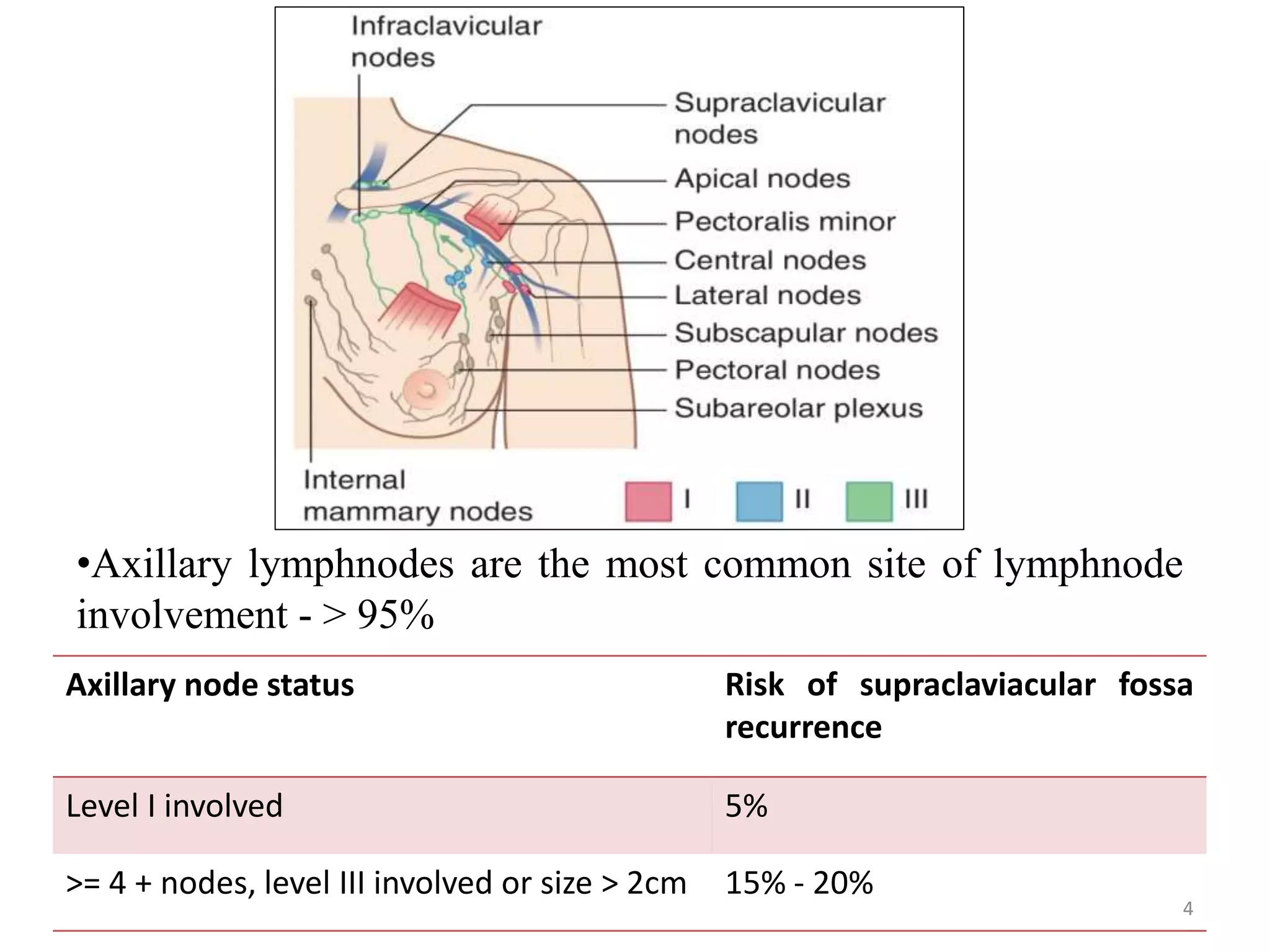
Management of Regional Lymph
Nodes in Breast Cancer
Dr. Shreya Singh
JR-III
Department of Radiation Oncology
IMS, BHU 1
Regional Lymph Nodes in Breast
Cancer
• Axillary group of lymph nodes
• Supra-clavicular lymph nodes
• Internal mammary chain of lymph nodes
2
Clinical Anatomy – Axillary and
Supraclavicular Lymph Nodes
Level Group Location
I Anterior, Posterior & Lateral
groups
Inferior and lateral to the Pectoralis
minor
II Central group Behind the Pectoralis minor
III Apical group Medial to the Pectoralis minor
3
Axillary node status Risk of supraclaviacular fossa
recurrence
Level I involved 5%
>= 4 + nodes, level III involved or size > 2cm 15% - 20%
•Axillary lymphnodes are the most common site of lymphnode
involvement - > 95%
4
Risk Factors for Axillary LN Involvement :
• Tumor size ≥ 5 cm
• Positive margins
• positive familial cancer history
• Age < 40 years
• Nipple involvement
• skin involvement
• perineural invasion
• lymphatic vessel invasion
• presence of an extensive intraductal component
5
Risk Factors for SCF Nodes Involvement :
• Tumor size ≥ 5 cm
• High histologic grade
• >4 positive nodes
• Level II or III involved nodes
• Angiolymphatic invasion
• Young age (< or =40 years)
6
Tumor size Lymphnode
involvement
< 0.5 cm 3 – 7 %
0.6 – 1 cm 12 – 17 %
1.1 – 2 cm 20 – 30%
2.1 – 3 cm 35 – 45%
3.1 – 5 cm 40 – 60%
> 5 cm 60 – 85%
Gunderson 4th edition 20157
Clinical Anatomy - Internal Mammary
Chain
• Lymphatics can also drain
into the internal mammary
lymph node chain (IMC).
• Not visualized on a CT scan
but their anatomical region
can be determined by the
internal mammary vessels,
which are easily seen on CT &
usually lie 3 to 4 cm lateral to
the midline.
8
• Breast cancer which develops in the medial, central or lower
breast more commonly drain to the IMC in addition to the
axillary lymph nodes.
• The frequency of IMC involvement depends on the tumor
location and axillary lymph node status.
Lower inner tumors(43%) > lower outer tumors(32%) >
central tumors(25%)
(Huang et al in China)
9
10
Assessment of the Axilla
11
Assessment of the Axilla
• Clinical Assessment
• Axillary Ultrasound
• Sentinel Lymph Node Biopsy
• Axillary Dissection
12
Clinical Assessment
• Physical examination of regional nodal basins
• Determine the size and whether the nodes
are matted or not
• Unfortunately, physical examination is
impacted by body habitus, making it highly
unreliable with a false-negative rate of as high
as 45%
13
Axillary Ultrasound
• Preoperative differentiation between limited and
advanced nodal disease can provide relevant
information regarding surgical planning and guiding
adjuvant radiation therapy.
• Ultrasound - preferred nodal assessment before therapy
• Benefit - needle biopsy for pathologic confirmation
14
Axillary Ultrasound
• Axillary US with fine-needle
aspiration (FNA) of abnormal
nodes have
o Sensitivity - 86.4% (93% if
metastatic deposits > 5 mm
vs44% for < 5 mm)
o Specificity - 100%
• Place clips to mark nodes with
biopsy - confirmed disease.
15
Sentinel Lymph Node Biopsy
• Sentinel lymph node – The hypothetical first
lymph node or group of nodes draining a cancer
• Most likely to contain metastasis if tumor has
metastasized
• First introduced for melanoma in early 1990s
• Done in-
o Malignant melanoma
o Breast cancer
o Also employed for staging- penile cancer
16
Sentinel Lymph Node Biopsy
• Accuracy increases when both blue dye and
radioactive colloid (Tc 99m sulphur colloid) are
used together
• Uptake is detected by Gamma probe
• Rationale is to identify subset of patients with
negative sentinel lymph nodes
• These patients can avoid subsequent axillary
dissection & morbidity
17
Sentinel Lymph Node Biopsy
18
Landmark Trials
19
ACOSOG Z0011
20
ACOSOG Z0011
• Eligibility Criteria :
o T1, T2 tumor
o Patients with 1 or 2 positive sentinel LN
o Breast conserving surgery
o No pre-op chemotherapy
• Median follow up – 6.3 years
21
22
Clinically Node Negative Disease:
Ref : ACOSOG Z0011 (Giuliano AE et al, 2017)23
Clinically Node Positive Disease:
Ref : ACOSOG Z0011 (Giuliano AE et al, 2017)
24
AMAROS TRIAL :
25
AMAROS TRIAL :
• Designed for higher risk sentinel node +ve patients
who do not fit into ACOSOG Z0011 criteria ( eg: 3
sentinel node +ve )
• First trial to prospectively compare axillary LN
dissection with axillary RT in such patients
• RESULT-
Lymphoedema noted to be significantly higher after
axillary LN dissection than after axillary RT
26
NSABP B-04
27
NSABP B-04
• Eligibility criteria :
o tumors confined to the breast or breast and axilla
o tumors movable in relation to the underlying
muscle and the chest wall
o axillary nodes movable in relation to the
o chest wall and neurovascular bundle
o no arm edema
o patient’s consent to participate
28
NSABP B-04
Negative
Axillary Nodes
Positive Axillary
Nodes
Clinical Examination
Radical
Mastectomy
Radical
Mastectomy
Total
Mastectomy
Total
Mastectomy
+ RT
Total
Mastectomy +
RT
Axillary
Dissection for
Positive Axillary
Nodes
29
NSABP B-04
• 1,079 patients with clinically negative axillary LN randomized
to 1 of 3 arms: radical mastectomy vs. total mastectomy (TM)
without axillary dissection but with post-op RT vs. total
mastectomy plus axillary dissection if LN pathologically positive.
• Also 586 patients with clinically + axillary LN randomized to 1
of 2 arms (radical mastectomy vs. total mastectomy without
axillary dissection but with post-op RT).
• No systemic therapy.
30
Results :
• At 25-year follow-up, no significant differences in DFS, or OS
among the three groups of patients with clinically negative
LN or the two groups of patients with clinically + LN.
• Approximately 40% of cN0 patients were found to be pLN+
after ALND. Among cN0 patients, axillary failure was <4% if
addressed surgically or with RT vs. 19% in TM alone arm.
• Nodal control rates in radical mastectomy arms and total
mastectomy plus radiation were comparable.
31
Canadian MA-20 Trial:
32
Canadian MA-20 Trial :
Inclusion Criteria –
• Treated with breast conserving surgery and sentinel-lymph-node
biopsy or axillary-node dissection
• Positive axillary lymph nodes or negative axillary nodes with high-
risk features like -
o primary tumor measuring ≥5 cm
o primary tumor measuring ≥ 2 cm with fewer than 10 axillary
nodes removed and at least one of the following:
• grade 3 histology
• ER negativity
• lymphovascular invasion.
• All patients received adjuvant systemic therapy with chemotherapy,
endocrine therapy, or both.
33
Canadian MA-20 Trial :
34
• Enrolled 1832 women
• Most (85%) had 1 to 3 positive lymph nodes, and a smaller
proportion (10%) had high-risk, node- negative breast cancer
• BCS+adj.Chemo or endcrine therapy
• Randomised to WBI Vs WBI+Regional nodal irradiation
• 62 months follow up
• RESULT –
Regional node irradiation reduces the risk of locoregional
and distant recurrence and improves DFS after 5 years
EORTC trial 22922/10925 :
35
EORTC trial 22922/10925 :
• Eligibility criteria :
o Histologically confirmed breast adenocarcinoma
o Stage I, II, or III
o Centrally or medially located primary tumor,
irrespective of axillary involvement or an
externally located tumor with axillary involvement
o Eligible patients had undergone mastectomy or
breast conserving surgery and axillary dissection
36
EORTC trial 22922/10925 :
37
Patient Characterstics :
38
Results:
• At median follow-up of 11 years –
o Regional nodal irradiation improved DFS (72.1 %
vs. 69.1%)
o Regional nodal irradiation improved distant DFS
(78 % vs. 75%)
o Regional nodal irradiation improved breast cancer
mortality (12.5% vs. 14.4%)
39
Conclusion :
• The results were similar to those of the National
Cancer Institute of Canada Clinical Trials Group MA.20
trial.
• They concluded that regional nodal irradiation was
beneficial to women with early stage breast cancer.
• It improved the rates of disease-free and distant
disease-free survival.
• It reduced the rate of death from breast cancer among
patients with involved axillary nodes, a medially or
centrally located primary tumor, or both.
40
EBCTCG Meta-analysis 2014
41
EBCTCG Meta-analysis 2014
• It is a meta-analysis of individual data for 8135 women
randomly assigned to treatment groups during 1964–1986 in
22 trials of radiotherapy to the chest wall and regional lymph
nodes after mastectomy and axillary surgery versus the same
surgery but no radiotherapy.
• Follow-up lasted 10 years for recurrence
42
• For 700 women with axillary dissection and no positive nodes,
radiotherapy had no significant effect on locoregional recurrence,
overall recurrence or breast cancer mortality.
• For 1314 women with axillary dissection and one to three positive
nodes, radiotherapy reduced locoregional recurrence, overall
recurrence and breast cancer mortality.
• 1133 of these 1314 women were in trials in which systemic therapy
(CMF or tamoxifen) was given in both trial groups and, for them,
radiotherapy again reduced locoregional recurrence, overall
recurrence and breast cancer mortality.
• For 1772 women with axillary dissection and four or more positive
nodes, radiotherapy reduced locoregional recurrence, overall
recurrence and breast cancer mortality.
43
44
Interpretation :
• The beneficial effects of radiotherapy on recurrence and
breast cancer mortality remained apparent when women with
one to three involved lymph nodes who were in trials in which
the policy was to give systemic therapy were considered on
their own.
• By contrast, in women with node-negative disease who
received mastectomy and axillary dissection, among whom
the proportion of women who had a locoregional recurrence
before any distant recurrence was small, there was no
evidence that radiotherapy provided any benefit.
45
Current Recommendations For
Regional Lymph Node Irradiation in
Breast Cancer
46
Indications for Regional lymph nodes
Irradiation ( Irrespective of Disease Stage)
Axillary Group of Lymph Nodes –
o ALN + with extensive extra capsular extension
o Sentinel lymphnode + with no dissection
o Inadequate axillary dissection
o High risk with no dissection
47
Indications for Regional lymph nodes
Irradiation ( Irrespective of Disease Stage)
Supraclavicular nodes –
o >= 4 + ALN after axillary dissection
o 1-3 ALN with high risk features
o High risk no dissection
o Clinically N2 or N3
o Sentinel lymphnode + with no dissection
48
Indications for Regional lymph nodes
Irradiation ( Irrespective of Disease Stage)
Internal Mammary Chain-
Individualised but consider for :
o + Axillary LN with central and medial lesions
o Sentinel lymphnode + in IMC
o + Sentinel lymphnode in axilla with drainage to IMC
o Stage III
49
RT Techniques :
• Conventional (2D)
• 3D- CRT
• IMRT
50
Patient Positioning :
Supine position
• Breast tilt board, to make chest wall surface
horizontal (make slope of chest wall parallel to
the table).
• Arm abducted at 90⁰ & hand holds handle of
arm rest. When arm angled >90,axillary nodes
overlap head of humerus.
• Face turned towards opposite side
51
Supraclavicular Lymph Nodes :
Single anterior field is used.
• Upper border :thyrocricoid groove
• Medial border :1 cm across midline extending
upward following medial border of SCM muscle
to thyrocricoid groove
• Lateral border: Vertical line at the level of the
coracoid process, just medial to the humeral
head.
• Lower border : matched with upper border of
tangential fields just below the clavicular head.
• Dose : 40Gy at 2.66 Gy/# in 15 # (calculated at a
depth of 3 cm) in 3 weeks.
52
Axillary Lymph Nodes
• The supraclavicular field is extended laterally to
cover at least two thirds of the humeral head
• Ideal depth of treatment= 5cm
• Level I as well as a portion of level II nodes will often
be included in the tangential field; level III and
supraclavicular nodes are covered in the
supraclavicular field.
53
Posterior Axillary Boost
• Palpate apex of axilla
• Mark this point on the anterior
skin surface
• Using this point as centre, a 8 x 8
cm template is aligned such that
superior border lies along the
upper border of clavicle .
• Field is set up using these
anterior skin marks
• Gantry rotated through 180
degree to deliver treatment.
54
Internal Mammary Lymph Nodes
• Clinical failures at this site are very rare.
• The majority of patients at risk receive adjuvant therapy.
• IMNs are difficult to treat because their exact location is
often uncertain.
• Irradiation of normal tissue
• Dose prescription point at depths of 4 to 5 cm
55
Internal Mammary Lymph Nodes
FIELD BORDERS FOR DIRECT
ANTERIOR FIELD -
• Medial border : midline
• Lateral border : 5 to 6 cm lateral
to the midline
• Superior border: abuts the
inferior border of the
supraclavicular field
• Inferior border : at the xiphoid or
higher
56
Internal Mammary Lymph Nodes
PARTIALLY WIDE TANGENTIAL
FIELDS
• Avoids matching of fields
• The nodes in the first three
intercostal spaces are thought to be
most clinically significant.
• The medial border of the tangential
field is moved 3 to 5 cm across the
midline to cover the internal
mammary nodes in the first three
intercostal spaces
57
IMC – Electron Beam
• To spare underlying lung, mediastinum, and spinal cord
• Electrons in the range of 12 to 16 MeV are preferred for
a portion of the treatment
58
3D-CRT or IMRT
• Reduction in the volume of normal tissues receiving a high dose,
with an increase in dose to the target volume.
• Improved dose distribution between the target and nontarget
tissue.
• Drawback: increase the volume of tissue exposed to lower doses of
radiation.
• Significantly lower degrees of moist desquamation due to the
improved homogeneity, compared with standard tangential fields.
59
Complications of Regional Nodal Irradiation:
• Skin toxicity
• Lymphoedema
• Impaired shoulder mobility
• Brachial plexopathy
• Increased incidence of symptomatic
pneumonitis
60
61

Regional lymph node management in breast cancer

  • 1.
    Management of RegionalLymph Nodes in Breast Cancer Dr. Shreya Singh JR-III Department of Radiation Oncology IMS, BHU 1
  • 2.
    Regional Lymph Nodesin Breast Cancer • Axillary group of lymph nodes • Supra-clavicular lymph nodes • Internal mammary chain of lymph nodes 2
  • 3.
    Clinical Anatomy –Axillary and Supraclavicular Lymph Nodes Level Group Location I Anterior, Posterior & Lateral groups Inferior and lateral to the Pectoralis minor II Central group Behind the Pectoralis minor III Apical group Medial to the Pectoralis minor 3
  • 4.
    Axillary node statusRisk of supraclaviacular fossa recurrence Level I involved 5% >= 4 + nodes, level III involved or size > 2cm 15% - 20% •Axillary lymphnodes are the most common site of lymphnode involvement - > 95% 4
  • 5.
    Risk Factors forAxillary LN Involvement : • Tumor size ≥ 5 cm • Positive margins • positive familial cancer history • Age < 40 years • Nipple involvement • skin involvement • perineural invasion • lymphatic vessel invasion • presence of an extensive intraductal component 5
  • 6.
    Risk Factors forSCF Nodes Involvement : • Tumor size ≥ 5 cm • High histologic grade • >4 positive nodes • Level II or III involved nodes • Angiolymphatic invasion • Young age (< or =40 years) 6
  • 7.
    Tumor size Lymphnode involvement <0.5 cm 3 – 7 % 0.6 – 1 cm 12 – 17 % 1.1 – 2 cm 20 – 30% 2.1 – 3 cm 35 – 45% 3.1 – 5 cm 40 – 60% > 5 cm 60 – 85% Gunderson 4th edition 20157
  • 8.
    Clinical Anatomy -Internal Mammary Chain • Lymphatics can also drain into the internal mammary lymph node chain (IMC). • Not visualized on a CT scan but their anatomical region can be determined by the internal mammary vessels, which are easily seen on CT & usually lie 3 to 4 cm lateral to the midline. 8
  • 9.
    • Breast cancerwhich develops in the medial, central or lower breast more commonly drain to the IMC in addition to the axillary lymph nodes. • The frequency of IMC involvement depends on the tumor location and axillary lymph node status. Lower inner tumors(43%) > lower outer tumors(32%) > central tumors(25%) (Huang et al in China) 9
  • 10.
  • 11.
  • 12.
    Assessment of theAxilla • Clinical Assessment • Axillary Ultrasound • Sentinel Lymph Node Biopsy • Axillary Dissection 12
  • 13.
    Clinical Assessment • Physicalexamination of regional nodal basins • Determine the size and whether the nodes are matted or not • Unfortunately, physical examination is impacted by body habitus, making it highly unreliable with a false-negative rate of as high as 45% 13
  • 14.
    Axillary Ultrasound • Preoperativedifferentiation between limited and advanced nodal disease can provide relevant information regarding surgical planning and guiding adjuvant radiation therapy. • Ultrasound - preferred nodal assessment before therapy • Benefit - needle biopsy for pathologic confirmation 14
  • 15.
    Axillary Ultrasound • AxillaryUS with fine-needle aspiration (FNA) of abnormal nodes have o Sensitivity - 86.4% (93% if metastatic deposits > 5 mm vs44% for < 5 mm) o Specificity - 100% • Place clips to mark nodes with biopsy - confirmed disease. 15
  • 16.
    Sentinel Lymph NodeBiopsy • Sentinel lymph node – The hypothetical first lymph node or group of nodes draining a cancer • Most likely to contain metastasis if tumor has metastasized • First introduced for melanoma in early 1990s • Done in- o Malignant melanoma o Breast cancer o Also employed for staging- penile cancer 16
  • 17.
    Sentinel Lymph NodeBiopsy • Accuracy increases when both blue dye and radioactive colloid (Tc 99m sulphur colloid) are used together • Uptake is detected by Gamma probe • Rationale is to identify subset of patients with negative sentinel lymph nodes • These patients can avoid subsequent axillary dissection & morbidity 17
  • 18.
  • 19.
  • 20.
  • 21.
    ACOSOG Z0011 • EligibilityCriteria : o T1, T2 tumor o Patients with 1 or 2 positive sentinel LN o Breast conserving surgery o No pre-op chemotherapy • Median follow up – 6.3 years 21
  • 22.
  • 23.
    Clinically Node NegativeDisease: Ref : ACOSOG Z0011 (Giuliano AE et al, 2017)23
  • 24.
    Clinically Node PositiveDisease: Ref : ACOSOG Z0011 (Giuliano AE et al, 2017) 24
  • 25.
  • 26.
    AMAROS TRIAL : •Designed for higher risk sentinel node +ve patients who do not fit into ACOSOG Z0011 criteria ( eg: 3 sentinel node +ve ) • First trial to prospectively compare axillary LN dissection with axillary RT in such patients • RESULT- Lymphoedema noted to be significantly higher after axillary LN dissection than after axillary RT 26
  • 27.
  • 28.
    NSABP B-04 • Eligibilitycriteria : o tumors confined to the breast or breast and axilla o tumors movable in relation to the underlying muscle and the chest wall o axillary nodes movable in relation to the o chest wall and neurovascular bundle o no arm edema o patient’s consent to participate 28
  • 29.
    NSABP B-04 Negative Axillary Nodes PositiveAxillary Nodes Clinical Examination Radical Mastectomy Radical Mastectomy Total Mastectomy Total Mastectomy + RT Total Mastectomy + RT Axillary Dissection for Positive Axillary Nodes 29
  • 30.
    NSABP B-04 • 1,079patients with clinically negative axillary LN randomized to 1 of 3 arms: radical mastectomy vs. total mastectomy (TM) without axillary dissection but with post-op RT vs. total mastectomy plus axillary dissection if LN pathologically positive. • Also 586 patients with clinically + axillary LN randomized to 1 of 2 arms (radical mastectomy vs. total mastectomy without axillary dissection but with post-op RT). • No systemic therapy. 30
  • 31.
    Results : • At25-year follow-up, no significant differences in DFS, or OS among the three groups of patients with clinically negative LN or the two groups of patients with clinically + LN. • Approximately 40% of cN0 patients were found to be pLN+ after ALND. Among cN0 patients, axillary failure was <4% if addressed surgically or with RT vs. 19% in TM alone arm. • Nodal control rates in radical mastectomy arms and total mastectomy plus radiation were comparable. 31
  • 32.
  • 33.
    Canadian MA-20 Trial: Inclusion Criteria – • Treated with breast conserving surgery and sentinel-lymph-node biopsy or axillary-node dissection • Positive axillary lymph nodes or negative axillary nodes with high- risk features like - o primary tumor measuring ≥5 cm o primary tumor measuring ≥ 2 cm with fewer than 10 axillary nodes removed and at least one of the following: • grade 3 histology • ER negativity • lymphovascular invasion. • All patients received adjuvant systemic therapy with chemotherapy, endocrine therapy, or both. 33
  • 34.
    Canadian MA-20 Trial: 34 • Enrolled 1832 women • Most (85%) had 1 to 3 positive lymph nodes, and a smaller proportion (10%) had high-risk, node- negative breast cancer • BCS+adj.Chemo or endcrine therapy • Randomised to WBI Vs WBI+Regional nodal irradiation • 62 months follow up • RESULT – Regional node irradiation reduces the risk of locoregional and distant recurrence and improves DFS after 5 years
  • 35.
  • 36.
    EORTC trial 22922/10925: • Eligibility criteria : o Histologically confirmed breast adenocarcinoma o Stage I, II, or III o Centrally or medially located primary tumor, irrespective of axillary involvement or an externally located tumor with axillary involvement o Eligible patients had undergone mastectomy or breast conserving surgery and axillary dissection 36
  • 37.
  • 38.
  • 39.
    Results: • At medianfollow-up of 11 years – o Regional nodal irradiation improved DFS (72.1 % vs. 69.1%) o Regional nodal irradiation improved distant DFS (78 % vs. 75%) o Regional nodal irradiation improved breast cancer mortality (12.5% vs. 14.4%) 39
  • 40.
    Conclusion : • Theresults were similar to those of the National Cancer Institute of Canada Clinical Trials Group MA.20 trial. • They concluded that regional nodal irradiation was beneficial to women with early stage breast cancer. • It improved the rates of disease-free and distant disease-free survival. • It reduced the rate of death from breast cancer among patients with involved axillary nodes, a medially or centrally located primary tumor, or both. 40
  • 41.
  • 42.
    EBCTCG Meta-analysis 2014 •It is a meta-analysis of individual data for 8135 women randomly assigned to treatment groups during 1964–1986 in 22 trials of radiotherapy to the chest wall and regional lymph nodes after mastectomy and axillary surgery versus the same surgery but no radiotherapy. • Follow-up lasted 10 years for recurrence 42
  • 43.
    • For 700women with axillary dissection and no positive nodes, radiotherapy had no significant effect on locoregional recurrence, overall recurrence or breast cancer mortality. • For 1314 women with axillary dissection and one to three positive nodes, radiotherapy reduced locoregional recurrence, overall recurrence and breast cancer mortality. • 1133 of these 1314 women were in trials in which systemic therapy (CMF or tamoxifen) was given in both trial groups and, for them, radiotherapy again reduced locoregional recurrence, overall recurrence and breast cancer mortality. • For 1772 women with axillary dissection and four or more positive nodes, radiotherapy reduced locoregional recurrence, overall recurrence and breast cancer mortality. 43
  • 44.
  • 45.
    Interpretation : • Thebeneficial effects of radiotherapy on recurrence and breast cancer mortality remained apparent when women with one to three involved lymph nodes who were in trials in which the policy was to give systemic therapy were considered on their own. • By contrast, in women with node-negative disease who received mastectomy and axillary dissection, among whom the proportion of women who had a locoregional recurrence before any distant recurrence was small, there was no evidence that radiotherapy provided any benefit. 45
  • 46.
    Current Recommendations For RegionalLymph Node Irradiation in Breast Cancer 46
  • 47.
    Indications for Regionallymph nodes Irradiation ( Irrespective of Disease Stage) Axillary Group of Lymph Nodes – o ALN + with extensive extra capsular extension o Sentinel lymphnode + with no dissection o Inadequate axillary dissection o High risk with no dissection 47
  • 48.
    Indications for Regionallymph nodes Irradiation ( Irrespective of Disease Stage) Supraclavicular nodes – o >= 4 + ALN after axillary dissection o 1-3 ALN with high risk features o High risk no dissection o Clinically N2 or N3 o Sentinel lymphnode + with no dissection 48
  • 49.
    Indications for Regionallymph nodes Irradiation ( Irrespective of Disease Stage) Internal Mammary Chain- Individualised but consider for : o + Axillary LN with central and medial lesions o Sentinel lymphnode + in IMC o + Sentinel lymphnode in axilla with drainage to IMC o Stage III 49
  • 50.
    RT Techniques : •Conventional (2D) • 3D- CRT • IMRT 50
  • 51.
    Patient Positioning : Supineposition • Breast tilt board, to make chest wall surface horizontal (make slope of chest wall parallel to the table). • Arm abducted at 90⁰ & hand holds handle of arm rest. When arm angled >90,axillary nodes overlap head of humerus. • Face turned towards opposite side 51
  • 52.
    Supraclavicular Lymph Nodes: Single anterior field is used. • Upper border :thyrocricoid groove • Medial border :1 cm across midline extending upward following medial border of SCM muscle to thyrocricoid groove • Lateral border: Vertical line at the level of the coracoid process, just medial to the humeral head. • Lower border : matched with upper border of tangential fields just below the clavicular head. • Dose : 40Gy at 2.66 Gy/# in 15 # (calculated at a depth of 3 cm) in 3 weeks. 52
  • 53.
    Axillary Lymph Nodes •The supraclavicular field is extended laterally to cover at least two thirds of the humeral head • Ideal depth of treatment= 5cm • Level I as well as a portion of level II nodes will often be included in the tangential field; level III and supraclavicular nodes are covered in the supraclavicular field. 53
  • 54.
    Posterior Axillary Boost •Palpate apex of axilla • Mark this point on the anterior skin surface • Using this point as centre, a 8 x 8 cm template is aligned such that superior border lies along the upper border of clavicle . • Field is set up using these anterior skin marks • Gantry rotated through 180 degree to deliver treatment. 54
  • 55.
    Internal Mammary LymphNodes • Clinical failures at this site are very rare. • The majority of patients at risk receive adjuvant therapy. • IMNs are difficult to treat because their exact location is often uncertain. • Irradiation of normal tissue • Dose prescription point at depths of 4 to 5 cm 55
  • 56.
    Internal Mammary LymphNodes FIELD BORDERS FOR DIRECT ANTERIOR FIELD - • Medial border : midline • Lateral border : 5 to 6 cm lateral to the midline • Superior border: abuts the inferior border of the supraclavicular field • Inferior border : at the xiphoid or higher 56
  • 57.
    Internal Mammary LymphNodes PARTIALLY WIDE TANGENTIAL FIELDS • Avoids matching of fields • The nodes in the first three intercostal spaces are thought to be most clinically significant. • The medial border of the tangential field is moved 3 to 5 cm across the midline to cover the internal mammary nodes in the first three intercostal spaces 57
  • 58.
    IMC – ElectronBeam • To spare underlying lung, mediastinum, and spinal cord • Electrons in the range of 12 to 16 MeV are preferred for a portion of the treatment 58
  • 59.
    3D-CRT or IMRT •Reduction in the volume of normal tissues receiving a high dose, with an increase in dose to the target volume. • Improved dose distribution between the target and nontarget tissue. • Drawback: increase the volume of tissue exposed to lower doses of radiation. • Significantly lower degrees of moist desquamation due to the improved homogeneity, compared with standard tangential fields. 59
  • 60.
    Complications of RegionalNodal Irradiation: • Skin toxicity • Lymphoedema • Impaired shoulder mobility • Brachial plexopathy • Increased incidence of symptomatic pneumonitis 60
  • 61.