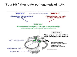
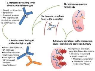
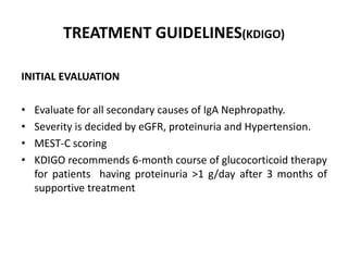
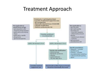
IgAN is the commonest GN worldwide with varied presentation without any concrete medical therapy to halt disease progression.So in this slide we will talk about pathogenesis & possible target of future therapies for IgAN
2. Introduction & Epidemiology
• Characterized by diffuse deposition of IgA in
mesangium with mesangial proliferation with varied
clinical spectrum from asymptomatic hematuria to
rapidly progressive glomerulonephritis.
• First described by Jean Berger and Nicole Hinglais in
1968
• Most prevalent Primary Glomerulonephritis
worldwide
• 30-40%,20%,10% of all biopsies performed in Asia,
Europe , North America respectively
3. • A clinicopathologic study of glomerular
disease: A single-center, five-year
retrospective study from Northwest India.
Saudi J Kidney Dis Transpl 2016;27:997-1005
Showed prevalence of 9.3%
• It affects individuals of all ages, but most
common in 2nd to 3rd decade, 80% of patients
are between age 16 to 35 at time of diagnosis.
• More common in males
• Initially thought to have a benign course, upto
40% of patients may progress to ESKD
• Overall, about 25% of patients develop ESKD
within 10 to 25 years from diagnosis
4. IgA Nephropathy types
• Primary IgA nephropathy or Idiopathic variety
• Secondary IgA nephropathy
• Familial IgA nephropathy
6. Pathogenesis
• In humans , IgA exists in two isoforms IgA1 and
IgA2 (84% and 16% respectively)
• Both isoforms can be in the monomeric and
polymeric forms, mIgA, and pIgA.
• Most pIgA1 and pIgA2 is produced in the mucosal
tissues, where pIgA molecules moves to the
mucosal surfaces by transcytosis .
• The BAFF/APRIL axis helps in class switch of
mucosal B cells into IgA producing plasmablasts and
plasma cells
• Some of these cells mistraffic to systemic circulation
during lymphocyte trafficking & these cells are
mainly responsible for increased GdIgA in IgAN
7. • IgAN is the result of the deposition of circulating immune
complexes leading to the activation of the complement
cascade via the alternate & lectin pathway.
• Previously it was thought that deposited IgA is
predominantly polymeric IgA1 due to the fact that
polymeric IgA1 is usually derived mainly from the mucosal
immune system
• This concept was supported by the clinical observation
that hematuria increases acutely in some patients at the
time of upper respiratory tract or gastrointestinal
infections.
8. • However, it has now been determined that the elevation in
polymeric IgA1 antibody synthesis does not occur in the mucosa
and polymeric IgA levels can be increased after systemic
immunization.
• In addition,Serum levels of IgA do not correlate with disease activity
or mesangial deposits; therefore, it is unlikely that the pathogenesis
of IgA nephropathy is related to a quantitative increase in serum
levels of polymeric IgA1.
• Rather, it relates to an anomaly in the IgA molecule itself, mainly in
its glycosylation.
• This is best exemplified by patients with IgA-secreting multiple
myeloma, only those patients with aberrant IgA glycosylation
develop glomerulonephritis.
• This leads to development of “Four Hit theory ”
10. 1. Increased circulating levels
of Galactose deficient-IgA1
2. Production of Anti-IgA1
antibodies (IgA or IgG)
3a. Immune complexes
form in the circulation
4. Immune complexes in the mesangium
cause local immune activation & injury
• Complement activation
• Cytokine/chemokine release
• Monocyte recruitment
• Matrix production
• Mesangial proliferation
• Glomerular sclerosis
• Interstitial Fibrosis
• Genetic predisposition
• Epigenetics
• Enzymatic variants
• Mis-trafficking of
B cells from mucosal
to systemic sites?
• Genetic predisposition,
HLA haplotype
• Germline mutations
• Molecular mimicry
• Viral infection
• Streptococcal
antigens
• Food antigens?
3b. Immune complexes
form in situ
11. Hit 1( Galactose deficient IgA1): GdIgA1
• In humans, the heavy chain of IgA1, but not that of IgA2, contains
an 18-amino acid hinge region that is rich in proline, serine, and
threonine residues.
• O-linked monosaccharides or oligosaccharides consisting of N-
acetylgalactosamine posttranslationally added to these amino acid
residues
• This N-acetylgalactosamine is usually substituted with a terminal
galactose by the enzyme core 1 β1,3-galactosyltransferase
(C1GALT1) and its chaperone(COSMC)(C1GALT1C1)
• The addition of a galactose residue to the glycosyl side chain is
blocked by premature sialylation of the N-acetylgalactosamine
residues on the hinge region of IgA1.
• So IgA galactosylation can be affected by excessive activity of α-2,6-
sialyltransferase, decreased activity of ß-1,3-galactosyltransferase,
and decreased stability of ß-1,3-galactosyltransferase due to
decreased activity of its chaperone & leads to formation of GdIgA1.
• Whether these abnormalities are acquired or genetically
determined remains unclear.
12.
13. Autoantibodies (Hit #2):
Galactose-deficient IgA1 (Gd-IgA1) reults in hinge region exposure of N-
acetylgalactosamine and neoepitope formation. This neoepitope is the target
of autoantibodies.
IgG is the predominant autoantibody isotype specific for Gd-IgA1
It is not known what triggers the production of autoantibodies
One hypothesis proposes that an infection by microorganisms carrying NAG
on their outer surfaces elicits production of NAG recognizing antibodies that
cross-react with Gd-IgA1.
Infection by Epstein–Barr virus, respiratory syncytial virus, herpes simplex
virus, and streptococci may induce production of such antibodies
14. Circulating Immune Complexes (Hit #3):
Circulating immune complexes (CICs) in IgAN patients consist of IgG
autoantibodies bound to Gd polymeric IgA1
The biological activity of the CICs is determined by composition and size
The CICs containing high content of Gd-IgA1 & Large-molecular-weight CICs
(800–900 kDa) are biologically active & can induce proliferation of mesangial
cells in culture, which was not observed for Gd-IgA1 alone and smaller
immune complexes
15. Deposition of Circulating Immune Complexes
and Renal Injury (Hit #4):
Circulating immune complexes that escape removal by the reticuloendothelial
system deposit in glomeruli via an interaction with a mesangial IgA receptor,
possibly the transferrin receptor.
Deposition of immuncomplexes results in local immune activation ,
inflammation & glomerular injury
16.
17. • In patients with familial and sporadic IgAN,
the serum level of Gd-IgA1 is a heritable trait;
many blood relatives have higher levels but
without any clinical manifestation of IgA
• Genome-wide association studies (GWAS) of
serum levels of Gd-IgA1 have identified two
novel loci in C1GALT1 and C1GALT1C1 .
• Reduced expression
of C1GALT1 and C1GALT1C1 manifested by
addition of less galactose to IgA
18. Secondary IgA
• Cirrhosis: due to impaired hepatic
clearance of abnormally glycosylated IgA.
• In Inflammatory Bowel Disease: due to
mucosal inflammation and increased
numbers or activity of IgA1-secreting cells.
19. • HIV : Due to IgA anti-HIV immune complexes
and several patients had circulating immune
complexes containing IgA idiotypic antibodies
directed against viral proteins, either anti-HIV
p24 or HIV gp41
• Mucosa-associated lymphoid tissue
lymphoma: Due to increase number of IgA
producing plasma cells resolved following
chemotherapy.
20. Pathology
Immunofluorescence Microscopy:
• glomerular immune deposits that stain dominantly or codominantly for IgA
• Staining is usually mesangial, but in patients with severe disease can have substantial capillary wall
staining.
• By definition, 100% of IgA nephropathy specimens stain for IgA. On a scale of 0 to 4+, the mean
intensity of IgA staining is approximately 3+
• IgM & IgG staining is observed in 84% & 62%of specimens respectively with a mean intensity
(when present) of only approximately 1+
• Almost all IgA nephropathy specimens have substantial staining for C3.
• Staining for C1q is rare and weak when present. If there is intense staining in a specimen that shows
substantial IgA and IgG, the possibility of lupus nephritis should be considered.
• Distinctive feature of IgA nephropathy is that unlike other immune complex disease ,more intense
staining of Gamma than Kappa light chains
21.
22. Electron Microscopy
Mesangial electrondense deposits
Immediately beneath the perimesangial basement membrane
Varying degrees of mesangial matrix expansion and hypercellularity.
Foot process effacement in patients with substantial proteinuria.
23. Light Microscopy :
Can cause any of the light microscopic phenotypes of proliferative
glomerulonephritis or normal glomeruli
Because of the episodic nature of IgAN, many patients have combinations of
focal sclerotic lesions and focal active proliferative lesions.
25. Oxford MEST Classification
• Histological features used to predict the progression of IgA
nephropathy.
• The study population was a multiethnic cohort of patients with IgA
nephropathy that included children (n = 59) and adults (n = 206)
who were followed for a mean of 69 months
• 2009-2012
• 4 parameters emerged as independently predictive of clinical
outcomes: mesangial hypercellularity(M), endocapillary
hypercellularity(E), segmental glomerulosclerosis(S), and tubular
atrophy–interstitial fibrosis(T).
• IgA Nephropathy Classification Working Group in 2017 also found
that the crescents are predictive of outcome, thereby changing the
MEST score to the MEST-C score (C for crescents)
26. MEST-C Score
Mesangial hypercellularity
(M)
≥4 mesangial cells in any
mesangial area of a
glomerulus
M0: mesangial
hypercellularity in ≤50% of
glomeruli M1: mesangial
hypercellularity in >50% of
glomeruli
Endocapillary hypercellularity
(E)
An increased number of cells
in glomerular capillary lumen
E0: absence E1: presence of
any glomeruli showing
endocapillary hypercellularity
Segmental
glomerulosclerosis (S)
Adhesion or sclerosis that
not involving the entire
glomerulus
S0: absence S1: presence of
any segmental
glomerulosclerosis
Tubular atrophy/ interstitial
fibrosis (T)
The percentage of tubular
atrophy/ interstitial fibrosis
of cortical area
T0: 0-25% T1: 26%-50% T2:
>50%
Cellular/ fibrocellular
crescents (C)
Extracapillary cell
proliferation > 2 cell layers
thick and < 50% matrix
C0: absence C1: crescents in
<25% of glomeruli C2:
crescents in ≥25% of
glomeruli
At least 8 viable glomeruli are required for MEST-C scoring.
27.
28. Interpretation of MEST-C Score :
• E and C scores are most responsive to treatment
• M, S, T, and C lesions (not E lesions) were strongly associated with
progression to kidney failure,
29. Clinical features
• Recurrent macroscopic hematuria 40 to 50% of patients which
usually coincides with mucosal infection .More often in
children than in young adults.
• Asymptomatic hematuria with or without proteinuria 30 to
40%
• Nephrotic Range proteinuria is unusual, only 5% of all IgAN.
30. Clinical Spectrum
1. Asymptomatic Hematuria, Varying Degree of
Proteinuria, With or Without Progressive Kidney
Disease: most Common
2. Synpharyngitic Macroscopic Hematuria: 10% to
15% of adult patients
3. Rapidly Progressive Glomerulonephritis
4. Nephrotic Syndrome
5. Acute Kidney Injury in IgA Nephropathy: RPGN
and acute tubular injury from red blood cell cast
obstruction and/or heme toxicity.
31. Investigation
• A typical finding is microscopic hematuria on
urinalysis
• Dysmorphic erythrocytes in the urine is
typical.
• Proteinuria is found in many patients mostly
less than 1 g/day
• Serum IgA level : Elevated in up to 50% of
patients but neither sensitive not specific
• Complement levels are normal
• Renal Biopsy for definitive diagnosis
33. Prognostic Factors
Good : Recurrent Macroscopic Hematuria
Poor : Hypertension
Persistent proteinuria
Impaired kidney function
Older age
Obesity
Tubulointerstitial disease on Biopsy
34. Differntial of IgA deposition
• IgA vasculitis
• Lupus nephritis
• IgA-dominant Staphylococcus-associated
glomerulonephritis (more common in
diabetics
35. TREATMENT GUIDELINES(KDIGO)
INITIAL EVALUATION
• Evaluate for all secondary causes of IgA Nephropathy.
• Severity is decided by eGFR, proteinuria and Hypertension.
• MEST-C scoring
• KDIGO recommends 6-month course of glucocorticoid therapy
for patients having proteinuria >1 g/day after 3 months of
supportive treatment
The current picture represents the sequence of events in the development of IgA Nephropathy. At the first step, there is increase in the circulating levels of the pathological IgA 1. This is usually due to a second hit in a genetically predisposed individual. The immune complex formed in-situ due to abnormal IgA1 and an auto antibody to it deposits in the glomuroli mainly in mesangium. The deposited immune complex activates the local immune system which relinquishes the kidney.
To be honest, the management of IgA nephropathy is not clear. However, the KDIGO guidelines have wonderfully summarized the existing literature and they suggest – please read 1,2,3.