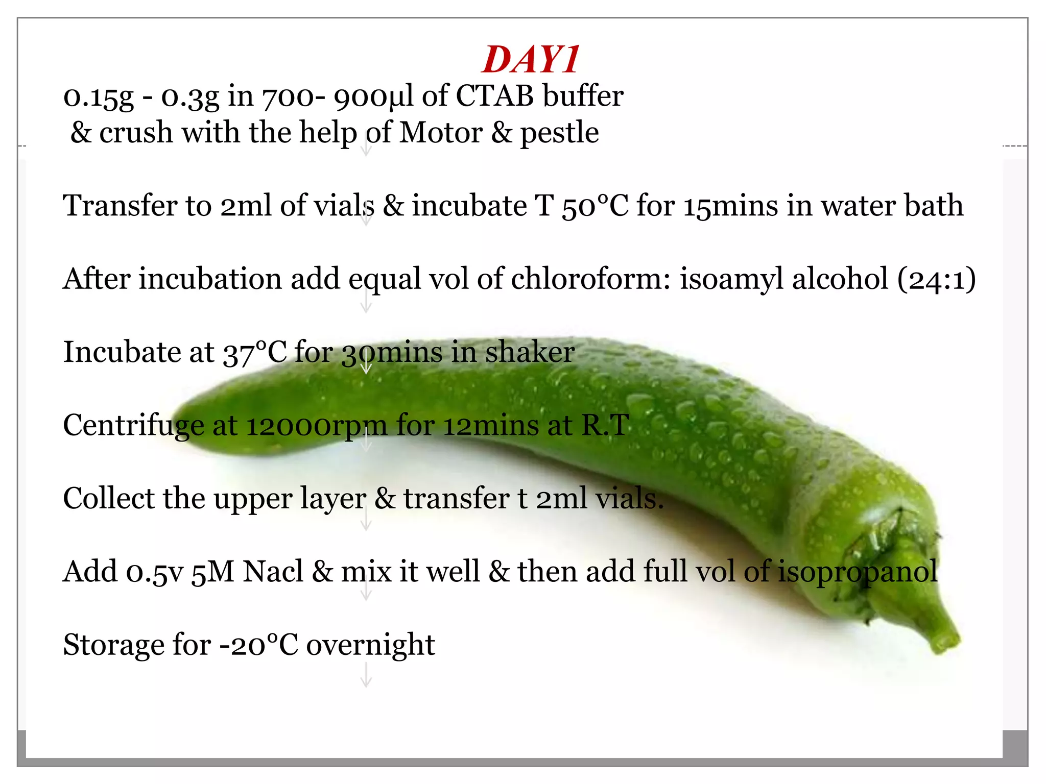
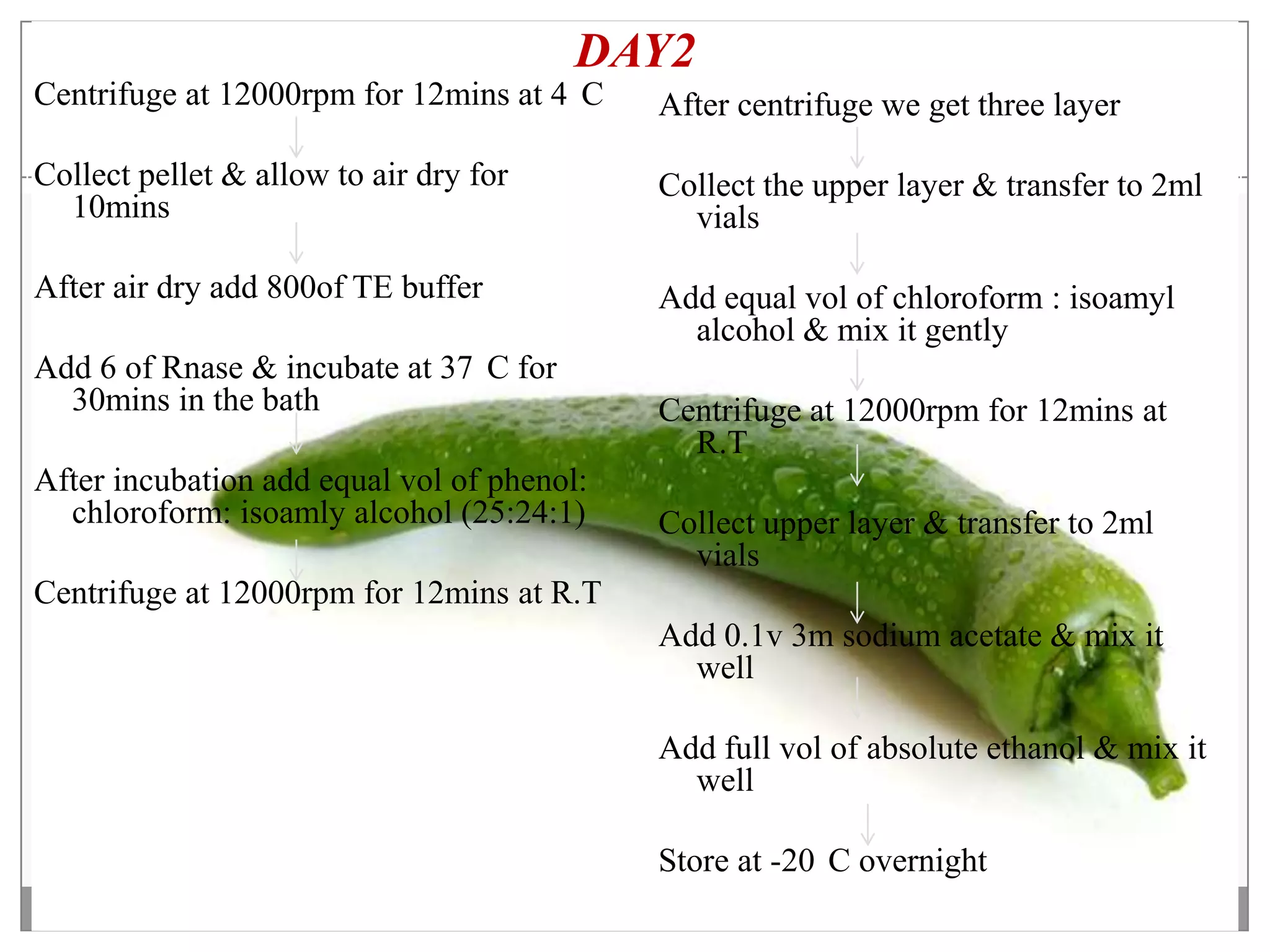
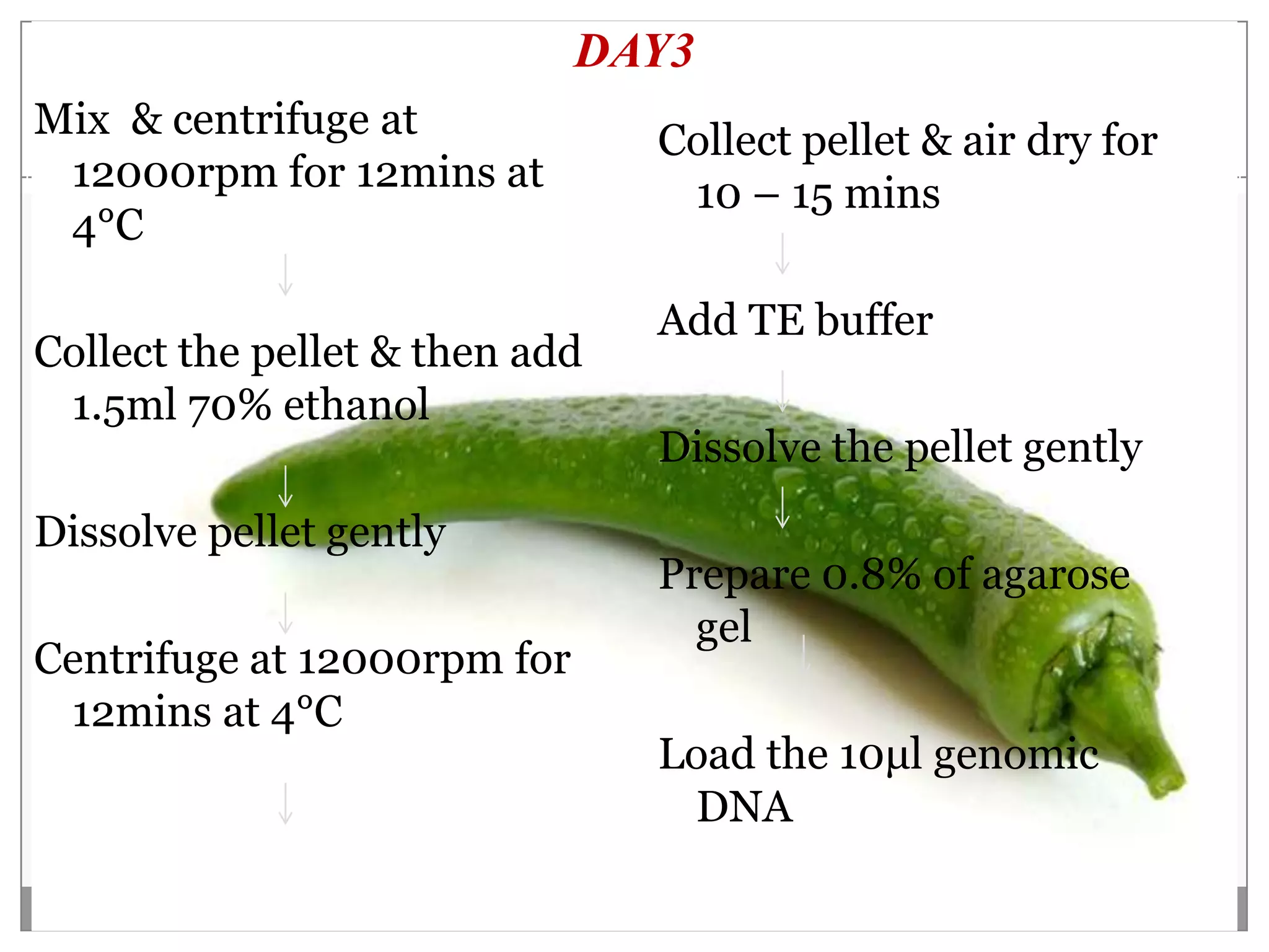
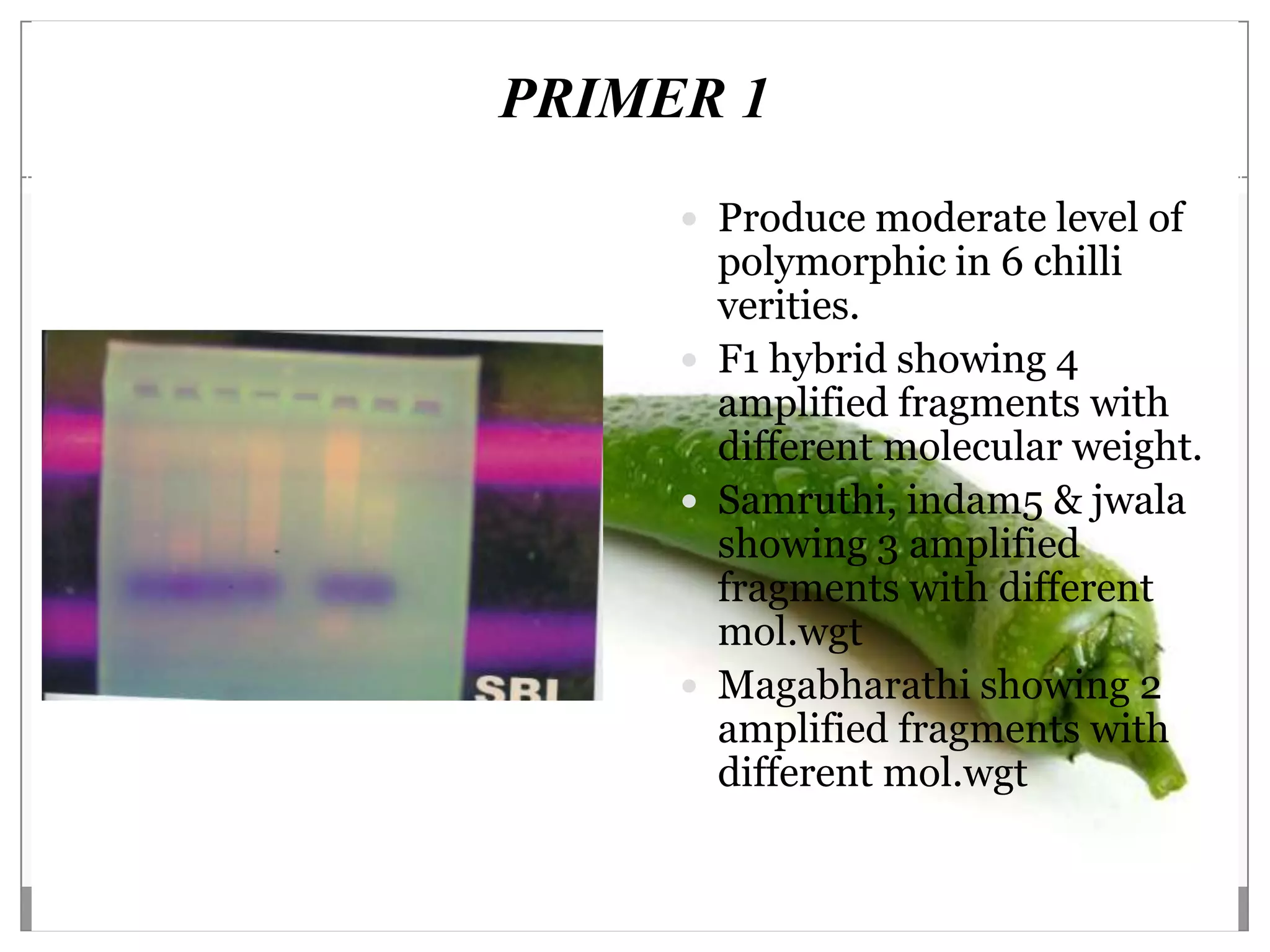
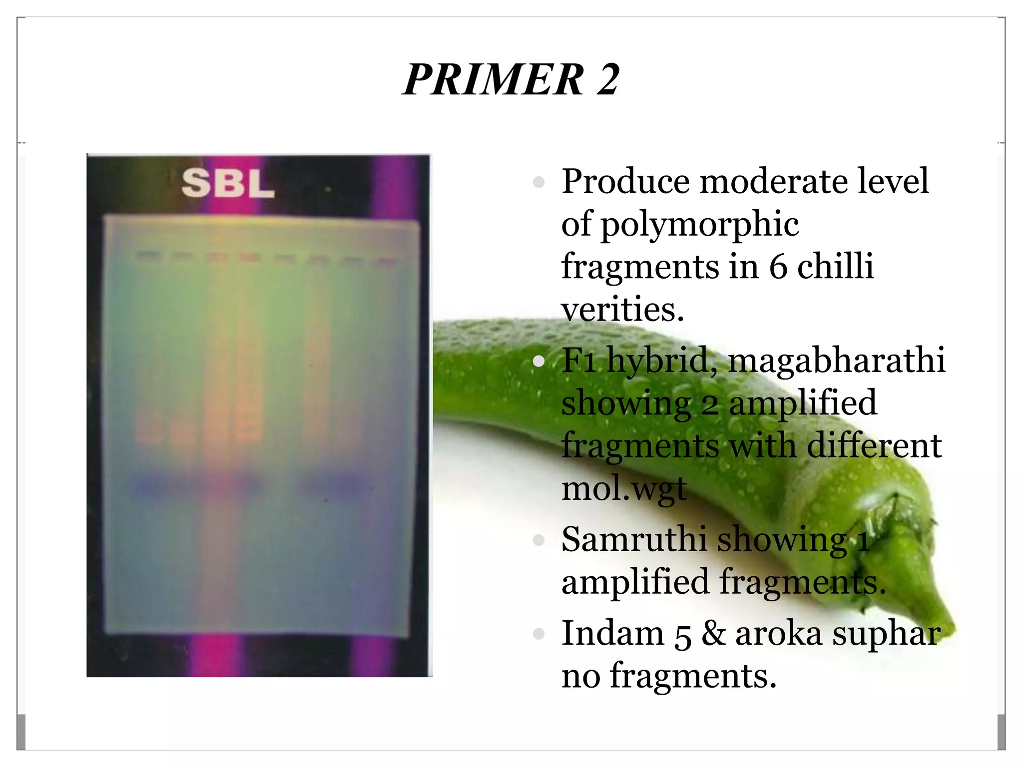
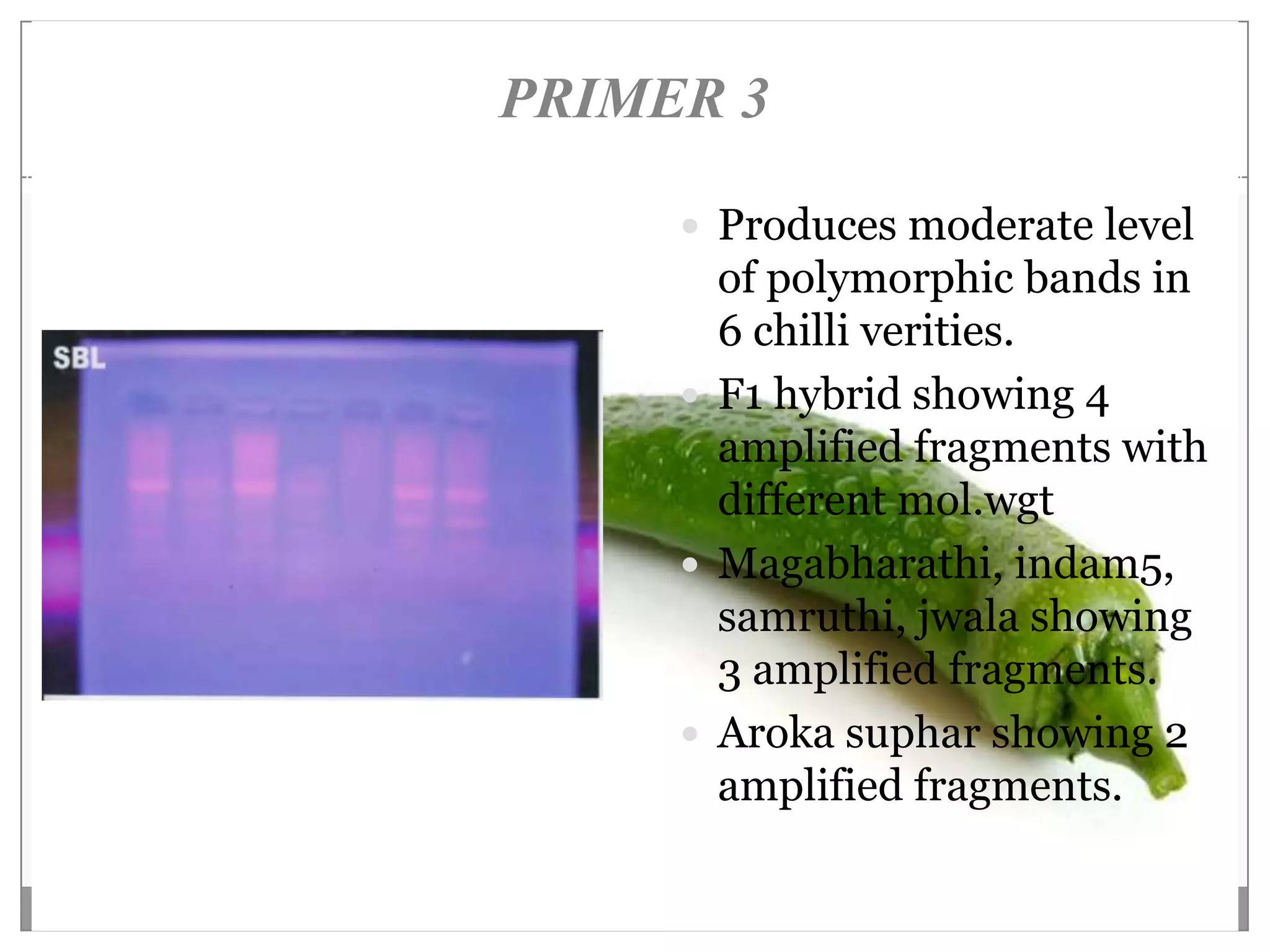
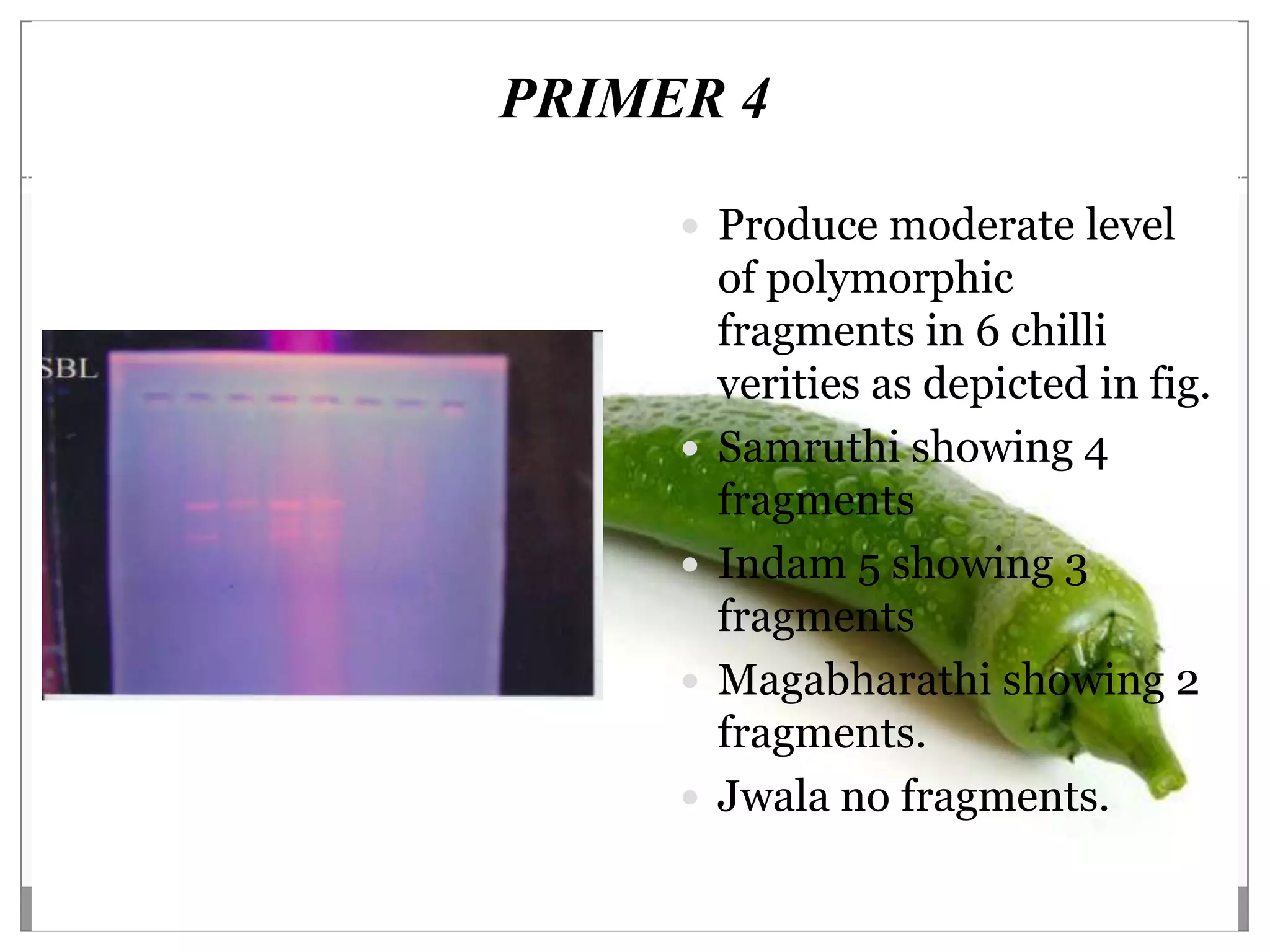
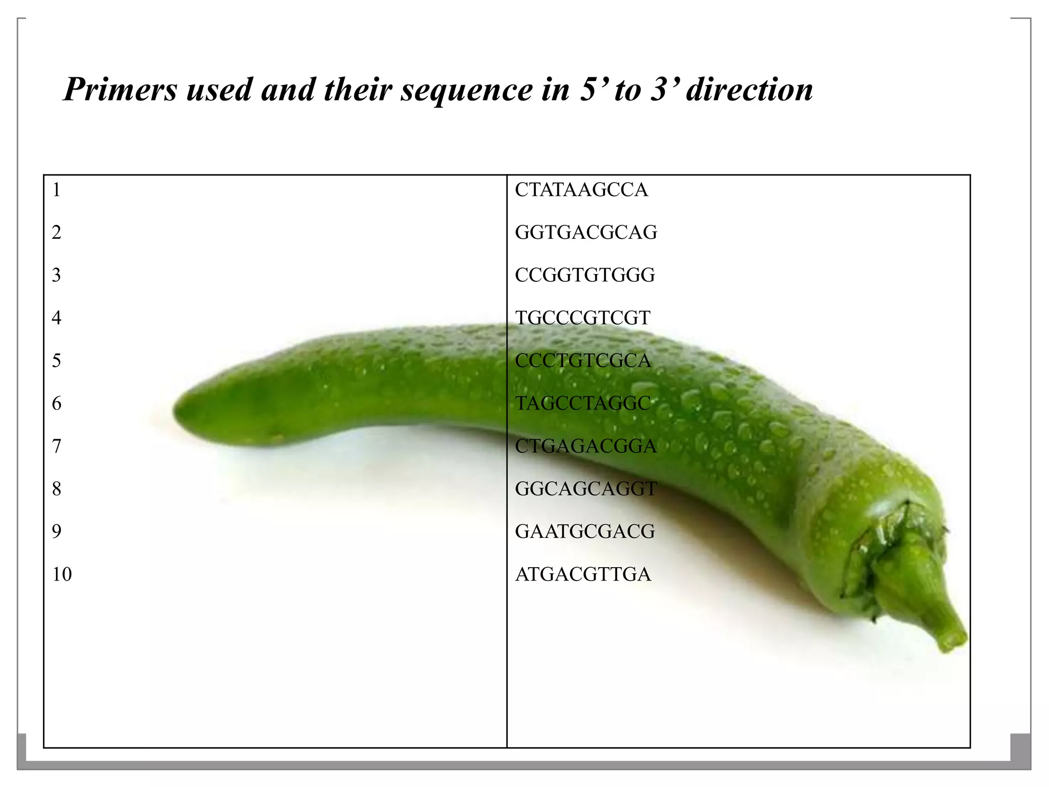
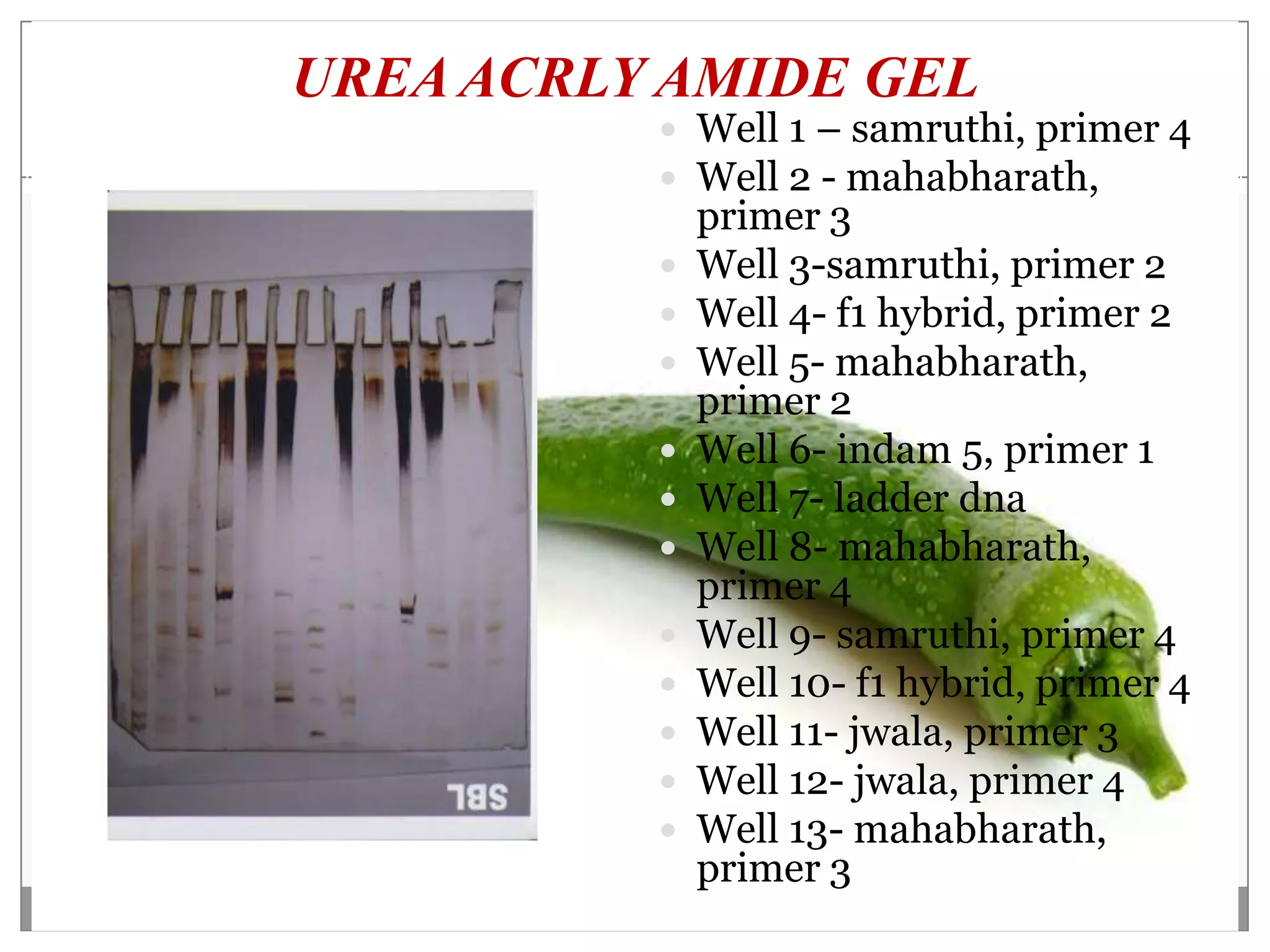
IDENTIFICATION OF POLYMORPHISM
  BY DNA FINGERPRINTING USING
         RAPD IN CHILLI

            Under the Guidance
             Dr. Deepak.R (HOD)
               External Guidance
                   Govind Rao
                 Submitted by
       Abhinav Varma (1CR08BT001)
       Sachin Subba (1CR09BT400)
       Syed Mubasir (1CR05BT026)
        Department of Biotechnology
             CMRIT Bangalore.
CONTENTS

 OBJECTIVE
 REVIEW OF LITERATURE
 INTRODUCTION
 MORPHOLOGY
 CLASSIFICATION
 CLIMATIC CONDITION
 MATERIALS AND METHODS
 RESULTS AND DISCUSSION
 CONCLUSION
OBJECTIVE
 The objective of this project is to examine the
    technology of DNA fingerprinting.
   Identification of polymorphism by DNA
    fingerprinting using RAPD in different variety of
    chilli considered for the present study.
   7 varieties of chillies were selected for the study.
    This work was carried out under the SBL
    bangalore.
   Isolation of genomic DNA.
   Agarose gel electrophoresis of PCR product.
   Spectrophotometric quantification.
   Techniques to detect genetic variation of DNA.
REVIEW OF LITERATURE
                               Sources of genetic variation
Genetic variation and its
importance
  Genetic variation is also    Mutation( changes in one
   called as genetic             or more letters of a DNA
   diversity.                    sequence)
  Genetic diversity is a       Mutation come in the form
   necessary for survival in     of single base pair (point
                                 mutation), deletion,
   a world full of changing      insertion, translocation or
   environmental stresses.       inverse of genes.
  Genetic diversity            Recombination is the
   provides the means for        shuffling of DNA segments
   species to adopt to          Transgenic, a tool of
   changing environmental        modern biotechnology.
   conditions in future.
TECHNIQUES TO DETECT GENETIC
             VARIATION
TRADATIONAL:
                                   CLONE/SEQUENCE BASED
 Studying the character tics of     MARKER :
  plants & animals that are easy    Single base pair polymorphic
  to observe & measure.              restriction site seq & measurable
                                     insertion or deletion
DNA BASED :
                                    It can be detected by
 Techniques to analyze DNA,         electrophoretic techniques
  developed within the last 20     FINGER PRINT MARKER :
  years, enabling to locate         Most commonly in use are the
  specific DNA chromosome.           minisatellites.
                                    Minisatellites is the
GENE FLOW :                          comparatively low upfront cost
 A fair degree of genetic           of detecting.
  modification occurs through      FUTURE BASED MARKER
                                     TECHNOLOGY :
  gene flow during sexual
                                    The generation of large insert
  reproduction, followed by          clone libraries for agriculture
  natural selection.                 animal species is already
                                     underway.
INTRODUCTION
ORIGIN:                                  DISTRIBUTION:
 The orgin of chillies is believed to    Capsicum was first introduced
  be as old as 7000 B.C.                   into Spain by Columbus in 1943.
 In 1888, experiments began for          Its cultivation spread from
  cross breeding of chili plant.           Mediterranean to England by
                                           1948.
 It was originated from mexico.
                                          Chilli is actually reported to
 Later new breed of chilli plants         nature of south america.
  were evolved by crossbreeding.
                                          Its cultivation was known to be
 New variety of chilli i.e. Anaheim       native of Peru.
  was grown on 1906.
                                          This crop was introduced to
 There are more than 400                  India by Portuguese towards the
  varieties of chilli grown all over       end of 15th century.
  the world
                                          It became popular in 17th century.
 The hottest chilli is “Naga
  Jolokia” which is cultivated in
  hilly terrain of Assam.
MORPHOLOGY

BRANCHING:                         FRUIT:
 Chilli pant is highly branched    Fruit is berry.
  herbaceous plant.                 Seeds are hot & embedded on
 Its height ranges from 50 –        pericarp.
  100cm                             Pericarp is leathery which
 Leaves are simple, alternative     turns from green to purple,
  with unequal margin.               purple to red.
                                    Fruit attain full maturity in
FLOWER:                              around 35 days.
 Flower sometimes occur in
  pairs.                           SEED:
 It is bell shaped, slender &      Seed start developing from 15
  terminal.                          days of anthesis.
                                    Diameter of seed varies from
                                     3-4 mm.
CLASSIFICATION


Kingdom :       Plantae
Division   :   Magnoliophyta
Class     :    Magnoliopsida
Subclass   :     Asteridae
Order    :      Solanales
Family     :      Solanaceae
Genus    :     Capsicum
Species  :      frutescens
CLIMATIC CONDITON


 It can be grown in both warm & cold climatic
    condition.
   The ideal temperature range is 20-25°C.
   The crop is killed in freezing temp and frost.
   Heavy continues rain during flowering results in
    poor fruit.
   High temp. & dry winds are injurious to plant.
MATERIALS AND METHODS
SAMPLE COLLECTION


SAMPLE COLLECTION
7 verities of chili plants were collected from I.I.H.R agri
  university, Bangalore. The samples are stored at 4⁰c.
 Mahabharath
 Sarca aroka
 Samruthi
 indam 5
 f1 hybrid
 Indam jwala
 SBL-C
PLASMID ISOLATION                                Add 0.3ml of solution 3 and mix it
                                                  gently & incubate in ice cold
                                                  condition for 8-10mins.
 100μl of B. glycerol stock was added into
   50ml of lb broth containing antibiotics
                                                  The centrifugation at 12000rpm for
                                                  12 mins
 The conical flasks were incubated
   overnight at 37 C on the orbital shaker.       collect supernatant and transfer to
                                                  2ml eppendorf tubes & 10-12μ was
                                                  added
 The cultures were transferred into sterile
   centrifuged tubes & chilled in an ice bath     collect the upper layer & transfer
   for 10mins at 4 C.                             to 2ml eppendorf tubes.
                                                  Add equal vol of
 The cells were harvested by centrifuge at       chloroform:isoamyl alcohol (24:1)
   6,000rpm for 6mins at 4 C.
                                                  vial was centrifuged at
                                                  10,000rpmfor 12mins
 Collect the pellet & then add 0.2ml of ice-
   cold solutin1 by using cyclo mixer.
                                                  pellet was washed with ice cold
                                                  70% ethanol
 Add 0.4ml of solution 2, mix it proper (RT)
                                                  the pellect was dried in speed
                                                  vacuum desiccators.
ISOLATION OF GENOMIC DNA
      (CTAB METHOD)
DAY1
0.15g - 0.3g in 700- 900μl of CTAB buffer
& crush with the help of Motor & pestle

Transfer to 2ml of vials & incubate T 50°C for 15mins in water bath

After incubation add equal vol of chloroform: isoamyl alcohol (24:1)

Incubate at 37°C for 30mins in shaker

Centrifuge at 12000rpm for 12mins at R.T

Collect the upper layer & transfer t 2ml vials.

Add 0.5v 5M Nacl & mix it well & then add full vol of isopropanol

Storage for -20°C overnight
DAY2
Centrifuge at 12000rpm for 12mins at 4 C    After centrifuge we get three layer

Collect pellet & allow to air dry for       Collect the upper layer & transfer to 2ml
  10mins                                      vials

After air dry add 800of TE buffer           Add equal vol of chloroform : isoamyl
                                              alcohol & mix it gently
Add 6 of Rnase & incubate at 37 C for
  30mins in the bath                        Centrifuge at 12000rpm for 12mins at
                                              R.T
After incubation add equal vol of phenol:
  chloroform: isoamly alcohol (25:24:1)     Collect upper layer & transfer to 2ml
                                              vials
Centrifuge at 12000rpm for 12mins at R.T
                                            Add 0.1v 3m sodium acetate & mix it
                                              well

                                            Add full vol of absolute ethanol & mix it
                                              well

                                            Store at -20 C overnight
DAY3
Mix & centrifuge at             Collect pellet & air dry for
 12000rpm for 12mins at          10 – 15 mins
 4°C

                                Add TE buffer
Collect the pellet & then add
 1.5ml 70% ethanol
                                Dissolve the pellet gently
Dissolve pellet gently
                                Prepare 0.8% of agarose
                                  gel
Centrifuge at 12000rpm for
 12mins at 4°C
                                Load the 10μl genomic
                                 DNA
SPECTROPHOTOMETRIC
        QUANTIFICATION OF DNA

REQUIREMENTS :
 UV spectrophotometer
  TE buffer
   DNA sample
   Micropipette
   Absolute Ethanol
PROCEDURE:
Prepare a known dilution of DNA sample in the TE
 buffer, which is used to dissolve the DNA sample.

Calibrate the spectrophotometer for blank using TE
 buffer.

Record the OD of the sample at 260nm and 280nm.

Calculate the concentration of DNA in the sample
 using the Relation
QUALITY PCR

REQUIREMENTS :
 Thermo stable Taq DNA polymerase
 dNTP mix (10mM)
 Chili genomic DNA
 Sterile distilled water
 PCR buffer (10x)
 Forward primers and reverse primer specific to
  positive control
 Micropipettes of different ranges
Reaction components:
S DNA      sample Positive       dNTPs (µl)   Buffer (10x)   Forward       Reverse       Taq          Sterile
. volume(µl)      control (µl)                (µl)           primer (µl)   primer (µl)   polymerase   water (µl)
N                                                                                        (µl)
o


1   8.0              3.0         2.25         2.5            0.75          0.75          0.75         7.0


2   8.0              3.0         2.25         2.5            0.75          0.75          0.75         7.0


3   8.0              3.0         2.25         2.5            0.75          0.75          0.75         7.0


4   8.0              3.0         2.25         2.5            0.75          0.75          0.75         7.0


5   8.0              3.0         2.25         2.5            0.75          0.75          0.75         7.0


6 8.0                3.0         2.25         2.5            0.75          0.75          0.75         7.0
QUALITY PCR PROGRAM
Heated lid 110ºC
     Pre- heated lid off
     Pause- off
   Initial denaturation- off
Loop 1 (initial denaturation)
   No. of cycles             1
   Segment        94ºC       3minutes
Loop 2
   No. of cycles              30
   Segment         94ºC       30sec
   Segment -       55ºC -      30sec
   Segment -       72ºC - 1minute
Final extention      72ºC - 5minutes
Final hold           10ºC
AMPLIFICATION OF DNA USING RAPD

REQUIREMENTS :
 Thermostable Taq DNA polymerase
 dNTP mix (10 mM)
 Template DNA
 Sterile distilled water
 PCR buffer (10x)
 Oligonucleotide primers
 Ice bucket
 Eppendorff vials
 Micropipettes of different ranges
 Thermal cycle
PROCEDURE:
Set up the following reaction mixture (25 l) in the same order.

             Ingredients                            Volume to be taken


            Template DNA                   10.0μl


                dNTPs                      2.5μl


              PCR buffer                   2.5μl


               Primers                     1.0μl


          Taq DNA polymerase               0.75μl


             Sterile water                 8.25μl


                Total                      25μl
All those mentioned ingredients are mixed and prepared for the
total no of reactns including a blank with a particular primer
excluding template

The calculated volume of masters mix ix then transferred to
labeled PCR tubes with template source and primer.

Finally 0.33µl of Taq DNA polymerase is added to each tube

The contents of the tube are mixed with a brief spin and
transferred to PTC 200 thermal cycler

The program with following conditions is selected for the
amplification
Number cycles           30
Segment 94.0ºC          1minute
Segment 35.0ºC          1minute
Segment 72.0ºC          1minute
UREA POLYACRYLAMIDE GEL
           ELECTROPHORESIS

REQUIREMENTS :
 Vertical electrophoresis unit
 Urea 7M
 Acrylamide 40%
 10x TBE (Tris Borate EDTA) buffer
 10%Ammonium Per Sulphate (APS)
 Tetra Ethyl Methylene Diamine (TEMED)
 Gel loading dye
 Autoclaved distilled water
PROCEDURE:
Preparation of gel (50ml)
 Weigh 9.08g of urea and dissolved by heating in about 15ml
  autoclaved distilled water.


 Add 6.25ml of 40% acrylamide and 5ml of 10x TBE buffer.

 Make up the volume to 50ml with autoclaved distilled water.

 Add 350μl of APS and 35l of TEMED and mix well.

 Immediately transfer the gel into the previously arranged
   vertical electrophoresis unit.
Electrophoresis of the DNA
Pre-run the gel for about one hour at 100V.

To the PCR sample add 4.2l of gel loading dye.(Xylene
  Cyanol).

Boil the samples for 10minutes at 85-90C.

Immediately chill the sample in ice for 2minutes.

Spin the sample at 3000rpm for 2minutes and load in top the
  gel.

The electrophoresis is carried out at 150V tll the dye front
  reaches the bottom of the base plate, the plates are cooled
  with an ice pack during the run to prevent over-heating
SILVER STAINING

REQUIREMENTS :
 Gel container
 Shaker incubator
 10% acetic acid
 de-ionised water
 autoclaved double distilled water
 silver nitrate solution
 2.5% sodium carbonate and 0.02% formaldehyde.
PROCEDURE:                         Incubate for 10minutes at room
Place the gel in 5 volumes of a      temperatures with shaking.
  mixture of 30% ethanol and         Repeat this step twice.
  10% acetic acid.
                                   Remove the deionised water and
Incubate the gel for 3 hours or      add 5 gel volume of 0.1%silver
  overnight with shaking at room     nitrate solution.
  temperatures.
                                   Incubate for 30minutes at room
Remove the ethanol / acetic acid     temperatures with shaking.
  solution and add 5 gel volume
  of 30% ethanol.                  Remove the silver nitrate
                                     solution and wash the gel for
Incubate for 30minutes at room       20seconds under a stream of
  temperatures with shaking.         deionised water.
  Repeat this step twice.
                                   Add 5 gel volume of a mixture of
Remove the ethanol solution and      2.5%sodium carbonate and
  add 10 gel volume of               0.02% formaldehyde
  deinonised water.
Incubate at room temperature with shaking. Bands will start
  appearing slowly.

Incubate until band appears.

Stop the reaction by washing with 1% acetic acid.

Wash several times with deionised water for 10 minutes each

The gel might now be observed over an illuminating source of
  white light for better result and documented.

For preserving the gel, place it in 20ml of a 20% glycerol
  solution.

Keep the gel between two layers of gelatin [aper and dry for 3
  days at 37ºC.
RESULTS AND DISCUSSION
GENOMIC DNA ISOLATION         AGAROSE GEL
 In the present study DNA      ELECTROPHORESIS
  was isolated from chilli     After isolation of G.DNA
  leaves following the CTAB     from chilli leaves sample
  method.                       were loaded into 0.8%
 Method described by Doyle     agarose gel.
  (1987) with few              To cross check the presence
  modification.                 or absence of G.DNA in
 About 320 g of pure G.DNA     isolated sample.
  could be isolated by this    In agarose gel we observed
  method.                       respective G.DNA bands
                                with little streaking.
                               These streaking might be
                                because breakage of DNA.
QUALITY PCR
       To cross check quality of
          G.DNA in test sample
         We arrange quality PCR
          using test chilli G.DNA
          sample.
         +ve control along with
          specific primer, loaded into
          0.8% agarose gel.
         Out of 7 chilli varities only
          6 varities were amplified
          throughout along with +ve
          control
         Indicates only 6 chilli
          G.DNA quality was good.
RAPD PCR
     The RAPD technique was
      standardize by adapting
      various temp, primer conc.
     After standardize of RAPD
      program routine analysis
      was done with a PCR.
     Program having 2mins
      initial denauration (94⁰c) .
    1min denauration (94⁰C)
    1min annealing (35⁰c)
    1min extension (72⁰c)
    For about 30 cycles, this was
      followed by one final
      extension 72⁰c for about
      5mins.
SCREENING OF RAPD PRIMERS

 RAPD analysis of isolated chilli plant G.DNA was
  carried out with 10 different oligonucleotide random
  primer.
 Out of 10 random primers tested for chilli only 4
  primers showing amplification with test sample.
 So that we use only 4 selected primers to study
  polymorphism in 6 chilli varities.
PRIMER 1
      Produce moderate level of
       polymorphic in 6 chilli
       verities.
      F1 hybrid showing 4
       amplified fragments with
       different molecular weight.
      Samruthi, indam5 & jwala
       showing 3 amplified
       fragments with different
       mol.wgt
      Magabharathi showing 2
       amplified fragments with
       different mol.wgt
PRIMER 2

     Produce moderate level
      of polymorphic
      fragments in 6 chilli
      verities.
     F1 hybrid, magabharathi
      showing 2 amplified
      fragments with different
      mol.wgt
     Samruthi showing 1
      amplified fragments.
     Indam 5 & aroka suphar
      no fragments.
PRIMER 3

      Produces moderate level
       of polymorphic bands in
       6 chilli verities.
      F1 hybrid showing 4
       amplified fragments with
       different mol.wgt
      Magabharathi, indam5,
       samruthi, jwala showing
       3 amplified fragments.
      Aroka suphar showing 2
       amplified fragments.
PRIMER 4

      Produce moderate level
         of polymorphic
         fragments in 6 chilli
         verities as depicted in fig.
        Samruthi showing 4
         fragments
        Indam 5 showing 3
         fragments
        Magabharathi showing 2
         fragments.
        Jwala no fragments.
Primers used and their sequence in 5’ to 3’ direction

1                                  CTATAAGCCA

2                                  GGTGACGCAG

3                                  CCGGTGTGGG

4                                  TGCCCGTCGT

5                                  CCCTGTCGCA

6                                  TAGCCTAGGC

7                                  CTGAGACGGA

8                                  GGCAGCAGGT

9                                  GAATGCGACG

10                                 ATGACGTTGA
UREA ACRLY AMIDE GEL
           Well 1 – samruthi, primer 4
           Well 2 - mahabharath,
              primer 3
             Well 3-samruthi, primer 2
             Well 4- f1 hybrid, primer 2
             Well 5- mahabharath,
              primer 2
             Well 6- indam 5, primer 1
             Well 7- ladder dna
             Well 8- mahabharath,
              primer 4
             Well 9- samruthi, primer 4
             Well 10- f1 hybrid, primer 4
             Well 11- jwala, primer 3
             Well 12- jwala, primer 4
             Well 13- mahabharath,
              primer 3
CONCLUSION

 Polymorphism between genotypes is due to either a
  nucleotide base change that alters the ability of the
  primer to anneal to the DNA template within the
  amplified fragment.
 All the primers cannot amplify all the verities of
  chilies. Variation was seen as certain primers could
  separate.
 Our study reflected the tremendous genetic diversity
  available among the genotypes.
 The rich genetic diversity in which breeding efforts
  depend can be utilized for current & future.
THANK YOU
Identification of polymorphism by dna fingerprinting using rapd 2

Identification of polymorphism by dna fingerprinting using rapd 2

  • 1.
    IDENTIFICATION OF POLYMORPHISM BY DNA FINGERPRINTING USING RAPD IN CHILLI Under the Guidance Dr. Deepak.R (HOD) External Guidance Govind Rao Submitted by Abhinav Varma (1CR08BT001) Sachin Subba (1CR09BT400) Syed Mubasir (1CR05BT026) Department of Biotechnology CMRIT Bangalore.
  • 2.
    CONTENTS  OBJECTIVE  REVIEWOF LITERATURE  INTRODUCTION  MORPHOLOGY  CLASSIFICATION  CLIMATIC CONDITION  MATERIALS AND METHODS  RESULTS AND DISCUSSION  CONCLUSION
  • 3.
    OBJECTIVE  The objectiveof this project is to examine the technology of DNA fingerprinting.  Identification of polymorphism by DNA fingerprinting using RAPD in different variety of chilli considered for the present study.  7 varieties of chillies were selected for the study. This work was carried out under the SBL bangalore.  Isolation of genomic DNA.  Agarose gel electrophoresis of PCR product.  Spectrophotometric quantification.  Techniques to detect genetic variation of DNA.
  • 4.
    REVIEW OF LITERATURE Sources of genetic variation Genetic variation and its importance  Genetic variation is also  Mutation( changes in one called as genetic or more letters of a DNA diversity. sequence)  Genetic diversity is a  Mutation come in the form necessary for survival in of single base pair (point mutation), deletion, a world full of changing insertion, translocation or environmental stresses. inverse of genes.  Genetic diversity  Recombination is the provides the means for shuffling of DNA segments species to adopt to  Transgenic, a tool of changing environmental modern biotechnology. conditions in future.
  • 5.
    TECHNIQUES TO DETECTGENETIC VARIATION TRADATIONAL: CLONE/SEQUENCE BASED  Studying the character tics of MARKER : plants & animals that are easy  Single base pair polymorphic to observe & measure. restriction site seq & measurable insertion or deletion DNA BASED :  It can be detected by  Techniques to analyze DNA, electrophoretic techniques developed within the last 20 FINGER PRINT MARKER : years, enabling to locate  Most commonly in use are the specific DNA chromosome. minisatellites.  Minisatellites is the GENE FLOW : comparatively low upfront cost  A fair degree of genetic of detecting. modification occurs through FUTURE BASED MARKER TECHNOLOGY : gene flow during sexual  The generation of large insert reproduction, followed by clone libraries for agriculture natural selection. animal species is already underway.
  • 6.
    INTRODUCTION ORIGIN: DISTRIBUTION:  The orgin of chillies is believed to  Capsicum was first introduced be as old as 7000 B.C. into Spain by Columbus in 1943.  In 1888, experiments began for  Its cultivation spread from cross breeding of chili plant. Mediterranean to England by 1948.  It was originated from mexico.  Chilli is actually reported to  Later new breed of chilli plants nature of south america. were evolved by crossbreeding.  Its cultivation was known to be  New variety of chilli i.e. Anaheim native of Peru. was grown on 1906.  This crop was introduced to  There are more than 400 India by Portuguese towards the varieties of chilli grown all over end of 15th century. the world  It became popular in 17th century.  The hottest chilli is “Naga Jolokia” which is cultivated in hilly terrain of Assam.
  • 7.
    MORPHOLOGY BRANCHING: FRUIT:  Chilli pant is highly branched  Fruit is berry. herbaceous plant.  Seeds are hot & embedded on  Its height ranges from 50 – pericarp. 100cm  Pericarp is leathery which  Leaves are simple, alternative turns from green to purple, with unequal margin. purple to red.  Fruit attain full maturity in FLOWER: around 35 days.  Flower sometimes occur in pairs. SEED:  It is bell shaped, slender &  Seed start developing from 15 terminal. days of anthesis.  Diameter of seed varies from 3-4 mm.
  • 8.
    CLASSIFICATION Kingdom : Plantae Division : Magnoliophyta Class : Magnoliopsida Subclass : Asteridae Order : Solanales Family : Solanaceae Genus : Capsicum Species : frutescens
  • 9.
    CLIMATIC CONDITON  Itcan be grown in both warm & cold climatic condition.  The ideal temperature range is 20-25°C.  The crop is killed in freezing temp and frost.  Heavy continues rain during flowering results in poor fruit.  High temp. & dry winds are injurious to plant.
  • 10.
  • 11.
    SAMPLE COLLECTION SAMPLE COLLECTION 7verities of chili plants were collected from I.I.H.R agri university, Bangalore. The samples are stored at 4⁰c.  Mahabharath  Sarca aroka  Samruthi  indam 5  f1 hybrid  Indam jwala  SBL-C
  • 12.
    PLASMID ISOLATION  Add 0.3ml of solution 3 and mix it gently & incubate in ice cold condition for 8-10mins.  100μl of B. glycerol stock was added into 50ml of lb broth containing antibiotics The centrifugation at 12000rpm for 12 mins  The conical flasks were incubated overnight at 37 C on the orbital shaker. collect supernatant and transfer to 2ml eppendorf tubes & 10-12μ was added  The cultures were transferred into sterile centrifuged tubes & chilled in an ice bath collect the upper layer & transfer for 10mins at 4 C. to 2ml eppendorf tubes. Add equal vol of  The cells were harvested by centrifuge at chloroform:isoamyl alcohol (24:1) 6,000rpm for 6mins at 4 C. vial was centrifuged at 10,000rpmfor 12mins  Collect the pellet & then add 0.2ml of ice- cold solutin1 by using cyclo mixer. pellet was washed with ice cold 70% ethanol  Add 0.4ml of solution 2, mix it proper (RT) the pellect was dried in speed vacuum desiccators.
  • 13.
    ISOLATION OF GENOMICDNA (CTAB METHOD)
  • 14.
    DAY1 0.15g - 0.3gin 700- 900μl of CTAB buffer & crush with the help of Motor & pestle Transfer to 2ml of vials & incubate T 50°C for 15mins in water bath After incubation add equal vol of chloroform: isoamyl alcohol (24:1) Incubate at 37°C for 30mins in shaker Centrifuge at 12000rpm for 12mins at R.T Collect the upper layer & transfer t 2ml vials. Add 0.5v 5M Nacl & mix it well & then add full vol of isopropanol Storage for -20°C overnight
  • 15.
    DAY2 Centrifuge at 12000rpmfor 12mins at 4 C After centrifuge we get three layer Collect pellet & allow to air dry for Collect the upper layer & transfer to 2ml 10mins vials After air dry add 800of TE buffer Add equal vol of chloroform : isoamyl alcohol & mix it gently Add 6 of Rnase & incubate at 37 C for 30mins in the bath Centrifuge at 12000rpm for 12mins at R.T After incubation add equal vol of phenol: chloroform: isoamly alcohol (25:24:1) Collect upper layer & transfer to 2ml vials Centrifuge at 12000rpm for 12mins at R.T Add 0.1v 3m sodium acetate & mix it well Add full vol of absolute ethanol & mix it well Store at -20 C overnight
  • 16.
    DAY3 Mix & centrifugeat Collect pellet & air dry for 12000rpm for 12mins at 10 – 15 mins 4°C Add TE buffer Collect the pellet & then add 1.5ml 70% ethanol Dissolve the pellet gently Dissolve pellet gently Prepare 0.8% of agarose gel Centrifuge at 12000rpm for 12mins at 4°C Load the 10μl genomic DNA
  • 17.
    SPECTROPHOTOMETRIC QUANTIFICATION OF DNA REQUIREMENTS :  UV spectrophotometer  TE buffer  DNA sample  Micropipette  Absolute Ethanol
  • 18.
    PROCEDURE: Prepare a knowndilution of DNA sample in the TE buffer, which is used to dissolve the DNA sample. Calibrate the spectrophotometer for blank using TE buffer. Record the OD of the sample at 260nm and 280nm. Calculate the concentration of DNA in the sample using the Relation
  • 19.
    QUALITY PCR REQUIREMENTS : Thermo stable Taq DNA polymerase  dNTP mix (10mM)  Chili genomic DNA  Sterile distilled water  PCR buffer (10x)  Forward primers and reverse primer specific to positive control  Micropipettes of different ranges
  • 20.
    Reaction components: S DNA sample Positive dNTPs (µl) Buffer (10x) Forward Reverse Taq Sterile . volume(µl) control (µl) (µl) primer (µl) primer (µl) polymerase water (µl) N (µl) o 1 8.0 3.0 2.25 2.5 0.75 0.75 0.75 7.0 2 8.0 3.0 2.25 2.5 0.75 0.75 0.75 7.0 3 8.0 3.0 2.25 2.5 0.75 0.75 0.75 7.0 4 8.0 3.0 2.25 2.5 0.75 0.75 0.75 7.0 5 8.0 3.0 2.25 2.5 0.75 0.75 0.75 7.0 6 8.0 3.0 2.25 2.5 0.75 0.75 0.75 7.0
  • 21.
    QUALITY PCR PROGRAM Heatedlid 110ºC Pre- heated lid off Pause- off Initial denaturation- off Loop 1 (initial denaturation) No. of cycles 1 Segment 94ºC 3minutes Loop 2 No. of cycles 30 Segment 94ºC 30sec Segment - 55ºC - 30sec Segment - 72ºC - 1minute Final extention 72ºC - 5minutes Final hold 10ºC
  • 22.
    AMPLIFICATION OF DNAUSING RAPD REQUIREMENTS :  Thermostable Taq DNA polymerase  dNTP mix (10 mM)  Template DNA  Sterile distilled water  PCR buffer (10x)  Oligonucleotide primers  Ice bucket  Eppendorff vials  Micropipettes of different ranges  Thermal cycle
  • 23.
    PROCEDURE: Set up thefollowing reaction mixture (25 l) in the same order. Ingredients Volume to be taken Template DNA 10.0μl dNTPs 2.5μl PCR buffer 2.5μl Primers 1.0μl Taq DNA polymerase 0.75μl Sterile water 8.25μl Total 25μl
  • 24.
    All those mentionedingredients are mixed and prepared for the total no of reactns including a blank with a particular primer excluding template The calculated volume of masters mix ix then transferred to labeled PCR tubes with template source and primer. Finally 0.33µl of Taq DNA polymerase is added to each tube The contents of the tube are mixed with a brief spin and transferred to PTC 200 thermal cycler The program with following conditions is selected for the amplification Number cycles 30 Segment 94.0ºC 1minute Segment 35.0ºC 1minute Segment 72.0ºC 1minute
  • 25.
    UREA POLYACRYLAMIDE GEL ELECTROPHORESIS REQUIREMENTS :  Vertical electrophoresis unit  Urea 7M  Acrylamide 40%  10x TBE (Tris Borate EDTA) buffer  10%Ammonium Per Sulphate (APS)  Tetra Ethyl Methylene Diamine (TEMED)  Gel loading dye  Autoclaved distilled water
  • 26.
    PROCEDURE: Preparation of gel(50ml) Weigh 9.08g of urea and dissolved by heating in about 15ml autoclaved distilled water. Add 6.25ml of 40% acrylamide and 5ml of 10x TBE buffer. Make up the volume to 50ml with autoclaved distilled water. Add 350μl of APS and 35l of TEMED and mix well. Immediately transfer the gel into the previously arranged vertical electrophoresis unit.
  • 27.
    Electrophoresis of theDNA Pre-run the gel for about one hour at 100V. To the PCR sample add 4.2l of gel loading dye.(Xylene Cyanol). Boil the samples for 10minutes at 85-90C. Immediately chill the sample in ice for 2minutes. Spin the sample at 3000rpm for 2minutes and load in top the gel. The electrophoresis is carried out at 150V tll the dye front reaches the bottom of the base plate, the plates are cooled with an ice pack during the run to prevent over-heating
  • 28.
    SILVER STAINING REQUIREMENTS : Gel container  Shaker incubator  10% acetic acid  de-ionised water  autoclaved double distilled water  silver nitrate solution  2.5% sodium carbonate and 0.02% formaldehyde.
  • 29.
    PROCEDURE: Incubate for 10minutes at room Place the gel in 5 volumes of a temperatures with shaking. mixture of 30% ethanol and Repeat this step twice. 10% acetic acid. Remove the deionised water and Incubate the gel for 3 hours or add 5 gel volume of 0.1%silver overnight with shaking at room nitrate solution. temperatures. Incubate for 30minutes at room Remove the ethanol / acetic acid temperatures with shaking. solution and add 5 gel volume of 30% ethanol. Remove the silver nitrate solution and wash the gel for Incubate for 30minutes at room 20seconds under a stream of temperatures with shaking. deionised water. Repeat this step twice. Add 5 gel volume of a mixture of Remove the ethanol solution and 2.5%sodium carbonate and add 10 gel volume of 0.02% formaldehyde deinonised water.
  • 30.
    Incubate at roomtemperature with shaking. Bands will start appearing slowly. Incubate until band appears. Stop the reaction by washing with 1% acetic acid. Wash several times with deionised water for 10 minutes each The gel might now be observed over an illuminating source of white light for better result and documented. For preserving the gel, place it in 20ml of a 20% glycerol solution. Keep the gel between two layers of gelatin [aper and dry for 3 days at 37ºC.
  • 31.
  • 32.
    GENOMIC DNA ISOLATION AGAROSE GEL  In the present study DNA ELECTROPHORESIS was isolated from chilli  After isolation of G.DNA leaves following the CTAB from chilli leaves sample method. were loaded into 0.8%  Method described by Doyle agarose gel. (1987) with few  To cross check the presence modification. or absence of G.DNA in  About 320 g of pure G.DNA isolated sample. could be isolated by this  In agarose gel we observed method. respective G.DNA bands with little streaking.  These streaking might be because breakage of DNA.
  • 33.
    QUALITY PCR  To cross check quality of G.DNA in test sample  We arrange quality PCR using test chilli G.DNA sample.  +ve control along with specific primer, loaded into 0.8% agarose gel.  Out of 7 chilli varities only 6 varities were amplified throughout along with +ve control  Indicates only 6 chilli G.DNA quality was good.
  • 34.
    RAPD PCR  The RAPD technique was standardize by adapting various temp, primer conc.  After standardize of RAPD program routine analysis was done with a PCR.  Program having 2mins initial denauration (94⁰c) . 1min denauration (94⁰C) 1min annealing (35⁰c) 1min extension (72⁰c) For about 30 cycles, this was followed by one final extension 72⁰c for about 5mins.
  • 35.
    SCREENING OF RAPDPRIMERS  RAPD analysis of isolated chilli plant G.DNA was carried out with 10 different oligonucleotide random primer.  Out of 10 random primers tested for chilli only 4 primers showing amplification with test sample.  So that we use only 4 selected primers to study polymorphism in 6 chilli varities.
  • 36.
    PRIMER 1  Produce moderate level of polymorphic in 6 chilli verities.  F1 hybrid showing 4 amplified fragments with different molecular weight.  Samruthi, indam5 & jwala showing 3 amplified fragments with different mol.wgt  Magabharathi showing 2 amplified fragments with different mol.wgt
  • 37.
    PRIMER 2  Produce moderate level of polymorphic fragments in 6 chilli verities.  F1 hybrid, magabharathi showing 2 amplified fragments with different mol.wgt  Samruthi showing 1 amplified fragments.  Indam 5 & aroka suphar no fragments.
  • 38.
    PRIMER 3  Produces moderate level of polymorphic bands in 6 chilli verities.  F1 hybrid showing 4 amplified fragments with different mol.wgt  Magabharathi, indam5, samruthi, jwala showing 3 amplified fragments.  Aroka suphar showing 2 amplified fragments.
  • 39.
    PRIMER 4  Produce moderate level of polymorphic fragments in 6 chilli verities as depicted in fig.  Samruthi showing 4 fragments  Indam 5 showing 3 fragments  Magabharathi showing 2 fragments.  Jwala no fragments.
  • 40.
    Primers used andtheir sequence in 5’ to 3’ direction 1 CTATAAGCCA 2 GGTGACGCAG 3 CCGGTGTGGG 4 TGCCCGTCGT 5 CCCTGTCGCA 6 TAGCCTAGGC 7 CTGAGACGGA 8 GGCAGCAGGT 9 GAATGCGACG 10 ATGACGTTGA
  • 41.
    UREA ACRLY AMIDEGEL  Well 1 – samruthi, primer 4  Well 2 - mahabharath, primer 3  Well 3-samruthi, primer 2  Well 4- f1 hybrid, primer 2  Well 5- mahabharath, primer 2  Well 6- indam 5, primer 1  Well 7- ladder dna  Well 8- mahabharath, primer 4  Well 9- samruthi, primer 4  Well 10- f1 hybrid, primer 4  Well 11- jwala, primer 3  Well 12- jwala, primer 4  Well 13- mahabharath, primer 3
  • 42.
    CONCLUSION  Polymorphism betweengenotypes is due to either a nucleotide base change that alters the ability of the primer to anneal to the DNA template within the amplified fragment.  All the primers cannot amplify all the verities of chilies. Variation was seen as certain primers could separate.  Our study reflected the tremendous genetic diversity available among the genotypes.  The rich genetic diversity in which breeding efforts depend can be utilized for current & future.
  • 43.