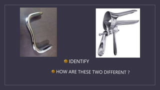
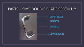
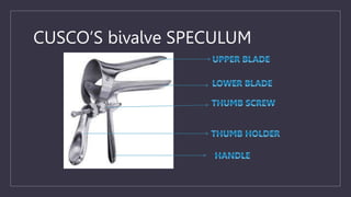
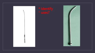
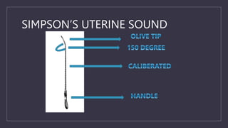
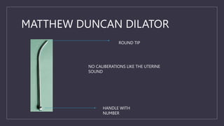
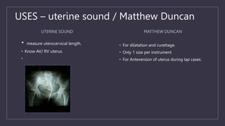
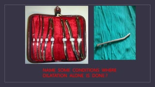
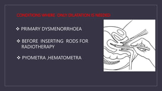
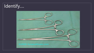
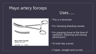
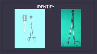
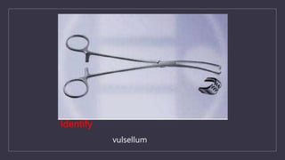
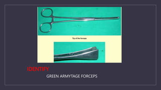
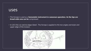
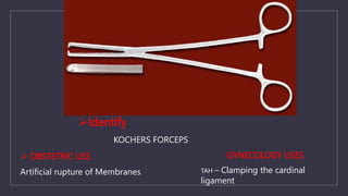
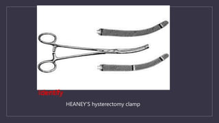
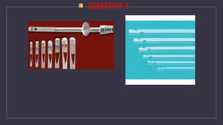
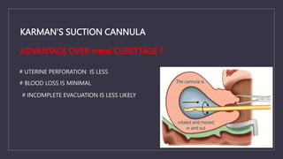
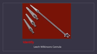
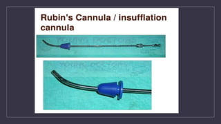
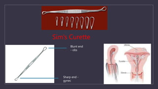
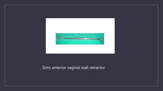
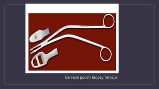
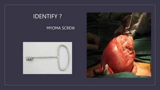
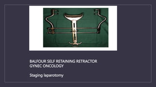
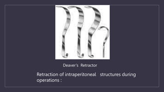
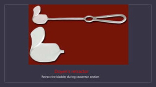
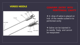
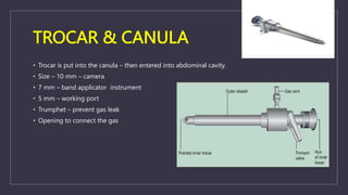
C
GYNEC OSCE
INSTRUMENTS
SPECIMENS
CHARTS
X RAY
DR ALKA, Dr MIRUNALINI
ASSISTANT PROFESSOR
OBGYN, SRIHER
C
INSTRUMENTS
IDENTIFY
J.MARION SIMS
PARTS – SIMS DOUBLE BLADE SPECULUM
CUSCO’S bivalve SPECULUM
USES - BOTH
OBSTETRIC USES
• 1) To confirm diagnosis of PROM.
• 2) During cervical encirclage procedure.
GYNEC USES
• 1) to take Pap smear
• 2) during vaginal hysterectomy.
• 3) To exam the anterior vaginal wall for diagnosis
of vesico-vaginal fistula.
• 4) To diagnose pelvic organ prolapse.
ADVANTAGES & DISADVANTAGES –
OVER SIMS SPECULUM
• Self retaining instrument – needs no
assistant
• Cannot be used for procedures involving
the anterior and posterior vaginal wall.
* Identify
* uses?
SIMPSON’S UTERINE SOUND
MATTHEW DUNCAN DILATOR
ROUND TIP
HANDLE WITH
NUMBER
NO CALIBERATIONS LIKE THE UTERINE
SOUND
USES – uterine sound / Matthew Duncan
UTERINE SOUND
• measure uterocervical length.
• Know AV/ RV uterus.
•
MATTHEW DUNCAN
• For dilatation and curettage.
• Only 1 size per instrument
• For Anteversion of uterus during lap cases.
C
ANY OTHER DILATOR
?
NAME SOME CONDITIONS WHERE
DILATATION ALONE IS DONE ?
CONDITIONS WHERE ONLY DILATATION IS NEEDED
 PRIMARY DYSMENORRHOEA
 BEFORE INSERTING RODS FOR
RADIOTHERAPY
 PYOMETRA ,HEMATOMETRA
Identify….
Mayo artery forceps
Uses……
• This is a hemostat.
• For clamping bleeding vessels.
• For grasping tissue at the time of
operation. (Opening and closing
peritoneum) .
• To hold stay sutures.
• 2 types- straight and curved.
IDENTIFY
USES
SPONGE HOLDING FORCEPS
• Preparation of parts with antiseptic solution
• To hold a sponge to mop from a distance
• For blunt dissection with a gauze
• To hold cervix in pregnancy during – cerclage,
diagnosis & repair of cervical tear, D&E
OVUM FORCEPS
1. To remove products of conception
2. To remove uterine polyp
3. To remove foreign body in vagina .
IDENTIFY
USES
ALLIS TISSUE HOLDING FORCEPS
– Hold edges of vaginal wall during
• Colporrhaphy
• TAH
• Vaginal wall cyst excision
– Myomectomy
– Hold the rectus sheath
– Hold the uterine edges in ceaserean
section
BABCOCK’S
• To hold soft tissues during surgery
• Fallopian tubes
• Bladder
• Bowel
• appendix
Identify
vulsellum
In which procedures post lip of cervix held
To diagnose enterocele
 Colpotomy
 Culdoscentesis
IDENTIFY
GREEN ARMYTAGE FORCEPS
• This forceps is used as a hemostatic instrument in caesarean operation. As the tips are
broad wide area can be compressed.
• In LSCS the cut uterine edges bleed . This forceps is applied to the two angles and lower and
upper edge of the incision.
uses
Identify
KOCHERS FORCEPS
 OBSTETRIC USE
Artificial rupture of Membranes
GYNECOLOGY USES
TAH – Clamping the cardinal
ligament
Identify
HEANEY’S hysterectomy clamp
• USE – TAH
• Serrations are oblique – risk of tissues slipping is less
• No tooth at the tip – risk of tissue trauma is less
Advantages over kochers
IDENTIFY ?
KARMAN’S SUCTION CANNULA
# UTERINE PERFORATION IS LESS
# BLOOD LOSS IS MINIMAL
# INCOMPLETE EVACUATION IS LESS LIKELY
ADVANTAGE OVER metal CURETTAGE ?
Identify
Leech Wilkinsons Cannula
HYSTERO SALPINGO GRAPHY
 Which day of cycle ?
6 to 8th day
 What DYE ?
Uro graffin / 60% Sodium Iotalamate or
(60%methylglucaminediatrizoate(water soluble)
 Alternatives to HSG ?
Laparoscopic chromotubation
 Advantage over lap chromotubation
Site of block
Contraindications
 SUSPECTED ECTOPIC GESTATION
 INTRA UTERINE GESTATION
 PELVIC INFECTION
 SENSITIVITY TO CONTRAST MEDIUM
C
ANY OTHER
CANNULA ??
• Ayre’s Spatula – for taking the smear from cervix,
posterior vaginal wall, upper 1/3 of lateral vaginal wall
• Cyto Brush – used to take smear from the cervical canal
• Solution used is 95% ethanol
• Indications of Pap smear
• CIN/Ca Cx
• Follow up after Wertheim’s hysterectomy
• Hormonal cytology from upper 1/3 of lateral vaginal wall
• Buccal smear for Barr bodies
• Liquid based cytology
• suspension of cells from the sample and this is used to
produce a thin layer of cells on a slide.
• The ThinPrep method requires an instrument and
special polycarbonate filters.
• After the instrument immerses the filter into the
vial, the filter is rotated to homogenize the sample.
Cells are collected on the surface of the filter when
a vacuum is applied.
• The filter is then pressed against a slide to
transfer the cells into a 20 mm diameter circle.
Urinary catheters
• Metal catheter
• Intermittent bladder drainage
• Uses
• Prior to any vaginal procedures
• Foleys catheter
• Size – 16 F
• Uses –
• Gynec –
• Continous bladder drainage – major surgical
procedures.
• Obs –
• Control haemorrhage (balloon Tamponade)
• Induction of labour.
• Bakri balloon tamponade
• Mechanical method of management of PPH.
• Maximum instilled water – 350 ml
• 24 F catheter.
• Cryo probe
• Treatment of LSIL or CIN 1
• Freeze thaw freeze technique. -70 C
• Cryotherapy – mechanism of action
• Side effects – profuse vaginal discharge
post procedure
• Loop electro excision procedure (LEEP)
• Conisation – surgical technique wherein
cone shaped cervix removed with the help
of the cautery.
• 8 – 10 mm cervical tissue
C
RAPID FIRE
Sim’s Curette
Blunt end
- obs
Sharp end -
gynec
Sims anterior vaginal wall retractor
•
Cervical punch biopsy forceps
MYOMA SCREW
IDENTIFY ?
BALFOUR SELF RETAINING RETRACTOR
GYNEC ONCOLOGY
Staging laparotomy
Retraction of intraperitoneal structures during
operations :
Deaver’s Retractor
Doyen’s retractor
Retract the bladder during ceaserean section
LANDONS BLADDER RETRACTOR
Retract skin / other structures
IDENTIFY
ENDOMETRIAL BIOPSY( PLASTIC CANNULA)
C
LAPAROSCOPE
 A drop of saline is placed on
hub of the needle sucked in to
peritoneal cavity
 Saline can be injected in
to needle freely and cannot
be reaspirated
CONFIRM ENTRY INTO
PERITONEAL CAVITY
VEREES NEEDLE
TROCAR & CANULA
• Trocar is put into the canula – then entered into abdominal cavity.
• Size – 10 mm – camera
• 7 mm – band applicator instrument
• 5 mm – working port
• Trumphet – prevent gas leak
• Opening to connect the gas
C
UROGYNAEC
IDENTIFY
TVT –O (Trans
Obturator Tape)
C
SPECIMENS
Basic principles
• Describe gross appearance based on
• Shape
• Pathological findings
• Cut section
• Diagnosis
Identify
Fibroid uterus
Probable questions ?
Medical management
Mifepristone
GnRH
How does GnRH act?
Hypogonadal state
• Medical oophorectomy
• Medical menopause
1. Identify
• Ca. endometrium
2. Corpus cancer syndrome ?
3. Familial hereditary syndrome
associated with Ca
endometrium.
4. How will you diagnose?
Fractional curettage
Identify
• TYPES
• Tumour marker
• RMI score
• Investigations
• treatment
BENIGN OVARIAN CYST
Identify
• Type
• Components
• Tumour marker
• treatment
MATURE CYSTIC TERATOMA
• IDENTIFY
• Ca Ovary
• Staging laparotomy
• Tumour marker
• RMI score
• chemotherapy
IDENTIFY
TUBAL Ectopic pregnancy
.What is SAM?
Surgically Administered Medical therapy
.Pre-requisites for methotrexate?
beta HCG < 1500 IU
Tubal mass <3.5cms
FH ABSENT
MOA – methotrexate
Side effects
Multi dose regimine
Department of Obstetrics and Gynaecology, SRMC&RI
IDENTIFY
Hydatiform mole
Karyotype of partial mole – Triploid
Treatment - S & E
How will you follow up this patient?
Weekly till normal for three weeks
Monthly for six months.
C
CHARTS
• A 27 year nulliparous lady (marital life 5 yrs) had come for infertility treatment
• Her usg shows this picture
• Comment on this
• Any other tests for the same..?
ANTRAL FOLLICULAR COUNT
• No. of visible follicles(2-8mm) on day 2/3
• Better predictor than AMH
PCOS Criteria
ROTTERDAM
ESHRE/ASRM
• 24 Years unmarried girl with
irregular cycles
• USG picture as given, comment
• Criteria for diagnosing this
condition
• Clinical implications and treatment
30 yrs , primary infertility for 3yrs , undergone ovulation induction and now has come with pain
abdomen and vomiting and her USG showed this picture
Diagnosis
Classification
Management and prevention
• Comment on this picture
• Endometrial grading
• Gonen and casper in 1990.
• Type a: entirely homogeneous, hyperechogenic pattern, without a central echogenic line
• Type b: intermediate iso-echogenic pattern, with the same reflectivity as the surrounding myometrium and a non-
myometrium and a non-prominent or absent central echogenic line
• Type c: multilayered ‘triple-line’ endometrium consisting of a prominent outer and central hyperechogenic line and inner
hyperechogenic line and inner hypo-echogenic or black region
• APPLEBAUM SCORING
• The endometrial and periendometrial areas are divided into the following four zones
• Zone 1: A 2-mm thick area surrounding the hyperechoic outer layer of the endometrium.
• Zone 2: The hyperechoic outer layer of the endometrium.
• Zone 3: The hypoechoic inner layer of the endometrium.
• Zone 4: The endometrial cavity.
Ovarian cysts
IOTA SIMPLE USG RULES FOR OVARIAN MASSES
• 44 yr old multiparous lady came with heavy menstrual bleeding and pain abdomen.
• Examination revealed bulky and tender uterus, bilateral fornices free
• Her USG showed this pic
• Diagnosis
• Management
• USG criteria for ADENOMYOSIS
• Diffuse uterine enlargement
• Diffusely heterogenous myometrium
• Asymmetrical thickening of myometrium
• Poor defining of endometrial –myometrial borders
• Inhomogenous hypoechoic areas
• Focal probe tenderness
• Identify this picture
• What modality of investigation is better for the diagnosis
• Specific complaints
• Management
• Identify
• FIGO classification
• STEP-W….???
• 47 yrs, multiparous women with heavy menstrual bleeding
• Imaging shows this picture
• Diagnosis
• Treatment
• Role of medical management
• Prerequisites for myomectomy
• Comment on this picture
• Indications and contraindications
• Procedure
Comment on these pictures
• Uterine anomalies
• Classification
• AFS(1988)- lacked specific diagnostic criteria
• ESHRE (2013) - based primarily on uterine anatomy with cervical vaginal anomalies.
Classes: U0–U6 , C0–C4, V0–V4. 3D-based diagnostic criteria for septate and bicornuate
uterus
• ASRM(2021)- Nine classes: Mullerian agenesis, cervical agenesis, unicornuate, uterus
didelphys, bicornuate, septate, longitudinal vaginal septum, transverse vaginal septum,
complex anomalies.
• Screening tests
• Diagnostic tests - 3D TVS and MRI
• The diagnostic accuracy of 3D - 97.6%
• Sensitivity- 98.3% and specificity -99.4%
• Angle of divergence: <75
• External contour normal or
mild indentation
• Angle of divergence >105
• Indentation>10mm
• Intervening cleft >1cm
• Intercornual distance >5cm
Bicornuate uterus
Septate uterus
Limitation
• External uterine contour cannot be evaluated
• In cases of non-communicating rudimentary uterine horn may be missed.
1- interostial line
2- Parallel line over the fundus
3-uterine thickness
4-septal indentation
Troiano and mccarthy formula
A line joining both the horns
• If it cosses the fundus or
<5mm- bicornuate
• >5mm- septate
Tvs pelvis
•
Uterine
morphology
Internal contour External contour
Normal Straight or convex Uniformly convex or with
indentation < 10 mm
Arcuate Concave fundal indentation with
central point of indentation at
obtuse angle (>90◦ )
Uniformly convex or with
indentation < 10 mm
Subseptate Presence of septum, which does
not extend to cervix, with central
point of septum at an acute angle
(<90)
Uniformly convex or with
indentation < 10 mm
Septate Presence of uterine septum that
completely divides cavity from
fundus to cervix
Uniformly convex or with
indentation < 10 mm
Unicornuate Single well-formed uterine cavity
with a single interstitial portion
of fallopian tube and concave
fundal contour
Indentation >10mm if
rudimentary horn is present
Bicornuate Two well-formed uterine cornua Indentation>10mm dividing
2 cornu
T shaped T-shaped uterine cavity
CLINICAL IMPLICATIONS OF SEPTATE UTERUS
• Infertility
• Frequency of ectopic 27.34% as compared to 13.3% otherwise
• Abortions
• First trimester – 28-45%
• Second trimester-5%
• Preterm deliveries
Comment on this picture
How to manage..?
HSG findings in Genital TB
FALLOPIAN TUBES
Specific findings
• Beaded tube
• Golf club appearance
• Pipestem app
• Floral app
• Leopard skin app
Non specific findings
• Hydrosalphinx
• Mucosal thickening
• Peritubal adhesions(tobacco pouch app, loculated spill, cockscrew app
PARARECTAL SPACE
• Medial- rectum
• Lateral- internal iliac artery
• Anterior- uterine artery
• Roof- posterior leaf of broad ligament
• Floor- levator ani
• The ureter further divides the pararectal
space into the medial and lateral pararectal
spaces
• Medial pararectal space- OKABAYASHI
SPACE
• Lateral pararectal - LATZKO SPACE
• Clinical implications : (superior hypogastric
plexus)nerve-sparing radical hysterectomy,
as well for nerve-sparing fertility preserving
procedures (endometriosis)
PARAVESICAL SPACE
• Medially- bladder,
• Laterally- pelvic walls
• Inferiorly -uterine artery
• The paravesical space is divided into
medial and lateral paravesical spaces by
the obliterated hypogastric artery or the
lateral umbilical ligament
• medial paravesical space dissection -
optimum oncological clearance
• lateral paravesical space - obturator and
pelvic lymph nodes(pelvic
lymphadenectomy)
• Criteria for conservative management, medical management
• SAM
• RUBINS CRITERIA
• STUDDFORD CRITERIA
• SPIEGELBERG CRITERIA
Identify this image
• CDC Criteria for diagnosis
• 1st line regimen for outpatient and inpatient treatment
• Complications / clinical implications
• Identify
• Clinical symptoms and management…?
C
THANK YOU

PG OGSSI revision course.pptx

  • 1.
    C GYNEC OSCE INSTRUMENTS SPECIMENS CHARTS X RAY DRALKA, Dr MIRUNALINI ASSISTANT PROFESSOR OBGYN, SRIHER
  • 2.
  • 3.
  • 4.
  • 5.
    PARTS – SIMSDOUBLE BLADE SPECULUM
  • 6.
  • 7.
    USES - BOTH OBSTETRICUSES • 1) To confirm diagnosis of PROM. • 2) During cervical encirclage procedure. GYNEC USES • 1) to take Pap smear • 2) during vaginal hysterectomy. • 3) To exam the anterior vaginal wall for diagnosis of vesico-vaginal fistula. • 4) To diagnose pelvic organ prolapse.
  • 8.
    ADVANTAGES & DISADVANTAGES– OVER SIMS SPECULUM • Self retaining instrument – needs no assistant • Cannot be used for procedures involving the anterior and posterior vaginal wall.
  • 9.
  • 10.
  • 11.
    MATTHEW DUNCAN DILATOR ROUNDTIP HANDLE WITH NUMBER NO CALIBERATIONS LIKE THE UTERINE SOUND
  • 12.
    USES – uterinesound / Matthew Duncan UTERINE SOUND • measure uterocervical length. • Know AV/ RV uterus. • MATTHEW DUNCAN • For dilatation and curettage. • Only 1 size per instrument • For Anteversion of uterus during lap cases.
  • 13.
  • 14.
    NAME SOME CONDITIONSWHERE DILATATION ALONE IS DONE ?
  • 15.
    CONDITIONS WHERE ONLYDILATATION IS NEEDED  PRIMARY DYSMENORRHOEA  BEFORE INSERTING RODS FOR RADIOTHERAPY  PYOMETRA ,HEMATOMETRA
  • 16.
  • 17.
    Mayo artery forceps Uses…… •This is a hemostat. • For clamping bleeding vessels. • For grasping tissue at the time of operation. (Opening and closing peritoneum) . • To hold stay sutures. • 2 types- straight and curved.
  • 18.
  • 19.
    USES SPONGE HOLDING FORCEPS •Preparation of parts with antiseptic solution • To hold a sponge to mop from a distance • For blunt dissection with a gauze • To hold cervix in pregnancy during – cerclage, diagnosis & repair of cervical tear, D&E OVUM FORCEPS 1. To remove products of conception 2. To remove uterine polyp 3. To remove foreign body in vagina .
  • 20.
  • 21.
    USES ALLIS TISSUE HOLDINGFORCEPS – Hold edges of vaginal wall during • Colporrhaphy • TAH • Vaginal wall cyst excision – Myomectomy – Hold the rectus sheath – Hold the uterine edges in ceaserean section BABCOCK’S • To hold soft tissues during surgery • Fallopian tubes • Bladder • Bowel • appendix
  • 22.
  • 23.
    In which procedurespost lip of cervix held To diagnose enterocele  Colpotomy  Culdoscentesis
  • 24.
  • 25.
    • This forcepsis used as a hemostatic instrument in caesarean operation. As the tips are broad wide area can be compressed. • In LSCS the cut uterine edges bleed . This forceps is applied to the two angles and lower and upper edge of the incision. uses
  • 26.
    Identify KOCHERS FORCEPS  OBSTETRICUSE Artificial rupture of Membranes GYNECOLOGY USES TAH – Clamping the cardinal ligament
  • 27.
  • 28.
    • USE –TAH • Serrations are oblique – risk of tissues slipping is less • No tooth at the tip – risk of tissue trauma is less Advantages over kochers
  • 29.
  • 30.
    KARMAN’S SUCTION CANNULA #UTERINE PERFORATION IS LESS # BLOOD LOSS IS MINIMAL # INCOMPLETE EVACUATION IS LESS LIKELY ADVANTAGE OVER metal CURETTAGE ?
  • 31.
  • 32.
    HYSTERO SALPINGO GRAPHY Which day of cycle ? 6 to 8th day  What DYE ? Uro graffin / 60% Sodium Iotalamate or (60%methylglucaminediatrizoate(water soluble)  Alternatives to HSG ? Laparoscopic chromotubation  Advantage over lap chromotubation Site of block
  • 33.
    Contraindications  SUSPECTED ECTOPICGESTATION  INTRA UTERINE GESTATION  PELVIC INFECTION  SENSITIVITY TO CONTRAST MEDIUM
  • 34.
  • 36.
    • Ayre’s Spatula– for taking the smear from cervix, posterior vaginal wall, upper 1/3 of lateral vaginal wall • Cyto Brush – used to take smear from the cervical canal • Solution used is 95% ethanol • Indications of Pap smear • CIN/Ca Cx • Follow up after Wertheim’s hysterectomy • Hormonal cytology from upper 1/3 of lateral vaginal wall • Buccal smear for Barr bodies • Liquid based cytology • suspension of cells from the sample and this is used to produce a thin layer of cells on a slide. • The ThinPrep method requires an instrument and special polycarbonate filters. • After the instrument immerses the filter into the vial, the filter is rotated to homogenize the sample. Cells are collected on the surface of the filter when a vacuum is applied. • The filter is then pressed against a slide to transfer the cells into a 20 mm diameter circle.
  • 37.
    Urinary catheters • Metalcatheter • Intermittent bladder drainage • Uses • Prior to any vaginal procedures • Foleys catheter • Size – 16 F • Uses – • Gynec – • Continous bladder drainage – major surgical procedures. • Obs – • Control haemorrhage (balloon Tamponade) • Induction of labour.
  • 38.
    • Bakri balloontamponade • Mechanical method of management of PPH. • Maximum instilled water – 350 ml • 24 F catheter.
  • 39.
    • Cryo probe •Treatment of LSIL or CIN 1 • Freeze thaw freeze technique. -70 C • Cryotherapy – mechanism of action • Side effects – profuse vaginal discharge post procedure • Loop electro excision procedure (LEEP) • Conisation – surgical technique wherein cone shaped cervix removed with the help of the cautery. • 8 – 10 mm cervical tissue
  • 40.
  • 41.
    Sim’s Curette Blunt end -obs Sharp end - gynec
  • 42.
    Sims anterior vaginalwall retractor
  • 43.
  • 44.
  • 45.
    BALFOUR SELF RETAININGRETRACTOR GYNEC ONCOLOGY Staging laparotomy
  • 46.
    Retraction of intraperitonealstructures during operations : Deaver’s Retractor
  • 47.
    Doyen’s retractor Retract thebladder during ceaserean section
  • 48.
    LANDONS BLADDER RETRACTOR Retractskin / other structures
  • 49.
  • 50.
  • 51.
     A dropof saline is placed on hub of the needle sucked in to peritoneal cavity  Saline can be injected in to needle freely and cannot be reaspirated CONFIRM ENTRY INTO PERITONEAL CAVITY VEREES NEEDLE
  • 52.
    TROCAR & CANULA •Trocar is put into the canula – then entered into abdominal cavity. • Size – 10 mm – camera • 7 mm – band applicator instrument • 5 mm – working port • Trumphet – prevent gas leak • Opening to connect the gas
  • 53.
  • 54.
  • 55.
  • 56.
  • 57.
    Basic principles • Describegross appearance based on • Shape • Pathological findings • Cut section • Diagnosis
  • 58.
    Identify Fibroid uterus Probable questions? Medical management Mifepristone GnRH How does GnRH act? Hypogonadal state • Medical oophorectomy • Medical menopause
  • 59.
    1. Identify • Ca.endometrium 2. Corpus cancer syndrome ? 3. Familial hereditary syndrome associated with Ca endometrium. 4. How will you diagnose? Fractional curettage
  • 60.
    Identify • TYPES • Tumourmarker • RMI score • Investigations • treatment BENIGN OVARIAN CYST
  • 61.
    Identify • Type • Components •Tumour marker • treatment MATURE CYSTIC TERATOMA
  • 62.
    • IDENTIFY • CaOvary • Staging laparotomy • Tumour marker • RMI score • chemotherapy
  • 63.
    IDENTIFY TUBAL Ectopic pregnancy .Whatis SAM? Surgically Administered Medical therapy .Pre-requisites for methotrexate? beta HCG < 1500 IU Tubal mass <3.5cms FH ABSENT MOA – methotrexate Side effects Multi dose regimine
  • 64.
    Department of Obstetricsand Gynaecology, SRMC&RI IDENTIFY Hydatiform mole Karyotype of partial mole – Triploid Treatment - S & E How will you follow up this patient? Weekly till normal for three weeks Monthly for six months.
  • 65.
  • 66.
    • A 27year nulliparous lady (marital life 5 yrs) had come for infertility treatment • Her usg shows this picture • Comment on this • Any other tests for the same..?
  • 67.
    ANTRAL FOLLICULAR COUNT •No. of visible follicles(2-8mm) on day 2/3 • Better predictor than AMH
  • 68.
    PCOS Criteria ROTTERDAM ESHRE/ASRM • 24Years unmarried girl with irregular cycles • USG picture as given, comment • Criteria for diagnosing this condition • Clinical implications and treatment
  • 69.
    30 yrs ,primary infertility for 3yrs , undergone ovulation induction and now has come with pain abdomen and vomiting and her USG showed this picture Diagnosis Classification Management and prevention
  • 70.
    • Comment onthis picture • Endometrial grading
  • 71.
    • Gonen andcasper in 1990. • Type a: entirely homogeneous, hyperechogenic pattern, without a central echogenic line • Type b: intermediate iso-echogenic pattern, with the same reflectivity as the surrounding myometrium and a non- myometrium and a non-prominent or absent central echogenic line • Type c: multilayered ‘triple-line’ endometrium consisting of a prominent outer and central hyperechogenic line and inner hyperechogenic line and inner hypo-echogenic or black region
  • 72.
    • APPLEBAUM SCORING •The endometrial and periendometrial areas are divided into the following four zones • Zone 1: A 2-mm thick area surrounding the hyperechoic outer layer of the endometrium. • Zone 2: The hyperechoic outer layer of the endometrium. • Zone 3: The hypoechoic inner layer of the endometrium. • Zone 4: The endometrial cavity.
  • 73.
  • 74.
    IOTA SIMPLE USGRULES FOR OVARIAN MASSES
  • 75.
    • 44 yrold multiparous lady came with heavy menstrual bleeding and pain abdomen. • Examination revealed bulky and tender uterus, bilateral fornices free • Her USG showed this pic • Diagnosis • Management
  • 76.
    • USG criteriafor ADENOMYOSIS • Diffuse uterine enlargement • Diffusely heterogenous myometrium • Asymmetrical thickening of myometrium • Poor defining of endometrial –myometrial borders • Inhomogenous hypoechoic areas • Focal probe tenderness
  • 77.
    • Identify thispicture • What modality of investigation is better for the diagnosis • Specific complaints • Management
  • 78.
    • Identify • FIGOclassification • STEP-W….???
  • 79.
    • 47 yrs,multiparous women with heavy menstrual bleeding • Imaging shows this picture • Diagnosis • Treatment • Role of medical management • Prerequisites for myomectomy
  • 80.
    • Comment onthis picture • Indications and contraindications • Procedure
  • 82.
  • 83.
    • Uterine anomalies •Classification • AFS(1988)- lacked specific diagnostic criteria • ESHRE (2013) - based primarily on uterine anatomy with cervical vaginal anomalies. Classes: U0–U6 , C0–C4, V0–V4. 3D-based diagnostic criteria for septate and bicornuate uterus • ASRM(2021)- Nine classes: Mullerian agenesis, cervical agenesis, unicornuate, uterus didelphys, bicornuate, septate, longitudinal vaginal septum, transverse vaginal septum, complex anomalies.
  • 85.
    • Screening tests •Diagnostic tests - 3D TVS and MRI • The diagnostic accuracy of 3D - 97.6% • Sensitivity- 98.3% and specificity -99.4%
  • 86.
    • Angle ofdivergence: <75 • External contour normal or mild indentation • Angle of divergence >105 • Indentation>10mm • Intervening cleft >1cm • Intercornual distance >5cm Bicornuate uterus Septate uterus Limitation • External uterine contour cannot be evaluated • In cases of non-communicating rudimentary uterine horn may be missed.
  • 87.
    1- interostial line 2-Parallel line over the fundus 3-uterine thickness 4-septal indentation Troiano and mccarthy formula A line joining both the horns • If it cosses the fundus or <5mm- bicornuate • >5mm- septate
  • 88.
    Tvs pelvis • Uterine morphology Internal contourExternal contour Normal Straight or convex Uniformly convex or with indentation < 10 mm Arcuate Concave fundal indentation with central point of indentation at obtuse angle (>90◦ ) Uniformly convex or with indentation < 10 mm Subseptate Presence of septum, which does not extend to cervix, with central point of septum at an acute angle (<90) Uniformly convex or with indentation < 10 mm Septate Presence of uterine septum that completely divides cavity from fundus to cervix Uniformly convex or with indentation < 10 mm Unicornuate Single well-formed uterine cavity with a single interstitial portion of fallopian tube and concave fundal contour Indentation >10mm if rudimentary horn is present Bicornuate Two well-formed uterine cornua Indentation>10mm dividing 2 cornu T shaped T-shaped uterine cavity
  • 90.
    CLINICAL IMPLICATIONS OFSEPTATE UTERUS • Infertility • Frequency of ectopic 27.34% as compared to 13.3% otherwise • Abortions • First trimester – 28-45% • Second trimester-5% • Preterm deliveries
  • 91.
    Comment on thispicture How to manage..?
  • 92.
    HSG findings inGenital TB FALLOPIAN TUBES Specific findings • Beaded tube • Golf club appearance • Pipestem app • Floral app • Leopard skin app Non specific findings • Hydrosalphinx • Mucosal thickening • Peritubal adhesions(tobacco pouch app, loculated spill, cockscrew app
  • 97.
    PARARECTAL SPACE • Medial-rectum • Lateral- internal iliac artery • Anterior- uterine artery • Roof- posterior leaf of broad ligament • Floor- levator ani • The ureter further divides the pararectal space into the medial and lateral pararectal spaces • Medial pararectal space- OKABAYASHI SPACE • Lateral pararectal - LATZKO SPACE • Clinical implications : (superior hypogastric plexus)nerve-sparing radical hysterectomy, as well for nerve-sparing fertility preserving procedures (endometriosis) PARAVESICAL SPACE • Medially- bladder, • Laterally- pelvic walls • Inferiorly -uterine artery • The paravesical space is divided into medial and lateral paravesical spaces by the obliterated hypogastric artery or the lateral umbilical ligament • medial paravesical space dissection - optimum oncological clearance • lateral paravesical space - obturator and pelvic lymph nodes(pelvic lymphadenectomy)
  • 99.
    • Criteria forconservative management, medical management • SAM • RUBINS CRITERIA • STUDDFORD CRITERIA • SPIEGELBERG CRITERIA
  • 100.
  • 101.
    • CDC Criteriafor diagnosis • 1st line regimen for outpatient and inpatient treatment • Complications / clinical implications
  • 102.
    • Identify • Clinicalsymptoms and management…?
  • 103.