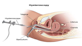
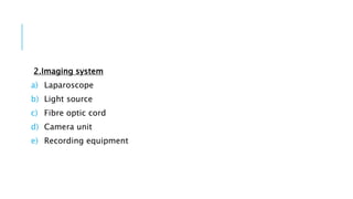
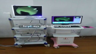
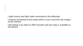
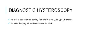
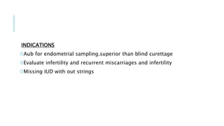
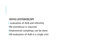
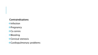
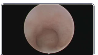
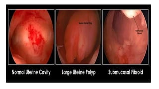
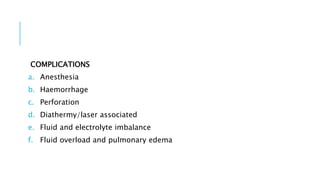
HYSTEROSCOPY
Akshai George Paul
Direct visual inspection of the cervical canal and uterine cavity
First described by Panteleoni in 1869
It used for diagnostic as well as for therapeutic purposes
EQUIPMENTS
1.Hysteroscopes
It can be flexible or rigid
Majority of it are rigid with a 4 mm diameter scope and outer sheath
OPERATING SHEATH MAY HAVE ABOUT 6-7 mm in diameter
One of the smallest ones are Bettochi hysteroscope 2.8 mm scope
with 4mm sheath
OPERATING HYSTEROSCOPE
oIt include telescope,the inflow sheath and the outflow sheath
oInflow sheath brings the distension media,while the out flow
withdraws the media
DIAGNOSTIC HYSTEROSCOPE
A single sheath inflow is sufficient
This enables smaller outer diameter so it is possible as outpatient
procedure with out anesthesaia
Oblique angled telescopes are available may be of varying angles
12,30 etc.
Flexible telescopes have an advantage with no touch technique
Discomfort is minimal and anesthesia is not required
2.Imaging system
a) Laparoscope
b) Light source
c) Fibre optic cord
d) Camera unit
e) Recording equipment
Light source and light cable connected to the telescope
Camera connected to the scope which in turn transmits the images
to the monitor
Recording is by video or DVD recorder and one copy is available to
the patient
3.Distension media
€As the anterior and posterior walls of the uterus are opposed
together
€Inorder to get good view uterine cavity should be distended with
fluid or gas
media indication Risks
Carbon dioxide Diagnostic only Gas embolism
1.5 % glycine Operative with
monopolar
diathermy
Volume overload
Hyponatremia
Normal saline Operative with bipolar
diathermy
Volume overload
Others 3 % sorbitol
†These solutions does not disperse current except NS in monopolar
†NS is safe in bipolar diathermy
DIAGNOSTIC HYSTEROSCOPY
ΩTo evaluate uterine cavity for anomalies , polyps ,fibroids
ΩTo take biopsy of endometrium in AUB
INDICATIONS
◊Aub for endometrial sampling.superior than blind curettage
◊Evaluate infertility and recurrent miscarriages and infertility
◊Missing IUD with out strings
PROCEDURE
Patient in modified lithotomy position
Parts painted and draped
Many doesnot need anesthesia
Prostaglandin E1(misoprostol) tablets can be used to dilate cervix
prior to procedure
Some time there is need for anesthesia GA/SA
Usually procedure is planned immediately for post menstrual
phase,ie endometrium is least vascular and thinnest
After local cleansing sims speculum is inserted and anterior lip of
the cervix is held with a volsellum forceps
Then the hysteroscope is inserted
Dilatation is done only if absolutely needed
Next cervical canal and endometrial cavity is visualised
Uterine distension allows good visualisation
Scope is advanced to fundus and rotated to inspect ostias
Each wall is systematically inspected for polyps and fibroids
Septums may be visualised
Adhesions may be visualised
Missing IUD may be seen
OFFICE HYSTEROSCOPY
 evaluation of AUB and infertility
No anesthesia is required
Endometrial samplings can be done
All evaluation of AUB in a single visit
Contraindications
Infection
Pregnancy
Ca cervix
Bleeding
Cervical stenosis
Cardiopulmonary problems
OPERATIVE HYSTEROSCOPY
A.Polypectomy
Polyps can be removed by using grasping forceps,but larger ones
need resectoscope
B.Myomectomy
Best removed hysteroscopically
Better toAssess the myomas sonohysterographically prior
Myomas less than 5 cm can be removed
Resectoscopes are best used for large myomas
GnRH agonists are administered to shrink the myomas
3.Septal resection
©It is done to improve the reproductive outcome with recurrent
pregnancy loss
©Usually septae is avascular
©End point is reached when both ostias are visible
4.Asherman syndrome
To remove the adhesions
Adhesiolysis continued till the tubal ostia is visible
5.Endometrial ablation
o for menorrhagia that does not respond to medical management
oTranscervical resection of endometrium using electrosurgery
resectoscope
oRoller ball electrodes are also used to coagulate the endometrium
oLaser ablation can also be done
6.Sterilisation
Not much performed
Occlude tubes using plugs/sclerosing agents
7.Tubal cannulation
Novy tubal cannulation catheter system is threaded through an
operating port of hysteroscope
Under direct vision outer catheter is advanced and placed at one of
the tubal ostia
Inner catheter is threaded approximately 2 cm into the proximal
fallopian tube
Inner catheter is flushed with water soluble dye
Using laparoscope presence or absence of spill is visualised
COMPLICATIONS
a. Anesthesia
b. Haemorrhage
c. Perforation
d. Diathermy/laser associated
e. Fluid and electrolyte imbalance
f. Fluid overload and pulmonary edema
THANK YOU

Hysteroscopy agp

  • 1.
  • 2.
    Direct visual inspectionof the cervical canal and uterine cavity First described by Panteleoni in 1869 It used for diagnostic as well as for therapeutic purposes
  • 4.
    EQUIPMENTS 1.Hysteroscopes It can beflexible or rigid Majority of it are rigid with a 4 mm diameter scope and outer sheath OPERATING SHEATH MAY HAVE ABOUT 6-7 mm in diameter One of the smallest ones are Bettochi hysteroscope 2.8 mm scope with 4mm sheath
  • 5.
    OPERATING HYSTEROSCOPE oIt includetelescope,the inflow sheath and the outflow sheath oInflow sheath brings the distension media,while the out flow withdraws the media DIAGNOSTIC HYSTEROSCOPE A single sheath inflow is sufficient This enables smaller outer diameter so it is possible as outpatient procedure with out anesthesaia
  • 10.
    Oblique angled telescopesare available may be of varying angles 12,30 etc. Flexible telescopes have an advantage with no touch technique Discomfort is minimal and anesthesia is not required
  • 11.
    2.Imaging system a) Laparoscope b)Light source c) Fibre optic cord d) Camera unit e) Recording equipment
  • 13.
    Light source andlight cable connected to the telescope Camera connected to the scope which in turn transmits the images to the monitor Recording is by video or DVD recorder and one copy is available to the patient
  • 14.
    3.Distension media €As theanterior and posterior walls of the uterus are opposed together €Inorder to get good view uterine cavity should be distended with fluid or gas media indication Risks Carbon dioxide Diagnostic only Gas embolism 1.5 % glycine Operative with monopolar diathermy Volume overload Hyponatremia Normal saline Operative with bipolar diathermy Volume overload Others 3 % sorbitol
  • 15.
    †These solutions doesnot disperse current except NS in monopolar †NS is safe in bipolar diathermy
  • 16.
    DIAGNOSTIC HYSTEROSCOPY ΩTo evaluateuterine cavity for anomalies , polyps ,fibroids ΩTo take biopsy of endometrium in AUB
  • 17.
    INDICATIONS ◊Aub for endometrialsampling.superior than blind curettage ◊Evaluate infertility and recurrent miscarriages and infertility ◊Missing IUD with out strings
  • 18.
    PROCEDURE Patient in modifiedlithotomy position Parts painted and draped Many doesnot need anesthesia Prostaglandin E1(misoprostol) tablets can be used to dilate cervix prior to procedure Some time there is need for anesthesia GA/SA Usually procedure is planned immediately for post menstrual phase,ie endometrium is least vascular and thinnest
  • 19.
    After local cleansingsims speculum is inserted and anterior lip of the cervix is held with a volsellum forceps Then the hysteroscope is inserted Dilatation is done only if absolutely needed Next cervical canal and endometrial cavity is visualised Uterine distension allows good visualisation Scope is advanced to fundus and rotated to inspect ostias Each wall is systematically inspected for polyps and fibroids
  • 20.
    Septums may bevisualised Adhesions may be visualised Missing IUD may be seen
  • 21.
    OFFICE HYSTEROSCOPY  evaluationof AUB and infertility No anesthesia is required Endometrial samplings can be done All evaluation of AUB in a single visit
  • 22.
  • 25.
  • 26.
    A.Polypectomy Polyps can beremoved by using grasping forceps,but larger ones need resectoscope B.Myomectomy Best removed hysteroscopically Better toAssess the myomas sonohysterographically prior Myomas less than 5 cm can be removed Resectoscopes are best used for large myomas
  • 27.
    GnRH agonists areadministered to shrink the myomas 3.Septal resection ©It is done to improve the reproductive outcome with recurrent pregnancy loss ©Usually septae is avascular ©End point is reached when both ostias are visible 4.Asherman syndrome To remove the adhesions Adhesiolysis continued till the tubal ostia is visible
  • 28.
    5.Endometrial ablation o formenorrhagia that does not respond to medical management oTranscervical resection of endometrium using electrosurgery resectoscope oRoller ball electrodes are also used to coagulate the endometrium oLaser ablation can also be done 6.Sterilisation Not much performed Occlude tubes using plugs/sclerosing agents
  • 29.
    7.Tubal cannulation Novy tubalcannulation catheter system is threaded through an operating port of hysteroscope Under direct vision outer catheter is advanced and placed at one of the tubal ostia Inner catheter is threaded approximately 2 cm into the proximal fallopian tube Inner catheter is flushed with water soluble dye Using laparoscope presence or absence of spill is visualised
  • 30.
    COMPLICATIONS a. Anesthesia b. Haemorrhage c.Perforation d. Diathermy/laser associated e. Fluid and electrolyte imbalance f. Fluid overload and pulmonary edema
  • 32.