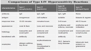
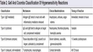
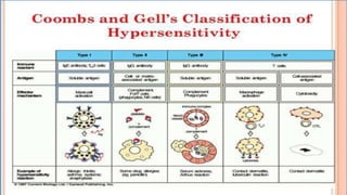
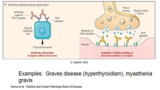
ATEKANIZA DOREEN VU-BPC-2209-0430-DAY
14
KAWALYA STEVEN VU-BPC-2307-0639-DAY
OMONG INNOCENT VU-BPC-2307-0167-DAY
WEGOSASA EVELYN VU-BPC-2209-1312-DAY
GROUP 7
HYPERSENSITIVITY REACTIONS
&
AUTOIMMUNE DISEASES
INTRODUCTION
. Hypersensitivity is a state of altered immune response against an antigen
characterized by hyper reactivity leading to immunopathology.
. Hypersensitivity reactions require a pre-sensitized (immune) state of the host.
. There are two categories of adaptive hypersensitivities:
– Immediate hypersensitivities refer to humoral
immunity (antigen/antibody reactions)
– Delayed hypersensitivities refer to cell-mediated immunity(cytotoxic
T-lymphocytes, macrophages, and cytokines)
Type I Hypersensitivity
(IgE mediated anaphylactic hypersensitivity)
Characteristics of Type-I hypersensitivity
• It is also known as immediate or anaphylactic hypersensitivity and is
mediated by IgE.
• The reaction occurs on exposure to allergen second time. The first
exposure (sensitizing dose) results in sensitization of host to allergen
and subsequent exposure (s) (shocking dose) will cause reaction
• Anaphylactic shock occurs in sensitized animals within seconds
to minutes (15-30after exposure to the allergen) after exposure
to the antigen, (allergen).
Sometimes the reaction may have a delayed onset (10-12 hours).
• Diagnostic tests for immediate hypersensitivity include skin (prick
and intradermal) tests resulting in wheal and flare reaction,
measurement of total IgE and specific IgE antibodies against the
suspected allergens by ELISA, Radioallergosorbent test (RAST)
Mechanism of Type-I hypersensitivity
• The mechanism of reaction involves preferential production of IgE, in
response to certain antigens (allergens).
• Individuals prone to type-I hypersensitivity preferentially produce
IL-4 and IL-13 that favor IgE class switch.
• IgE has very high affinity for its receptor (FcεIII; CD23) expressed on
surface of mast cells and basophils; the Fc portion of IgE binds to these
receptors.
• A subsequent exposure to the same allergen cross links the cell- bound IgE
and triggers the release of various pharmacologically active substances by
a process called as ‘degranulation ’; mast cell degranulation is preceded
by increased Ca++ influx, which is acrucial process.
• These agents cause the early phase of allergic reactions that appears
within minutes after exposure to the antigen.
Cross linking of IgE by allergen triggers mast cell degranulation
Mechanism of Type-I hypersensitivity
• Degranulation of cells result in the synthesis and secretion of
inflammatory mediators such as platelet-activating factor, leukotreins,
bradykinins, prostaglandins, and cytokines that contribute to
inflammation.
• The reaction is amplified by PAF (platelet activation factor) that causes platelet
aggregation and release of histamine, heparin and vasoactive amines. Eosinophil
chemotactic factor of anaphylaxis (ECF-A) and neutrophil chemotactic
factors attract eosinophils and neutrophils, respectively, which release
various hydrolytic enzymes that cause necrosis
• These agents cause the early phase of allergic reactions that appears within
minutes after exposure to the antigen.
• Late phase allergic reactions may begin several hours after exposure to antigen.
• Cell-bound IgE on the surface of basophils of sensitive individuals binds a
substance called histamine releasing factor (possibly produced by macrophages
and B-lymphocytes) causing further histamine release
Induction and effecter mechanisms in Type-I Hypersensitivity
Effects of Type-I Hypersensitivity
. The inflammatory agents released or produced cause the following:
– dilation of bloodvessels, which causes local redness (erythema) at the
site of allergen delivery. If dilation is widespread, this can contribute to
decreased vascular resistance, a drop in blood pressure, and shock.
– increased capillary permeability, which causes swelling of local tissues
(edema). If widespread, it can contribute to decreased blood volume
and shock.
– constriction of bronchial airways, which leads to wheezing and
difficulty in breathing.
– stimulation of mucous secretion, which leads to congestion of airways.
– stimulation of nerve endings, which leads to itching and pain in the
skin.
Pathology of Type-I hypersensitivity
• The primary cellular component in Type-I is mast cell or basophil. The
reaction is amplified and/or modified by platelets,
neutrophils and eosinophils.
• The reaction may involve skin (urticaria and eczema), eyes
(conjunctivitis), nasopharynx (rhinorrhea, rhinitis),
bronchopulmonary tissues (asthma) and gastrointestinal tract
(gastroenteritis).
The reaction may cause from minor inconvenience to death.
Pathology of Type-I hypersensitivity
• The IgE mediated diseases in humans include systemic
anaphylactic shock, asthma, Hay fever (allergic rhinitis), tropical pulmonary
eosionophila, allergic conjunctivitis, skin reactions (urticaria, eczema), food
allergies.
Wheal and Flare reaction
• Is the characteristic local cutaneous reaction, developing within 10 to
15 minutes of injecting an allergen into the skin.
• It consists of an elevated, blanched “wheal” surrounded by a
spreading by a spreading “flare” of erythema caused by release of
histamine from mast cells.
• Wheal is a raised, itchy (pruritic) area of skin. It reflect
circumscribed dermal edema (fluid collection in the layer of skin
below the surface).
• A wheal is also sometimes called a welt and often a hive.
• Flare is an area of redness surrounding wheal
• The larger the wheal and flare, the greater the sensitivity.
Mechanism of Wheal and Flare reaction
The mechanism of wheal and flare reaction is expressed by Triple response of
Lewis:
Red spot: due to capillary dilatation
Flare: redness in the surrounding area due to arteriolar dilatation mediated by axon
reflex.
Wheal: due to exudation of fluid from capillaries and venules
Type II Hypersensitivity
(Antibody-mediated cytotoxic hypersensitivity)
Characteristics of Type II Hypersensitivity
• Type II hypersensitivity are antibody mediated cytotoxic reactions occurring
when an antibody binds to antigens located on the surface of cells
(usually RBCs). The reaction time is minutes to hours.
• It is mediated, primarily, by antibodies of IgM or IgG class.
• The bound antibody can cause cell lysis by activating the classical
complement pathway, promoting phagocytosis (opsonization) or through
ADCC.
• Many different antigens may trigger this cell destruction, but an infection in
a genetically predisposed animal appears to be a major triggering pathway.
• Antigens are normally endogenous, although exogenous chemicals (haptens,
such as ivy or drugs) that can attach to cell membranes can also induce type II
reactions.
• Autoimmune haemolytic anaemia, Blood transfusion reactions,
Erythroblastosis fetalis, drug-induced hemolytic anemia,
Mechanism of Type II Hypersensitivity
. IgM or IgG antibodies are made against self antigens either as a result of failure
of immune tolerance or cross-reactive antibodies can develop during
infections, which may bind to normal tissue antigens and trigger antibody-
mediated cytotoxicity.
. Subsequent binding of these antibodies to the surface of host cells leads to:
– Opsonization of the host cells whereby phagocytes stick to host cells by way of
IgG, C3b, or C4b and discharge their lysosomes.
–Activation of the classical complement pathway
causing MAC induced lysis.
–ADCC mediated destruction of the host cells whereby NK cells attach to the Fc
portion of the antibodies.
Pathology of Type II Hypersensitivity
. Mediated by Abs directed towards antigens present on cell surfaces
or the extracellular matrix (type IIA) or abs with
agonistic/antagonistic properties (type IIB).
. The most common cells involved are blood cells. The outcome maybe:
– hemolytic anemia if RBCs are involved,
– leukopenia involving WBCs, or
– thrombocytopenia involving platelets.
. Under some circumstances, a cytotoxic attack on vascular epithelial
cells will cause avasculitis with local vascular leakage.
Examples of Type II Hypersensitivity
>AB and Rh blood group reactions (blood transfusion reactions,
erythroblastosis foetalis)
>Autoimmune diseases:
>Rheumatic fever where antibodies result in joint and heart valve
damage;
>Idiopathic thrombocytopenia purpura where antibodies result in
the destruction of platelets;
>Myasthenia gravis where antibodies bind to the acetylcholine
receptors on muscle cells causing faulty enervation of muscles;
>Goodpasture's syndrome where antibodies lead to destruction of
Examples of Type II Hypersensitivity
>Autoimmune diseases:
• Graves' disease where antibodies are made against thyroid-
stimulating hormone receptors of thyroid cells leading to faulty
thyroid function;
• Multiple sclerosis where antibodies are made against the
oligodendroglial cells that make myelin, the protein that forms
the myelin sheath that insulates the nerve fiber of neurons in
the brain and spinal cord;
>Some drug reactions,e.g. penicillin
> Bovine Neonatal Pancytopenia
Erythroblastosis Foetalis
TYPE - III HYPERSENSITIVITY
(Immune complex mediated hypersensitivity)
Characteristics of Type III Hypersensitivity
• In type III hypersensitivity, soluble immune complexes are formed
in blood and are deposited in various tissues (typically the skin, kidney
and joints), activate classical complement pathway and cause
inflammatory damage.
• It is mediated by soluble immune complexes. They are mostly of IgG
class, although IgM may also be involved.
• The reaction takes hours todays (3-10 hours) to develop.
• The antigen may be exogenous (chronic bacterial, viral or parasitic
infections), or endogenous (non-organ specific autoimmunity: e.g.,
Systemic Lupus Erythematosus-SLE).
• The antigen is soluble and not attached to the organ involved.
• The prerequisite for the development of immune-complex disease
is the persistent presence of soluble antigen and antibody.
Mechanism of Type III Hypersensitivity
Soluble antigen-antibody (IgG or IgM) complexes are normally removed by macrophages in the
spleen and liver.
On formation of excessive amount or large immune complexes these gets lodged
in capillaries, pass between endothelial cells of blood vessels – specially those in the
skin, joints, and kidneys - and become trapped on the surrounding basement membrane
beneath these cells.
The Ag-Ab complexes then activate the classical complement pathway; the damage being
caused by platelets and neutrophils :
• Massive inflammation, due to complement protein C5a;
• Influx of neutrophils, due to complement protein C5a , resulting in neutrophils
discharging their lysosomes and causing tissue destruction and further inflammation;
• MAC induced lysis of surrounding tissue cells; and
• Aggregation of platelets, resulting in more inflammation and the formation of
microthrombi that block capillaries.
TYPE - III HYPERSENSITIVITY
Pathology of Type III Hypersensitivity
. The affinity of antibody and size of immune complexes are important in production of
disease and determining the tissue involved.
. The lesion contains primarily neutrophils and deposits of immune complexes and
complement.
. Macrophages infiltrating in later stages may be involved in the healing process.
. The location of the immune complexes is largely determined by the route by which antigen
enters the body:
– Inhaled antigens give rise to apneumonitis,
– antigens that enter through the skin cause local skin lesions, and
– antigens that access the bloodstream form immune complexes that are deposited
in renal glomeruli or joints.
. Clinical signs are therefore variable but may include fever, cutaneous signs,
polyarthritis, ataxia, behavior change, or nonspecific signs such as vomiting, diarrhea, or
Examples of Type II Hypersensitivity
. Examples in human beings are:
• Serum sickness
• Arthus reaction
• Polyarteritis
• Rheumatoid arthritis
• Glomerulonephritis
• Systemic Lupus Erythromatosus
• Allergies to penicillin and sulfonamides
• Poststreptococcal Glomerulonephritis
• Meningitis
• Hepatitis
• Mononucleosis.
Arthus Reaction
. An Arthus reaction is a localized type III hypersensitivity reaction
characterized by immune complex– mediated small- vessel vasculitis
associated with deposition of immune complexes and activation of
complement, and consequent organ damage
. Immune complexes form following high local concentration of vaccine antigens
and high circulating antibody concentration.
. It was first observed by Arthus in 1903 following repeated injections of horse
serum into rabbits.
. The formation of immune complexes initiates complement activation and
an influx of inflammatory cells, followed by thrombus formation and
hemorrhagic infarction in the areas of most intense inflammation; the
types of mediators involved in Arthus reaction determines the
pathological outcome of disease.
Arthus Reaction
It is characterized by pain, swelling, induration, and edema beginning
several hours after immunization and usually reaching a peak 12 to
36 hours after immunization.
It is self-limiting, resolving over the course of a few days.
The frequency and severity can be lessened by spacing
immunizations more widely, as has been recommended for
tetanus-diphtheria toxoid booster injections.
Serum Sickness
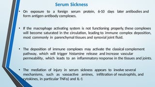
• Serum sickness is a systemic type III hypersensitivity reaction that
results from the injection of heterologous or foreign protein or
serum.
• Certain medications (eg, penicillin, nonsteroidal anti-
inflammatory drugs [NSAIDs]) have also been associated with
serum sickness– like reactions.
• These reactions typically occur 1 to 3 weeks after exposure to the drug,
but may occur as early as 1 to 24 hours afterward.
• Clinical manifestations include general malaise, fever, urticaria,
artheralgia, eosinophilia, splenomegaly and lymphadenopathy.
Serum Sickness
• On exposure to a foreign serum protein, 6-10 days later antibodies and
form antigen-antibody complexes.
• If the macrophage activating system is not functioning properly, these complexes
will become saturated in the circulation, leading to immune complex deposition,
most commonly in parenchymal tissues and synovial joint fluid.
• The deposition of immune complexes may activate the classical complement
pathway, which will trigger histamine release and increase vascular
permeability, which leads to an inflammatory response in the tissues and joints.
• The mediation of injury in serum sickness appears to involve several
mechanisms, such as vasoactive amines, infiltration of neutrophils, and
cytokines, in particular TNFα) and IL-1
Pathogenesis of Serum Sickness
Arthus reaction versus Serum Sickness
TYPE - IV HYPERSENSITIVITY
(Cell mediated hypersensitivity)
(Delayed type hypersensitivity)
Characteristics of Type IV Hypersensitivity
• Type IV HR is often called delayed type hypersensitivity as the
reaction takes more than 12 hours to develop. Typically the maximal reaction
time occurs between 48 to 72 hours
• It is mediated by cells that cause an inflammatory reaction to either exogenous
or autoantigens
• The major cells involved are T lymphocytes and
monocytes/macrophages.
• This reaction to exogenous antigens involves T cells and also antigen-presenting
cells (APC), all produce cytokines that stimulate a local inflammatory
response in a sensitized individual.
• DHR cannot be transferred from an animal to another by means of
antibodies or serum. However, it can be transferred by T cells, particularly CD4
Th1 cells.
Mechanism of Type IV Hypersensitivity
• CD8 cytotoxic T cells and CD4 helper T cells recognize antigen in a complex
with either type I or II MHC antigens
• The antigen-presenting cells in this case are macrophages and they release
interleukin 1, which further stimulates the proliferation of CD4 cells.
• These cells release IL-2 and IFN-λ, which together regulate the immune
reaction; other lymphokines involved in delayed hypersensitivity reaction
include monocyte chemotactic factor, TNF α etc.
• Cytokines produced by keratinocytes, APC, and T cells recruit antigen-
nonspecific T cells and macrophages to participate in a local inflammatory
reaction.
• Activated CD8 cells destroy target cells on contact, while activated
macrophages produce hydrolytic enzymes and, transform into
multinucleated giant cells.
Mechanism of Type IV Hypersensitivity
Pathology of Type IV Hypersensitivity
. It is involved in autoimmune-infectious diseases (tuberculosis, leprosy,
blastomycosis, histoplasmosis, toxoplasmosis, leishmaniasis, etc.) and
granulomas due to infections and foreign antigens.
. There are three variants of delayed hypersensitivity as listed below and
their maximal reaction time appears in brackets:
– Contact (48 to 72 hours)
– Tuberculin (48 to 72 hours)
– Granulomatous (21 to 28 days)
. The delayed hypersensitivity lesions mainly contain monocytes and a few T
cells.
– Contact: mononuclear cells infiltrates present in both dermis and
epidermis.
Delayed Hypersensitivity Reactions
Granuloma (T.B)
Tuberculin type
Contact Dermatitis
Comparison of Types of Hypersensitivity
COXSACKIE B VIRUSES
PATHOGENESIS CLINICAL MANIFESTATION
• After initial replication in the pharynx and
intestines,
• the virus can spread to various organs,
including the heart and pancreas.
• Coxsackie B viruses cause direct viral
cytolysis of myocardial cells,
• triggering an inflammatory response.
• The host immune response can further
damage cardiac tissue, exacerbating
myocarditis.
•Myocarditis: Chest pain, dyspnea, arrhythmias,
and signs of heart failure.
•Severe cases may progress to dilated
cardiomyopathy.
.
•Pericarditis: Inflammation of the pericardium,
presenting with chest pain, friction rub, and
evidence of pericardial effusion.
•Perimyocarditis is a combination of both.
•Pleurodynia (epidemic myalgia): characterized
by sudden, Stabbing chest pain and muscle
tenderness, often called "devil's grip."
CONTN……… Mechanism of Myocarditis
• It involves cardiotropic viruses (viruses with a high affinity for gaining
entry to cardiac muscle cells, usually via binding to a transmembrane
receptors).
• Coxsackie B, specifically B3 and B5, interact with coxsackievirus-
adenovirus receptor (CAR) and decay-accelerating factor (DAF) or
CD55 and enter into mycardial cells.
• Over first week: The cell damage by virus leads to release of
interleukins IL-6 and IL-8 and damage associated molecule patterns
(DAMP). These mediate the recruitment of inflammatory cells of the
innate immune system.
• Pro-inflammatory blood cells infiltration heart tissues: lymphocytes,
macrophages and, NK cells.
CONTN…………
• Later stimulate release of Tumor Necrosis Factor-α which binds on
TNF-receptor 1, inducing necrosis and apoptosis of myocytes.
• Additionally, any stress such as pain or anxiety triggers the medullary
cells to produce neutrophils, eosinophils and monocytes which
infiltrate into the myocardium.
• The spleen is also activated to replenish more pro-inflammatory cells.
• These mobilize and release lytic enzymes to further more damage the
myocardium, and release of interferon-gamma, that leads to further
recruitment.
Contn………….
• Over the next 2–4 weeks, viral replication continues with subsequent
activation of the acquired immune system by activating Helper T cells,
signaling leads to cytotoxic T cell infiltration in myocardium and signaling
B cells to form antibodies, including the auto-antibodies.
• Auto-antibodies destroy the cardiac myosin chains and myocardio-fibrosis
• CBV binds on Toll-Like receptors TLR; This exaggerates inflammatory
response to the viral damage of myocardium, leading to chronic
inflammation
• Over the next few months to years, this process either resolves and
concludes with viral clearance.
• Or it may progress to cause permanent heart damage such as heart
failure due to dilated cardiomyopathy and ventricular dysfunction or
cardiac arrest .
DIAGNOSIS ECG: Normal ECG wave
Perimyocarditis with
tachycardia: PR segment
depression diffuse ST
elevation
Lymphocytic myocarditis
H & E tissue biopsy, showing
lymphocytes and myocyte
coagulative necrosis (-hyper-
eosinophilic cytoplasm with loss of
striations) .
Echocar
diogra
m
showin
g
cardiac
tampon
ade

HYPERSENSITIVITY REACTIONS & AUTOIMMUNE DISEASES.pptx

  • 1.
    ATEKANIZA DOREEN VU-BPC-2209-0430-DAY 14 KAWALYASTEVEN VU-BPC-2307-0639-DAY OMONG INNOCENT VU-BPC-2307-0167-DAY WEGOSASA EVELYN VU-BPC-2209-1312-DAY GROUP 7
  • 2.
  • 3.
    INTRODUCTION . Hypersensitivity isa state of altered immune response against an antigen characterized by hyper reactivity leading to immunopathology. . Hypersensitivity reactions require a pre-sensitized (immune) state of the host. . There are two categories of adaptive hypersensitivities: – Immediate hypersensitivities refer to humoral immunity (antigen/antibody reactions) – Delayed hypersensitivities refer to cell-mediated immunity(cytotoxic T-lymphocytes, macrophages, and cytokines)
  • 6.
    Type I Hypersensitivity (IgEmediated anaphylactic hypersensitivity)
  • 7.
    Characteristics of Type-Ihypersensitivity • It is also known as immediate or anaphylactic hypersensitivity and is mediated by IgE. • The reaction occurs on exposure to allergen second time. The first exposure (sensitizing dose) results in sensitization of host to allergen and subsequent exposure (s) (shocking dose) will cause reaction • Anaphylactic shock occurs in sensitized animals within seconds to minutes (15-30after exposure to the allergen) after exposure to the antigen, (allergen). Sometimes the reaction may have a delayed onset (10-12 hours). • Diagnostic tests for immediate hypersensitivity include skin (prick and intradermal) tests resulting in wheal and flare reaction, measurement of total IgE and specific IgE antibodies against the suspected allergens by ELISA, Radioallergosorbent test (RAST)
  • 8.
    Mechanism of Type-Ihypersensitivity • The mechanism of reaction involves preferential production of IgE, in response to certain antigens (allergens). • Individuals prone to type-I hypersensitivity preferentially produce IL-4 and IL-13 that favor IgE class switch. • IgE has very high affinity for its receptor (FcεIII; CD23) expressed on surface of mast cells and basophils; the Fc portion of IgE binds to these receptors. • A subsequent exposure to the same allergen cross links the cell- bound IgE and triggers the release of various pharmacologically active substances by a process called as ‘degranulation ’; mast cell degranulation is preceded by increased Ca++ influx, which is acrucial process. • These agents cause the early phase of allergic reactions that appears within minutes after exposure to the antigen.
  • 9.
    Cross linking ofIgE by allergen triggers mast cell degranulation
  • 10.
    Mechanism of Type-Ihypersensitivity • Degranulation of cells result in the synthesis and secretion of inflammatory mediators such as platelet-activating factor, leukotreins, bradykinins, prostaglandins, and cytokines that contribute to inflammation. • The reaction is amplified by PAF (platelet activation factor) that causes platelet aggregation and release of histamine, heparin and vasoactive amines. Eosinophil chemotactic factor of anaphylaxis (ECF-A) and neutrophil chemotactic factors attract eosinophils and neutrophils, respectively, which release various hydrolytic enzymes that cause necrosis • These agents cause the early phase of allergic reactions that appears within minutes after exposure to the antigen. • Late phase allergic reactions may begin several hours after exposure to antigen. • Cell-bound IgE on the surface of basophils of sensitive individuals binds a substance called histamine releasing factor (possibly produced by macrophages and B-lymphocytes) causing further histamine release
  • 11.
    Induction and effectermechanisms in Type-I Hypersensitivity
  • 12.
    Effects of Type-IHypersensitivity . The inflammatory agents released or produced cause the following: – dilation of bloodvessels, which causes local redness (erythema) at the site of allergen delivery. If dilation is widespread, this can contribute to decreased vascular resistance, a drop in blood pressure, and shock. – increased capillary permeability, which causes swelling of local tissues (edema). If widespread, it can contribute to decreased blood volume and shock. – constriction of bronchial airways, which leads to wheezing and difficulty in breathing. – stimulation of mucous secretion, which leads to congestion of airways. – stimulation of nerve endings, which leads to itching and pain in the skin.
  • 13.
    Pathology of Type-Ihypersensitivity • The primary cellular component in Type-I is mast cell or basophil. The reaction is amplified and/or modified by platelets, neutrophils and eosinophils. • The reaction may involve skin (urticaria and eczema), eyes (conjunctivitis), nasopharynx (rhinorrhea, rhinitis), bronchopulmonary tissues (asthma) and gastrointestinal tract (gastroenteritis). The reaction may cause from minor inconvenience to death.
  • 14.
    Pathology of Type-Ihypersensitivity • The IgE mediated diseases in humans include systemic anaphylactic shock, asthma, Hay fever (allergic rhinitis), tropical pulmonary eosionophila, allergic conjunctivitis, skin reactions (urticaria, eczema), food allergies.
  • 15.
    Wheal and Flarereaction • Is the characteristic local cutaneous reaction, developing within 10 to 15 minutes of injecting an allergen into the skin. • It consists of an elevated, blanched “wheal” surrounded by a spreading by a spreading “flare” of erythema caused by release of histamine from mast cells. • Wheal is a raised, itchy (pruritic) area of skin. It reflect circumscribed dermal edema (fluid collection in the layer of skin below the surface). • A wheal is also sometimes called a welt and often a hive. • Flare is an area of redness surrounding wheal • The larger the wheal and flare, the greater the sensitivity.
  • 17.
    Mechanism of Whealand Flare reaction The mechanism of wheal and flare reaction is expressed by Triple response of Lewis: Red spot: due to capillary dilatation Flare: redness in the surrounding area due to arteriolar dilatation mediated by axon reflex. Wheal: due to exudation of fluid from capillaries and venules
  • 18.
  • 19.
    Characteristics of TypeII Hypersensitivity • Type II hypersensitivity are antibody mediated cytotoxic reactions occurring when an antibody binds to antigens located on the surface of cells (usually RBCs). The reaction time is minutes to hours. • It is mediated, primarily, by antibodies of IgM or IgG class. • The bound antibody can cause cell lysis by activating the classical complement pathway, promoting phagocytosis (opsonization) or through ADCC. • Many different antigens may trigger this cell destruction, but an infection in a genetically predisposed animal appears to be a major triggering pathway. • Antigens are normally endogenous, although exogenous chemicals (haptens, such as ivy or drugs) that can attach to cell membranes can also induce type II reactions. • Autoimmune haemolytic anaemia, Blood transfusion reactions, Erythroblastosis fetalis, drug-induced hemolytic anemia,
  • 20.
    Mechanism of TypeII Hypersensitivity . IgM or IgG antibodies are made against self antigens either as a result of failure of immune tolerance or cross-reactive antibodies can develop during infections, which may bind to normal tissue antigens and trigger antibody- mediated cytotoxicity. . Subsequent binding of these antibodies to the surface of host cells leads to: – Opsonization of the host cells whereby phagocytes stick to host cells by way of IgG, C3b, or C4b and discharge their lysosomes. –Activation of the classical complement pathway causing MAC induced lysis. –ADCC mediated destruction of the host cells whereby NK cells attach to the Fc portion of the antibodies.
  • 23.
    Pathology of TypeII Hypersensitivity . Mediated by Abs directed towards antigens present on cell surfaces or the extracellular matrix (type IIA) or abs with agonistic/antagonistic properties (type IIB). . The most common cells involved are blood cells. The outcome maybe: – hemolytic anemia if RBCs are involved, – leukopenia involving WBCs, or – thrombocytopenia involving platelets. . Under some circumstances, a cytotoxic attack on vascular epithelial cells will cause avasculitis with local vascular leakage.
  • 24.
    Examples of TypeII Hypersensitivity >AB and Rh blood group reactions (blood transfusion reactions, erythroblastosis foetalis) >Autoimmune diseases: >Rheumatic fever where antibodies result in joint and heart valve damage; >Idiopathic thrombocytopenia purpura where antibodies result in the destruction of platelets; >Myasthenia gravis where antibodies bind to the acetylcholine receptors on muscle cells causing faulty enervation of muscles; >Goodpasture's syndrome where antibodies lead to destruction of
  • 25.
    Examples of TypeII Hypersensitivity >Autoimmune diseases: • Graves' disease where antibodies are made against thyroid- stimulating hormone receptors of thyroid cells leading to faulty thyroid function; • Multiple sclerosis where antibodies are made against the oligodendroglial cells that make myelin, the protein that forms the myelin sheath that insulates the nerve fiber of neurons in the brain and spinal cord; >Some drug reactions,e.g. penicillin > Bovine Neonatal Pancytopenia
  • 26.
  • 27.
    TYPE - IIIHYPERSENSITIVITY (Immune complex mediated hypersensitivity)
  • 28.
    Characteristics of TypeIII Hypersensitivity • In type III hypersensitivity, soluble immune complexes are formed in blood and are deposited in various tissues (typically the skin, kidney and joints), activate classical complement pathway and cause inflammatory damage. • It is mediated by soluble immune complexes. They are mostly of IgG class, although IgM may also be involved. • The reaction takes hours todays (3-10 hours) to develop. • The antigen may be exogenous (chronic bacterial, viral or parasitic infections), or endogenous (non-organ specific autoimmunity: e.g., Systemic Lupus Erythematosus-SLE). • The antigen is soluble and not attached to the organ involved. • The prerequisite for the development of immune-complex disease is the persistent presence of soluble antigen and antibody.
  • 29.
    Mechanism of TypeIII Hypersensitivity Soluble antigen-antibody (IgG or IgM) complexes are normally removed by macrophages in the spleen and liver. On formation of excessive amount or large immune complexes these gets lodged in capillaries, pass between endothelial cells of blood vessels – specially those in the skin, joints, and kidneys - and become trapped on the surrounding basement membrane beneath these cells. The Ag-Ab complexes then activate the classical complement pathway; the damage being caused by platelets and neutrophils : • Massive inflammation, due to complement protein C5a; • Influx of neutrophils, due to complement protein C5a , resulting in neutrophils discharging their lysosomes and causing tissue destruction and further inflammation; • MAC induced lysis of surrounding tissue cells; and • Aggregation of platelets, resulting in more inflammation and the formation of microthrombi that block capillaries.
  • 30.
    TYPE - IIIHYPERSENSITIVITY
  • 31.
    Pathology of TypeIII Hypersensitivity . The affinity of antibody and size of immune complexes are important in production of disease and determining the tissue involved. . The lesion contains primarily neutrophils and deposits of immune complexes and complement. . Macrophages infiltrating in later stages may be involved in the healing process. . The location of the immune complexes is largely determined by the route by which antigen enters the body: – Inhaled antigens give rise to apneumonitis, – antigens that enter through the skin cause local skin lesions, and – antigens that access the bloodstream form immune complexes that are deposited in renal glomeruli or joints. . Clinical signs are therefore variable but may include fever, cutaneous signs, polyarthritis, ataxia, behavior change, or nonspecific signs such as vomiting, diarrhea, or
  • 32.
    Examples of TypeII Hypersensitivity . Examples in human beings are: • Serum sickness • Arthus reaction • Polyarteritis • Rheumatoid arthritis • Glomerulonephritis • Systemic Lupus Erythromatosus • Allergies to penicillin and sulfonamides • Poststreptococcal Glomerulonephritis • Meningitis • Hepatitis • Mononucleosis.
  • 33.
    Arthus Reaction . AnArthus reaction is a localized type III hypersensitivity reaction characterized by immune complex– mediated small- vessel vasculitis associated with deposition of immune complexes and activation of complement, and consequent organ damage . Immune complexes form following high local concentration of vaccine antigens and high circulating antibody concentration. . It was first observed by Arthus in 1903 following repeated injections of horse serum into rabbits. . The formation of immune complexes initiates complement activation and an influx of inflammatory cells, followed by thrombus formation and hemorrhagic infarction in the areas of most intense inflammation; the types of mediators involved in Arthus reaction determines the pathological outcome of disease.
  • 34.
    Arthus Reaction It ischaracterized by pain, swelling, induration, and edema beginning several hours after immunization and usually reaching a peak 12 to 36 hours after immunization. It is self-limiting, resolving over the course of a few days. The frequency and severity can be lessened by spacing immunizations more widely, as has been recommended for tetanus-diphtheria toxoid booster injections.
  • 36.
    Serum Sickness • Serumsickness is a systemic type III hypersensitivity reaction that results from the injection of heterologous or foreign protein or serum. • Certain medications (eg, penicillin, nonsteroidal anti- inflammatory drugs [NSAIDs]) have also been associated with serum sickness– like reactions. • These reactions typically occur 1 to 3 weeks after exposure to the drug, but may occur as early as 1 to 24 hours afterward. • Clinical manifestations include general malaise, fever, urticaria, artheralgia, eosinophilia, splenomegaly and lymphadenopathy.
  • 37.
    Serum Sickness • Onexposure to a foreign serum protein, 6-10 days later antibodies and form antigen-antibody complexes. • If the macrophage activating system is not functioning properly, these complexes will become saturated in the circulation, leading to immune complex deposition, most commonly in parenchymal tissues and synovial joint fluid. • The deposition of immune complexes may activate the classical complement pathway, which will trigger histamine release and increase vascular permeability, which leads to an inflammatory response in the tissues and joints. • The mediation of injury in serum sickness appears to involve several mechanisms, such as vasoactive amines, infiltration of neutrophils, and cytokines, in particular TNFα) and IL-1
  • 38.
  • 39.
    Arthus reaction versusSerum Sickness
  • 40.
    TYPE - IVHYPERSENSITIVITY (Cell mediated hypersensitivity) (Delayed type hypersensitivity)
  • 41.
    Characteristics of TypeIV Hypersensitivity • Type IV HR is often called delayed type hypersensitivity as the reaction takes more than 12 hours to develop. Typically the maximal reaction time occurs between 48 to 72 hours • It is mediated by cells that cause an inflammatory reaction to either exogenous or autoantigens • The major cells involved are T lymphocytes and monocytes/macrophages. • This reaction to exogenous antigens involves T cells and also antigen-presenting cells (APC), all produce cytokines that stimulate a local inflammatory response in a sensitized individual. • DHR cannot be transferred from an animal to another by means of antibodies or serum. However, it can be transferred by T cells, particularly CD4 Th1 cells.
  • 42.
    Mechanism of TypeIV Hypersensitivity • CD8 cytotoxic T cells and CD4 helper T cells recognize antigen in a complex with either type I or II MHC antigens • The antigen-presenting cells in this case are macrophages and they release interleukin 1, which further stimulates the proliferation of CD4 cells. • These cells release IL-2 and IFN-λ, which together regulate the immune reaction; other lymphokines involved in delayed hypersensitivity reaction include monocyte chemotactic factor, TNF α etc. • Cytokines produced by keratinocytes, APC, and T cells recruit antigen- nonspecific T cells and macrophages to participate in a local inflammatory reaction. • Activated CD8 cells destroy target cells on contact, while activated macrophages produce hydrolytic enzymes and, transform into multinucleated giant cells.
  • 43.
    Mechanism of TypeIV Hypersensitivity
  • 44.
    Pathology of TypeIV Hypersensitivity . It is involved in autoimmune-infectious diseases (tuberculosis, leprosy, blastomycosis, histoplasmosis, toxoplasmosis, leishmaniasis, etc.) and granulomas due to infections and foreign antigens. . There are three variants of delayed hypersensitivity as listed below and their maximal reaction time appears in brackets: – Contact (48 to 72 hours) – Tuberculin (48 to 72 hours) – Granulomatous (21 to 28 days) . The delayed hypersensitivity lesions mainly contain monocytes and a few T cells. – Contact: mononuclear cells infiltrates present in both dermis and epidermis.
  • 45.
  • 46.
  • 48.
    Comparison of Typesof Hypersensitivity
  • 51.
    COXSACKIE B VIRUSES PATHOGENESISCLINICAL MANIFESTATION • After initial replication in the pharynx and intestines, • the virus can spread to various organs, including the heart and pancreas. • Coxsackie B viruses cause direct viral cytolysis of myocardial cells, • triggering an inflammatory response. • The host immune response can further damage cardiac tissue, exacerbating myocarditis. •Myocarditis: Chest pain, dyspnea, arrhythmias, and signs of heart failure. •Severe cases may progress to dilated cardiomyopathy. . •Pericarditis: Inflammation of the pericardium, presenting with chest pain, friction rub, and evidence of pericardial effusion. •Perimyocarditis is a combination of both. •Pleurodynia (epidemic myalgia): characterized by sudden, Stabbing chest pain and muscle tenderness, often called "devil's grip."
  • 52.
    CONTN……… Mechanism ofMyocarditis • It involves cardiotropic viruses (viruses with a high affinity for gaining entry to cardiac muscle cells, usually via binding to a transmembrane receptors). • Coxsackie B, specifically B3 and B5, interact with coxsackievirus- adenovirus receptor (CAR) and decay-accelerating factor (DAF) or CD55 and enter into mycardial cells. • Over first week: The cell damage by virus leads to release of interleukins IL-6 and IL-8 and damage associated molecule patterns (DAMP). These mediate the recruitment of inflammatory cells of the innate immune system. • Pro-inflammatory blood cells infiltration heart tissues: lymphocytes, macrophages and, NK cells.
  • 53.
    CONTN………… • Later stimulaterelease of Tumor Necrosis Factor-α which binds on TNF-receptor 1, inducing necrosis and apoptosis of myocytes. • Additionally, any stress such as pain or anxiety triggers the medullary cells to produce neutrophils, eosinophils and monocytes which infiltrate into the myocardium. • The spleen is also activated to replenish more pro-inflammatory cells. • These mobilize and release lytic enzymes to further more damage the myocardium, and release of interferon-gamma, that leads to further recruitment.
  • 54.
    Contn…………. • Over thenext 2–4 weeks, viral replication continues with subsequent activation of the acquired immune system by activating Helper T cells, signaling leads to cytotoxic T cell infiltration in myocardium and signaling B cells to form antibodies, including the auto-antibodies. • Auto-antibodies destroy the cardiac myosin chains and myocardio-fibrosis • CBV binds on Toll-Like receptors TLR; This exaggerates inflammatory response to the viral damage of myocardium, leading to chronic inflammation • Over the next few months to years, this process either resolves and concludes with viral clearance. • Or it may progress to cause permanent heart damage such as heart failure due to dilated cardiomyopathy and ventricular dysfunction or cardiac arrest .
  • 55.
  • 56.
    Perimyocarditis with tachycardia: PRsegment depression diffuse ST elevation Lymphocytic myocarditis H & E tissue biopsy, showing lymphocytes and myocyte coagulative necrosis (-hyper- eosinophilic cytoplasm with loss of striations) .
  • 57.