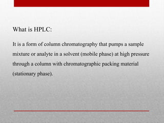
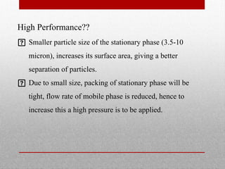
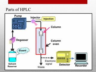
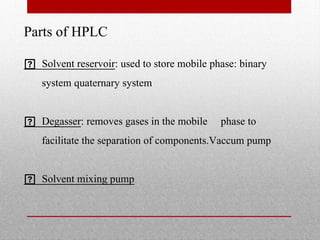
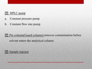
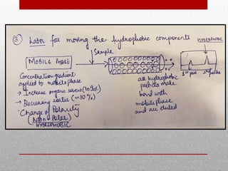
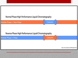
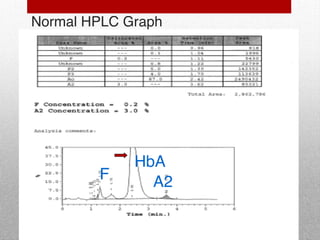
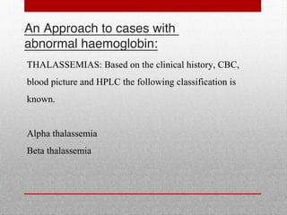
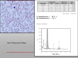
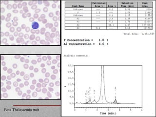
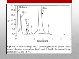
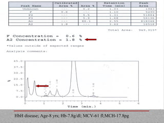
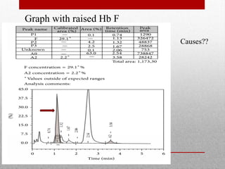
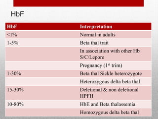
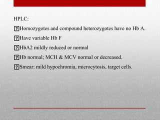
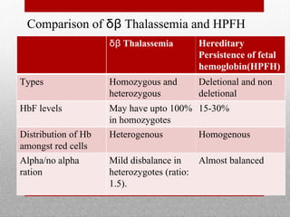
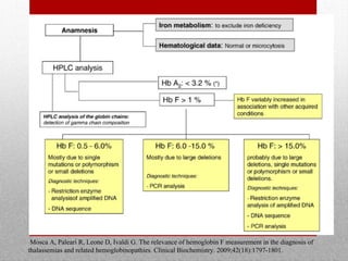
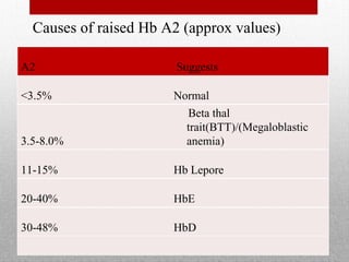
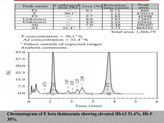
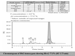
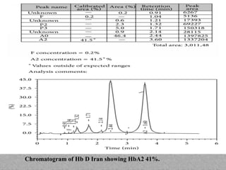
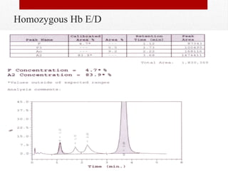
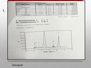
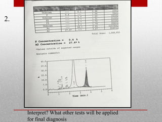
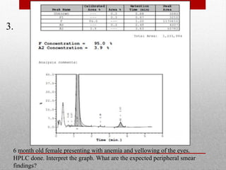
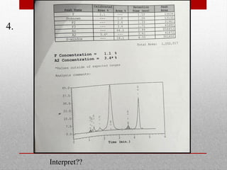
High Performance
Liquid
Chromatography
(HPLC)
By: Dr Amrita Talwar
Moderator: Dr Garima
Chromatography:
Is a laboratory technique for the separation of a mixture.
The mixture is dissolved in a fluid (gas, solvent, water,
...) called the mobile phase, which carries it through a
system (a column, a capillary tube, a plate, or a sheet) on
which is fixed a material called the stationary phase.
The different constituents of the mixture have different
affinities for the stationary phase.
Types of Chromatography:
Mechanism of separation:
• Adsorption: HPLC
• Partition: paper chromatography
• Ion exchange
• Size exclusion
Types of Chromatography:
Type of mobile phase
• Liquid chromatography
• Gas chromatography
• Super critical fluid: like CO2
Types of Chromatography:
Based on shape of chromatographic bed:
• Planar chromatography
• Column chromatography
High performance liquid chromatography:
 What is HPLC
 Principle
 Types
 Procedure
 Parts
 Advantages
What is HPLC:
It is a form of column chromatography that pumps a sample
mixture or analyte in a solvent (mobile phase) at high pressure
through a column with chromatographic packing material
(stationary phase).
High Performance??
 Smaller particle size of the stationary phase (3.5-10
micron), increases its surface area, giving a better
separation of particles.
 Due to small size, packing of stationary phase will be
tight, flow rate of mobile phase is reduced, hence to
increase this a high pressure is to be applied.
Principle of HPLC:
Mixture of molecules (such as normal and variant
haemoglobins) with a net positive charge are separated into
its components by their adsorption onto a negatively charged
stationary phase in a chromatography column, followed by
their elution by a mobile phase.
Parts of HPLC
Parts of HPLC
 Solvent reservoir: used to store mobile phase: binary
system quaternary system
 Degasser: removes gases in the mobile phase to
facilitate the separation of components.Vaccum pump
 Solvent mixing pump
 HPLC pump
a. Constant pressure pump
b. Constant flow rate pump
 Pre column(Guard column):removes contamination before
solvent enters the analytical column.
 Sample injector
Analytical column:
 Length:10-30 cm
 Internal diameter: 4-6mm.
 Stainless steel: withstands the high pressure
applied(1000-4000psi)
 Principle : normal phase
reverse phase
Detectors:
 Refractive index
 Conductivity
 UV visible detector
 Fluorescence detector
So what’s actually happening inside the analytical column?
(in a reverse phase chromatography)
Mobile phase:
 polar(hydrophillic)
Stationary phase: non polar (hydrophobic)
 Packed silica gel(SiO2)
 Due to C18/C8 group
• The stationary phase is hydrophobic,hence, hydrophobic
molecules in sample form bond with stationary
phase(adsorb) and are not eluted out.
• The hydrophillic molecules donot form bond with
hydrophobic stationary phase and are eluted first with the
hydrophillic mobile phase.
Types of HPLC:
1. Normal phase
2. Reverse phase
NORMAL PHASE REVERSE PHASE
Mobile phase Non-polar
(HYDROPHOBIC/ANIO
NIC)
Polar(HYDROPHILLIC/
CATIONIC)
Stationary phase Polar(HYDROPHILLIC/
CATIONIC)
Non-polar
(HYDROPHOBIC/ANIO
NIC)
Silica gel (SiO2)
Silanol group repsonsible
for polar nature
C18 or C8 silica is used
Advantage • For compounds that
are not soluble in
water.
• Used for separation of
isomers
• Easier to use
• Hydrophobic
stationary phase can
be applied to wide
range of molecules

The Variant II Chromatographic Station (VCS) dual pumps
deliver a programmed buffer gradient of increasing ionic
strength to the cartridge, where HbA2/F are separated based
on their ionic interaction with the cartridge material.

The separated HbA2/F then pass through the flow cell of
the filter photometer where the changes in absorbance at
415 nm are measured.

An additional filter at 690 nm corrects the background
absorbance.

The Variant II CDM (CDM) Software performs reduction of
raw data collected from each analysis.

To aid in the interpretation of results, windows have been
established for the most frequently occurring haemoglobins
based on the characteristic retention time.

Retention time is the elapsed time from the injection of the
sample to the apex of a hemoglobin peak.

A slower flow rate of the buffer improves the separation of
different haemoglobins with a similar retention time,
however, increases the processing time.

Haemoglobins eluted from the column are represented
graphically and automatically quantified by
spectroscopy.

The retention time is stated in relation to that expected
for a known normal or abnormal haemoglobin (usually
in relation to haemoglobin A, F, S, C or D).
ANALYSIS:
ANALYSIS: Normal retention time of
various hemoglobins:
ANALYSIS:
HbA
HbF
HbA2
P2window
P3window
ADVANTAGES:
Less labour intensive
A very small sample is adequate
Quantification of normal and variant haemoglobins is
available for each sample
ADVANTAGES:
As Hb A2 is quantified, b thalassaemia trait can be
diagnosed in a single procedure, replacing the combination
of haemoglobin electrophoresis and microcolumn
chromatography
Larger range of variant haemoglobins can be provisionally
identified
HEMOGLOBIN AND ITS
STRUCTURE:
B
B
A
A
heme
Hemoglobin structure
4 heme chains
4 polypeptide
chains
A
A
A
A A
A
B G D
B D
G
HbA HbF HbA2
95-98% ~1% <3.5%
Haemoglobins in normal adults
Hemoglobin Type Name Components
Adult A 22
A2 22
Fetal F 22
Embryonic Portland 22
Gower 1 22
Gower 2 22
Abnormal H 4
Bart’s 4
Normal levels
Types Quantity
HbA (α2 β2) >95%
HbA2 (α2 δ2) <3.5%
HbF (α2 γ2) <2.0%
Normal HPLC Graph
HbA
A2
F
Abnormal hemoglobins
TYPES OF THALASSEMIA:
An Approach to cases with
abnormal haemoglobin:
THALASSEMIAS: Based on the clinical history, CBC,
blood picture and HPLC the following classification is
known.
Alpha thalassemia
Beta thalassemia
Beta Thalassemia Major
Beta Thalassemia trait
Low A2
• Iron Deficiency Anemia
• Alpha Thalassemia trait
• HbH Disease
• Delta Thalassemia
• Delta –Beta Thalassemia
Peak before 1 min
• Sample Integrity
• Icteric sample
• HbH
HbH disease; Age-8 yrs; Hb-7.8g/dl; MCV-61 fl;MCH-17.8pg
Graph with raised Hb F
Causes??
HbF
HbF Interpretation
<1% Normal in adults
1-5% Beta thal trait
In association with other Hb
S/C/Lepore
Pregnancy (1st trim)
1-30% Beta thal Sickle heterozygote
Heterozygous delta beta thal
15-30% Deletional & non deletional
HPFH
10-80% HbE and Beta thalassemia
Homozygous delta beta thal
δβ Thalassemia:
Deletion of both δ and β gene.
Various mutations described for different subtypes like
Mediterranean, Senegalese , Indian and Chinese.
There is preservation of gamma genes.(preserved Hb F)

The phenotype of heterozygotes resembles that of beta
thalassemia trait without an increase of Hb A2 percentage.

Hb F is consistently elevated, varying from 5% to 30%.

Homozygotes for δβ thalassemia have 100% Hb F.

Heterozygotes usually have splenomegaly and a Hb
concentration of 8–13g/dl.

MCV and MCH may be reduced or low-normal.
Delta beta thalassaemia trait.
The red cell indices were RBC 6.04 million, Hb 14 g/dl,
Hct 0.42, MCV 69 fl, MCH 23.2 pg and MCHC
33.4g/dl;
Hereditary Persistence of fetal
hemoglobin(HPFH):
Inherited characteristic in which heterozygotes show an
increased proportion of hemoglobin F, persisting beyond
infancy.
Causes:
DELETIONAL: within the beta globin cluster.
NON-DELETIONAL: mutations or polymorphisms of
regulatory genes.
HPLC:
Homozygotes and compound heterozygotes have no Hb A.
Have variable Hb F
HbA2 mildly reduced or normal
Hb normal; MCH & MCV normal or decreased.
Smear: mild hypochromia, microcytosis, target cells.
Comparison of δβ Thalassemia and HPFH
δβ Thalassemia Hereditary
Persistence of fetal
hemoglobin(HPFH)
Types Homozygous and
heterozygous
Deletional and non
deletional
HbF levels May have upto 100%
in homozygotes
15-30%
Distribution of Hb
amongst red cells
Heterogenous Homogenous
Alpha/no alpha
ration
Mild disbalance in
heterozygotes (ratio:
1.5).
Almost balanced
Mosca A, Paleari R, Leone D, Ivaldi G. The relevance of hemoglobin F measurement in the diagnosis of
thalassemias and related hemoglobinopathies. Clinical Biochemistry. 2009;42(18):1797-1801.
Causes of raised Hb A2 (approx values)
A2 Suggests
<3.5% Normal
3.5-8.0%
Beta thal
trait(BTT)/(Megaloblastic
anemia)
11-15% Hb Lepore
20-40% HbE
30-48% HbD
Hemoglobin Lepore
Autosomal recessive mutation with in frame mutation between
5’ end of delta globin gene and 3’ end of beta globin gene
(unequal crossover); δβ hybrid globin chain is formed.
The variant δβ fusion chain is synthesized at a much reduced
rate in comparison with the normal β chain.
Three variants are known: Hb Boston/ Washinton
Hb Baltimore
Hb Hollandia
Hb Lepore is important because of the possibility of
interaction with haemoglobin S and with b thalassemias.
Hb Lepore has the same mobility as Hb S on cellulose
acetate electrophoresis at alkaline pH and moves with
HbA at acid pH.
On HPLC, it has the same retention time as HbA2
HOMOZYGOUS state: Hb A/A2 are absent
Hb F and Hb lepore are present.
Presents in the first 5 years of life, hepato-
splenomegaly, skeletal deformity.
HETEROZYGOTES : mild anemia with microcytosis and
hypochromia, with fetal hemoglobin levels moderately high
Hb Lepore 5-15%
Hb F: <5%
HPLC chromatogram in HbLepore trait; Hb Lepore was 13.4% and its
retention time was 3.49 min
Lepore trait. The red cell indices : RBC
5.36million, Hb 12 g/dl, Hct 0.351, MCV
66 fl, MCH 22.2 pg and MCHC 33.8g/dl.
Hemoglobin E:
Autosomal recessive.
Beta chain variant, a2 b26Glu Lys, common in South-East
Asia.
The betaE chain is synthesized at a reduced rate in comparison
with betaA.
Homozygosity for HbE produces a clinically mild condition.
Heterozygotes, compound heterozygotes and homozygotes
show some thalassemic features.
Hb E trait:
Hb: normal to mild fall
Red cell count (RBC);MCHC: normal or increased
MCV; MCH reduced
Hb electrophoresis at alkaline pH: variant Hb with
same mobility as HbC and A2.
Citrate agar or agarose gel at acid pH: same as that of
HbA and A2.
On HPLC, it is easily separated from HbA and C, but
co-elutes with HbA2.
Heterozygotes HbE comprises 30% or less of
total haemoglobin.
>39% of haemoglobin E: Suggests HbE/b
thalassemia
Hemoglobin E homozygous
Hemoglobin E trait
Chromatogram of E beta thalassemia showing elevated HbA2 51.4%, Hb F
30%.
Chromatogram of HbE homozygous showing HbA2 77.5% (RT 3.73 min)
Hemoglobin D Iran
Mutation of codon 22 of the beta globin chain with
transversion of GAA to CAA(substitution of glutamate with
glutamine)
Homozygous cases present with anaemia, poikilocytosis and
mild haemolysis.
Cases of Hb D Iran trait are generally asymptomatic.
It is commonly found co-existent with β-Thalassemia
Chromatogram of Hb D Iran showing HbA2 41%.
Homozygous Hb E/D
Haemoglobinopathies with separate
peaks on HPLC
Hemoglobin D Punjab:
Haemoglobin D-Punjab is a b chain variant,
a2b2121Glu Gln.
Incidence highest amongst Sikhs in the Punjab (prevalence of
2–3%).
Haemoglobin D-Punjab trait
Heterozygosity for HbD is of genetic but no other clinical
significance.
All parameters are normal.
On electrophoresis:
Cellulose acetate at alkaline pH, Hb D-Punjab has the same
electrophoretic mobility as HbS.
On citrate agar or agarose gel at acid pH, separate from S and
move with or near to Hb A.
On isoelectric focusing, D-Punjab is separated from HbA and S
Haemoglobin D-Punjab disease:
Homozygous; mild disease.
There may be mild haemolysis; mild haemolytic anaemia.
Some may have splenomegaly.
Hb concentration: 9–10g/dl up to normal levels.
Red cell count: elevated
MCV; MCH: reduced.
The reticulocyte count: normal or elevated to 2–4%.
Blood film: infrequent to many target cells and few
irregularly contracted cells.
Other tests: reduced osmotic fragility and red cell
survival.
Hb electrophoresis and HPLC normal HbA2 and F
Rest being Hb D.
Chromatogram of Hb D Punjab homozygous showing Hb D 87.9%.
Sickle cell anemia:
Sickle cell haemoglobin, Hb S, has a valine for glutamic acid
substitution at position 6 of the b chain.(a2b26Glu Val)
Partial and fully deoxygenated HbS can be incorporated into
a polymer.
Long polymers distort the red cell into a holly-leaf or into a
crescent or sickle shape, that hinder blood flow through
capillaries.
Presence of HbA,A2 and F retard the sickling.
Presence of Hb C, D-Punjab and O-Arab facilitate
sickling.
Sickle cell trait:
Blood film: may be completely normal or show
microcytosis or target cells. Classical sickle cells are
not seen.
HPLC: HbA: >50%, HbS around 40%
Sickle cell trait; both chromatograms show Hb A, A2 and S and chromatogram
(a) shows, in addition, increased HbF; HbA that has undergone post-
translational modification appears as two peaks to the left of haemoglobin A
Sickle cell disease(homozygous):
Blood film: in adults variable number of crescent or
sickle-shaped sickle cells
HbS homozygous with HbS> 50%;normal HbA2 and elevated HbF
Peaks co – eluting in the same retention time can be
differentiated by applying one or more supplementary
techniques like:
1. Electrophoresis:
b. Cellulose acetate electrophoresis
at alkaline pH
b. Citrate agar or agarose gel electrophoresis
at acid pH
2. Isoelectric focussing
QUESTIONS TO BE ANSWERED BY POST
GRADUATED
Interpret
1.
2.
Interpret? What other tests will be applied
for final diagnosis
3.
6 month old female presenting with anemia and yellowing of the eyes.
HPLC done. Interpret the graph. What are the expected peripheral smear
findings?
4.
Interpret??
5.
Interpret?
Thank you for
patient reading
Next activity
09.11.2020 Journal Club
Dr Ankita Y.
Moderator Dr Umesh Tiwari

HPLC Subject seminar.pdf

  • 1.
    High Performance Liquid Chromatography (HPLC) By: DrAmrita Talwar Moderator: Dr Garima
  • 2.
    Chromatography: Is a laboratorytechnique for the separation of a mixture. The mixture is dissolved in a fluid (gas, solvent, water, ...) called the mobile phase, which carries it through a system (a column, a capillary tube, a plate, or a sheet) on which is fixed a material called the stationary phase. The different constituents of the mixture have different affinities for the stationary phase.
  • 3.
    Types of Chromatography: Mechanismof separation: • Adsorption: HPLC • Partition: paper chromatography • Ion exchange • Size exclusion
  • 4.
    Types of Chromatography: Typeof mobile phase • Liquid chromatography • Gas chromatography • Super critical fluid: like CO2
  • 5.
    Types of Chromatography: Basedon shape of chromatographic bed: • Planar chromatography • Column chromatography
  • 6.
    High performance liquidchromatography:  What is HPLC  Principle  Types  Procedure  Parts  Advantages
  • 7.
    What is HPLC: Itis a form of column chromatography that pumps a sample mixture or analyte in a solvent (mobile phase) at high pressure through a column with chromatographic packing material (stationary phase).
  • 8.
    High Performance??  Smallerparticle size of the stationary phase (3.5-10 micron), increases its surface area, giving a better separation of particles.  Due to small size, packing of stationary phase will be tight, flow rate of mobile phase is reduced, hence to increase this a high pressure is to be applied.
  • 9.
    Principle of HPLC: Mixtureof molecules (such as normal and variant haemoglobins) with a net positive charge are separated into its components by their adsorption onto a negatively charged stationary phase in a chromatography column, followed by their elution by a mobile phase.
  • 10.
  • 11.
    Parts of HPLC Solvent reservoir: used to store mobile phase: binary system quaternary system  Degasser: removes gases in the mobile phase to facilitate the separation of components.Vaccum pump  Solvent mixing pump
  • 12.
     HPLC pump a.Constant pressure pump b. Constant flow rate pump  Pre column(Guard column):removes contamination before solvent enters the analytical column.  Sample injector
  • 13.
    Analytical column:  Length:10-30cm  Internal diameter: 4-6mm.  Stainless steel: withstands the high pressure applied(1000-4000psi)  Principle : normal phase reverse phase
  • 14.
    Detectors:  Refractive index Conductivity  UV visible detector  Fluorescence detector
  • 15.
    So what’s actuallyhappening inside the analytical column? (in a reverse phase chromatography) Mobile phase:  polar(hydrophillic) Stationary phase: non polar (hydrophobic)  Packed silica gel(SiO2)  Due to C18/C8 group
  • 18.
    • The stationaryphase is hydrophobic,hence, hydrophobic molecules in sample form bond with stationary phase(adsorb) and are not eluted out. • The hydrophillic molecules donot form bond with hydrophobic stationary phase and are eluted first with the hydrophillic mobile phase.
  • 19.
    Types of HPLC: 1.Normal phase 2. Reverse phase
  • 20.
    NORMAL PHASE REVERSEPHASE Mobile phase Non-polar (HYDROPHOBIC/ANIO NIC) Polar(HYDROPHILLIC/ CATIONIC) Stationary phase Polar(HYDROPHILLIC/ CATIONIC) Non-polar (HYDROPHOBIC/ANIO NIC) Silica gel (SiO2) Silanol group repsonsible for polar nature C18 or C8 silica is used Advantage • For compounds that are not soluble in water. • Used for separation of isomers • Easier to use • Hydrophobic stationary phase can be applied to wide range of molecules
  • 22.
     The Variant IIChromatographic Station (VCS) dual pumps deliver a programmed buffer gradient of increasing ionic strength to the cartridge, where HbA2/F are separated based on their ionic interaction with the cartridge material.  The separated HbA2/F then pass through the flow cell of the filter photometer where the changes in absorbance at 415 nm are measured.
  • 23.
     An additional filterat 690 nm corrects the background absorbance.  The Variant II CDM (CDM) Software performs reduction of raw data collected from each analysis.  To aid in the interpretation of results, windows have been established for the most frequently occurring haemoglobins based on the characteristic retention time.
  • 24.
     Retention time isthe elapsed time from the injection of the sample to the apex of a hemoglobin peak.  A slower flow rate of the buffer improves the separation of different haemoglobins with a similar retention time, however, increases the processing time.
  • 25.
     Haemoglobins eluted fromthe column are represented graphically and automatically quantified by spectroscopy.  The retention time is stated in relation to that expected for a known normal or abnormal haemoglobin (usually in relation to haemoglobin A, F, S, C or D).
  • 26.
  • 27.
    ANALYSIS: Normal retentiontime of various hemoglobins:
  • 28.
  • 29.
    ADVANTAGES: Less labour intensive Avery small sample is adequate Quantification of normal and variant haemoglobins is available for each sample
  • 30.
    ADVANTAGES: As Hb A2is quantified, b thalassaemia trait can be diagnosed in a single procedure, replacing the combination of haemoglobin electrophoresis and microcolumn chromatography Larger range of variant haemoglobins can be provisionally identified
  • 31.
  • 32.
    B B A A heme Hemoglobin structure 4 hemechains 4 polypeptide chains
  • 33.
    A A A A A A B GD B D G HbA HbF HbA2 95-98% ~1% <3.5% Haemoglobins in normal adults
  • 35.
    Hemoglobin Type NameComponents Adult A 22 A2 22 Fetal F 22 Embryonic Portland 22 Gower 1 22 Gower 2 22 Abnormal H 4 Bart’s 4
  • 36.
    Normal levels Types Quantity HbA(α2 β2) >95% HbA2 (α2 δ2) <3.5% HbF (α2 γ2) <2.0%
  • 37.
  • 38.
  • 39.
  • 40.
    An Approach tocases with abnormal haemoglobin: THALASSEMIAS: Based on the clinical history, CBC, blood picture and HPLC the following classification is known. Alpha thalassemia Beta thalassemia
  • 42.
  • 43.
  • 45.
    Low A2 • IronDeficiency Anemia • Alpha Thalassemia trait • HbH Disease • Delta Thalassemia • Delta –Beta Thalassemia
  • 46.
    Peak before 1min • Sample Integrity • Icteric sample • HbH
  • 48.
    HbH disease; Age-8yrs; Hb-7.8g/dl; MCV-61 fl;MCH-17.8pg
  • 49.
    Graph with raisedHb F Causes??
  • 50.
    HbF HbF Interpretation <1% Normalin adults 1-5% Beta thal trait In association with other Hb S/C/Lepore Pregnancy (1st trim) 1-30% Beta thal Sickle heterozygote Heterozygous delta beta thal 15-30% Deletional & non deletional HPFH 10-80% HbE and Beta thalassemia Homozygous delta beta thal
  • 51.
    δβ Thalassemia: Deletion ofboth δ and β gene. Various mutations described for different subtypes like Mediterranean, Senegalese , Indian and Chinese. There is preservation of gamma genes.(preserved Hb F)
  • 52.
     The phenotype ofheterozygotes resembles that of beta thalassemia trait without an increase of Hb A2 percentage.  Hb F is consistently elevated, varying from 5% to 30%.  Homozygotes for δβ thalassemia have 100% Hb F.  Heterozygotes usually have splenomegaly and a Hb concentration of 8–13g/dl.  MCV and MCH may be reduced or low-normal.
  • 53.
    Delta beta thalassaemiatrait. The red cell indices were RBC 6.04 million, Hb 14 g/dl, Hct 0.42, MCV 69 fl, MCH 23.2 pg and MCHC 33.4g/dl;
  • 54.
    Hereditary Persistence offetal hemoglobin(HPFH): Inherited characteristic in which heterozygotes show an increased proportion of hemoglobin F, persisting beyond infancy. Causes: DELETIONAL: within the beta globin cluster. NON-DELETIONAL: mutations or polymorphisms of regulatory genes.
  • 55.
    HPLC: Homozygotes and compoundheterozygotes have no Hb A. Have variable Hb F HbA2 mildly reduced or normal Hb normal; MCH & MCV normal or decreased. Smear: mild hypochromia, microcytosis, target cells.
  • 56.
    Comparison of δβThalassemia and HPFH δβ Thalassemia Hereditary Persistence of fetal hemoglobin(HPFH) Types Homozygous and heterozygous Deletional and non deletional HbF levels May have upto 100% in homozygotes 15-30% Distribution of Hb amongst red cells Heterogenous Homogenous Alpha/no alpha ration Mild disbalance in heterozygotes (ratio: 1.5). Almost balanced
  • 57.
    Mosca A, PaleariR, Leone D, Ivaldi G. The relevance of hemoglobin F measurement in the diagnosis of thalassemias and related hemoglobinopathies. Clinical Biochemistry. 2009;42(18):1797-1801.
  • 58.
    Causes of raisedHb A2 (approx values) A2 Suggests <3.5% Normal 3.5-8.0% Beta thal trait(BTT)/(Megaloblastic anemia) 11-15% Hb Lepore 20-40% HbE 30-48% HbD
  • 59.
    Hemoglobin Lepore Autosomal recessivemutation with in frame mutation between 5’ end of delta globin gene and 3’ end of beta globin gene (unequal crossover); δβ hybrid globin chain is formed. The variant δβ fusion chain is synthesized at a much reduced rate in comparison with the normal β chain.
  • 60.
    Three variants areknown: Hb Boston/ Washinton Hb Baltimore Hb Hollandia Hb Lepore is important because of the possibility of interaction with haemoglobin S and with b thalassemias. Hb Lepore has the same mobility as Hb S on cellulose acetate electrophoresis at alkaline pH and moves with HbA at acid pH.
  • 61.
    On HPLC, ithas the same retention time as HbA2 HOMOZYGOUS state: Hb A/A2 are absent Hb F and Hb lepore are present. Presents in the first 5 years of life, hepato- splenomegaly, skeletal deformity. HETEROZYGOTES : mild anemia with microcytosis and hypochromia, with fetal hemoglobin levels moderately high Hb Lepore 5-15% Hb F: <5%
  • 62.
    HPLC chromatogram inHbLepore trait; Hb Lepore was 13.4% and its retention time was 3.49 min Lepore trait. The red cell indices : RBC 5.36million, Hb 12 g/dl, Hct 0.351, MCV 66 fl, MCH 22.2 pg and MCHC 33.8g/dl.
  • 63.
    Hemoglobin E: Autosomal recessive. Betachain variant, a2 b26Glu Lys, common in South-East Asia. The betaE chain is synthesized at a reduced rate in comparison with betaA. Homozygosity for HbE produces a clinically mild condition. Heterozygotes, compound heterozygotes and homozygotes show some thalassemic features.
  • 64.
    Hb E trait: Hb:normal to mild fall Red cell count (RBC);MCHC: normal or increased MCV; MCH reduced Hb electrophoresis at alkaline pH: variant Hb with same mobility as HbC and A2. Citrate agar or agarose gel at acid pH: same as that of HbA and A2. On HPLC, it is easily separated from HbA and C, but co-elutes with HbA2.
  • 65.
    Heterozygotes HbE comprises30% or less of total haemoglobin. >39% of haemoglobin E: Suggests HbE/b thalassemia
  • 66.
  • 67.
    Chromatogram of Ebeta thalassemia showing elevated HbA2 51.4%, Hb F 30%.
  • 68.
    Chromatogram of HbEhomozygous showing HbA2 77.5% (RT 3.73 min)
  • 69.
    Hemoglobin D Iran Mutationof codon 22 of the beta globin chain with transversion of GAA to CAA(substitution of glutamate with glutamine) Homozygous cases present with anaemia, poikilocytosis and mild haemolysis. Cases of Hb D Iran trait are generally asymptomatic. It is commonly found co-existent with β-Thalassemia
  • 70.
    Chromatogram of HbD Iran showing HbA2 41%.
  • 71.
  • 72.
  • 73.
    Hemoglobin D Punjab: HaemoglobinD-Punjab is a b chain variant, a2b2121Glu Gln. Incidence highest amongst Sikhs in the Punjab (prevalence of 2–3%). Haemoglobin D-Punjab trait Heterozygosity for HbD is of genetic but no other clinical significance. All parameters are normal.
  • 74.
    On electrophoresis: Cellulose acetateat alkaline pH, Hb D-Punjab has the same electrophoretic mobility as HbS. On citrate agar or agarose gel at acid pH, separate from S and move with or near to Hb A. On isoelectric focusing, D-Punjab is separated from HbA and S
  • 75.
    Haemoglobin D-Punjab disease: Homozygous;mild disease. There may be mild haemolysis; mild haemolytic anaemia. Some may have splenomegaly. Hb concentration: 9–10g/dl up to normal levels. Red cell count: elevated MCV; MCH: reduced. The reticulocyte count: normal or elevated to 2–4%.
  • 76.
    Blood film: infrequentto many target cells and few irregularly contracted cells. Other tests: reduced osmotic fragility and red cell survival. Hb electrophoresis and HPLC normal HbA2 and F Rest being Hb D.
  • 77.
    Chromatogram of HbD Punjab homozygous showing Hb D 87.9%.
  • 78.
    Sickle cell anemia: Sicklecell haemoglobin, Hb S, has a valine for glutamic acid substitution at position 6 of the b chain.(a2b26Glu Val) Partial and fully deoxygenated HbS can be incorporated into a polymer. Long polymers distort the red cell into a holly-leaf or into a crescent or sickle shape, that hinder blood flow through capillaries.
  • 79.
    Presence of HbA,A2and F retard the sickling. Presence of Hb C, D-Punjab and O-Arab facilitate sickling. Sickle cell trait: Blood film: may be completely normal or show microcytosis or target cells. Classical sickle cells are not seen. HPLC: HbA: >50%, HbS around 40%
  • 80.
    Sickle cell trait;both chromatograms show Hb A, A2 and S and chromatogram (a) shows, in addition, increased HbF; HbA that has undergone post- translational modification appears as two peaks to the left of haemoglobin A
  • 81.
    Sickle cell disease(homozygous): Bloodfilm: in adults variable number of crescent or sickle-shaped sickle cells
  • 82.
    HbS homozygous withHbS> 50%;normal HbA2 and elevated HbF
  • 83.
    Peaks co –eluting in the same retention time can be differentiated by applying one or more supplementary techniques like: 1. Electrophoresis: b. Cellulose acetate electrophoresis at alkaline pH b. Citrate agar or agarose gel electrophoresis at acid pH 2. Isoelectric focussing
  • 84.
    QUESTIONS TO BEANSWERED BY POST GRADUATED
  • 85.
  • 86.
    2. Interpret? What othertests will be applied for final diagnosis
  • 87.
    3. 6 month oldfemale presenting with anemia and yellowing of the eyes. HPLC done. Interpret the graph. What are the expected peripheral smear findings?
  • 88.
  • 89.
  • 90.
  • 91.
    Next activity 09.11.2020 JournalClub Dr Ankita Y. Moderator Dr Umesh Tiwari