1. Life forms first evolved in oceans over 1 billion years ago and some of the earliest phytoplankton produced not only glucose through photosynthesis but also vitamin D2 when exposed to sunlight.
2. In the 1600s, the industrial revolution in Northern Europe led to more dense urban environments with lack of sun exposure, resulting in the bone deforming disease rickets in children.
3. In the early 20th century, physicians discovered that rickets improved in children exposed to ultraviolet lamps or sunlight, and that foods fortified with vitamin D or exposed to UV rays prevented rickets. This led to global programs that essentially eradicated rickets through vitamin D fortification and sun exposure recommendations.

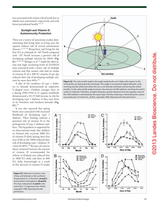
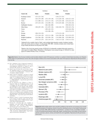
![©2013LandesBioscience.Donotdistribute.
58 Dermato-Endocrinology Volume 5 Issue 1
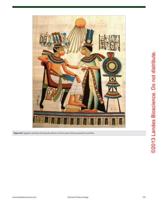
Skin pigmentation or the lack
thereof was important in the evo-
lution of humans as they migrated
North and South of the equator.
Africans such as the Maasai (Fig.
37) living outdoors exposed to sun-
light daily throughout the year have
robust circulating concentrations of
the major circulating form of vita-
min D, 25(OH)D, in the range of
46 ng/mL.53
Although there have been sev-
eral explanations for why skin pig-
ment devolved as humans migrated
North and South of the equator one
of the most likely explanations is
as humans migrated farther North
and South of the equator the zenith
angle of the sun increased resulting
in a decrease in the amount of solar
UVB radiation reaching the earth
thereby reducing vitamin D3
synthe-
sis. A decrease in the amount of skin
pigment resulted in a decrease in the
sun screening protection permitting
more of the UVB radiation to reach
the epidermal cells. This provided
an evolutionary advantage by being
more efficient in producing vitamin
D3
.49
It had long been speculated
that our Neanderthal ancestors were
heavily pigmented hairy creatures.
This however did not make a lot of
sense since having heavy pigmenta-
tion and excessive hair would markedly reduce cutaneous produc-
tion of vitamin D3
which was essential for maximizing skeletal
health throughout life thereby reducing risk of life-threatening
fractures. However, more important is the fact that vitamin D
deficiency in utero and during the first few years of life would
have caused infantile rickets resulting in a flat deformed pelvis
with a small pelvic outlet. Furthermore vitamin D is important
for muscle function which is also crucial for birthing.22,54
These
conditions caused by vitamin D deficiency would have made it
difficult for females to give birth. Therefore in order to survive
and procreate skin pigmentation had to markedly decrease in
order to permit more UVB photons to enter the skin to produce
sufficient amounts of previtamin D3
.54,55
Recent evidence has
suggested that Neanderthals had a mutation of their melanocyte
stimulating hormone receptor resulting in them being redheaded
and having Celtic-like fair skin.56,57
This is the likely explanation
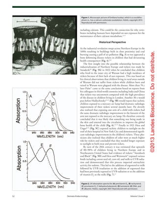
for why people in Northern Europe have skin types 1 and 2.
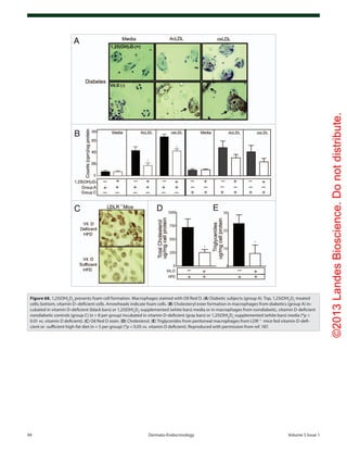
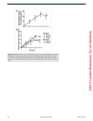
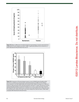
c) Aging. It was observed that 7-dehydrocholesterol concen-
trations in human epidermis were inversely related to age (Fig.
38).58
The effect of aging on the cutaneous production of vita-
min D3
was demonstrated in a study that exposed healthy young
adults and older adults to the same amount of UVB radiation
25-hydroxyvitamin D [25(OH)D] levels as found by Armas et
al.52
(Fig. 35). The associations between skin lightness, UVB
dose and 25(OH)D are documented in Figure 36.
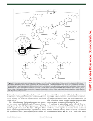
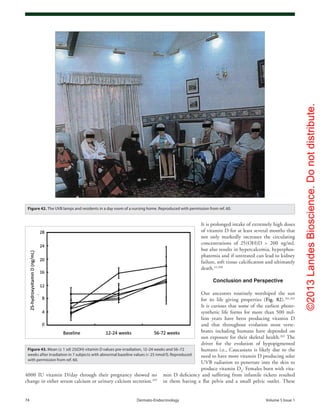
Figure 11. Photolysis of provitamin D3
(pro-D3
, 7-dehydrocholesterol) into previtamin D3
(pre-D3
) and its
thermal isomerization to vitamin D3
in hexane and in lizard skin at 25°C. In hexane pro-D3
is photolyzed
to s-cis,s-cis-pre-D3
. Once formed, this energetically unstable conformation undergoes a conformational
change to the s-trans,s-cis-pre-D3
. Only the s-cis,s-cis-pre-D3
can undergo thermal isomerization to
vitamin D3
. The s-cis,s-cis conformer of pre-D3
is stabilized in the phospholipid bilayer by hydrophilic
interactions between the 3β-hydroxl group and the polar head of the lipids, as well as by the van der
Waals interactions between the steroid ring and side-chain structure and the hydrophobic tail of the lip-
ids. These interactions significantly decrease the conversion of the s-cis,s-cis conformer to the s-trans,s-
cis conformer, thereby facilitating the thermal isomerization of s-cis,s-cis-pre-D3
to vitamin D3
. Holick,
copyright 1995. Reproduced with permission.
Figure 12. Thermal isomerization of previtamin D3
to vitamin D3
as a
function of time in lizard skin (●) and in hexane (□) at 25°C (left) and
5°C (right). Each point represents the mean value from three separate
analyses. Holick, copyright 1995. Reproduced with permission.](https://image.slidesharecdn.com/holickd-200529140344/85/Holick-vit-D-Health-8-320.jpg)
![©2013LandesBioscience.Donotdistribute.
70 Dermato-Endocrinology Volume 5 Issue 1
significantly lower serum concentrations of 25(OH)D than those
without depression.212
Approaches for Preventing and Treating
Vitamin D Deficiency
The Institute of Medicine using a population model defined vita-
min D deficiency for bone health as a circulating concentration
of 25(OH)D < 20 ng/mL. They recommended that to satisfy
97.5% of the United States population’s needs for vitamin D that
children 0–1 y, and adults 1–70 y and 70+ years require 400, 600
and 800 IUs of vitamin D daily respectively (Fig. 70).213
The
Endocrine Society used a medical model to make recommenda-
tions for the prevention and treatment of vitamin D deficiency
[25(OH)D < 20 ng/mL] and vitamin D insufficiency [25(OH)
D of 21–29 ng/mL] and concluded that a range rather than an
absolute amount of vitamin D could be recommended for chil-
dren 0–1 y, children 1–18 y and all adults of 400–1000 IUs,
600–1000 IUs and 1500–2000 IUs of vitamin D daily respec-
tively (Fig. 70).24
Both the IOM213
and The Endocrine Society24
concluded that
a circulating concentration of 25(OH)D up to 100 ng/mL was
safe. They also found that most but not all of the literature sup-
ports the concept that vitamin D2
is as effective as vitamin D3
in maintaining circulating concentrations of 25(OH)D.24,73,213-221
For almost 100 y a variety of strategies have been used to treat
and prevent vitamin D deficiency especially in children.5,68,73,114
From 1930 through 1950s parents purchased a lamp at their local
pharmacy that emitted vitamin D3
producing UVB radiation
(Fig. 71).70,222,223
Children wearing eye protection had their arms,
abdomen and legs were routinely exposed to a UV emitting lamp
several times a week (Fig. 6).12,13
In Russia children in school in
wintertime were routinely exposed to a mercury arc lamp placed
in the center of the school room that emitted UVB radiation to
prevent vitamin D deficiency rickets (Fig. 72).224
The Sperti lamp which originally was designed with a sin-
gle mercury arc lamp225,226
was commonly used in the United
States in the 1940s and 1950s to prevent rickets and children
(Fig. 71).8,70,114
This lamp was also effective in improving the
circulating concentrations of 25(OH)D in individuals who had
cystic fibrosis and who were unable to absorb vitamin D from
dietary and supplemental sources (Fig. 73A and B).227,228
Because
the lamp produced a lot of heat the Sperti lamp was redesigned
and the mercury arc lamp was replaced with 4 fluorescent lamps
(Fig. 74) that emitted UVB radiation and produced previtamin
D3
(Fig. 75).229
This lamp was effective in raising circulating
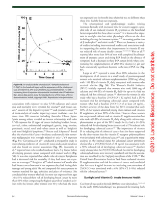
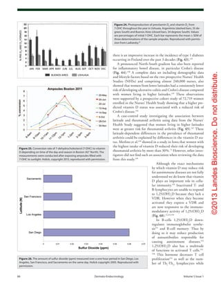
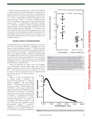
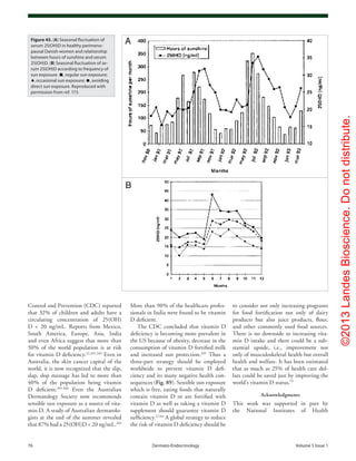
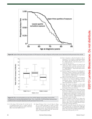
Figure 33. In two lightly pigmented Caucasian (A) and three heavily
pigmented Afroamerican subjects (B) after total body exposure to
0.054 J/cm2
of UVR. (C) Serial change in circulating vitamin D after re-
exposure of on Afroamerican subject (● in panel B) to a 0.32 J/cm2
dose
of UVR. Reproduced with permission from ref. 50.
Figure 34. The conversion of 7-dehydrocholesterol to previtamin D3
in an ampoule model, Type II and Type V skin after exposing to noon
sunlight in June at Boston (42°N), Massachusetts. The data represent the
means ± SEM of duplicate determinations. Reproduced with permission
from ref. 51.
Figure 35. Scatter plot of baseline serum 25-hydroxyvitamin D (25-OH-
D) on skin lightness (L*) score for unexposed skin, showing significant
positive correlation of serum 25-OH-D and L* (r2 = 0.1856). Heaney,
copyright 2006. Reproduced with permission.](https://image.slidesharecdn.com/holickd-200529140344/85/Holick-vit-D-Health-20-320.jpg)
![©2013LandesBioscience.Donotdistribute.
www.landesbioscience.com Dermato-Endocrinology 75
Clearly the paranoia about food fortification with vitamin D
causing toxicity needs to be reconsidered in light of observations
that infants who consumed 2000 IU vitamin D per day during
their first year of life not only did not have any evidence of toxicity
but for the ensuing 31 y markedly decreased their risk for type 1
diabetes.145
In the 1970s sunscreens were first introduced as a way to
prevent sunburning. The sunscreens contained UVB absorbing
chemicals such as paraaminobezoic acid because it was believed
that only UVB radiation damaged the skin and caused skin
cancer. It is now realized that UVA radiation not only alters
the immune system making it more immunotolerant but also
increases risk for non-melanoma and melanoma skin cancers.
Over the past four decades with very little thought as to its con-
sequences, several national and international health organiza-
tions have condemned any direct sun exposure. The American
Academy of Dermatology has taken the extreme position of rec-
ommending that no one should ever be exposed to direct sunlight
without sun protection. This radical view of sunlight and UVB
radiation has led to its designation as a carcinogen. To suggest
that one should never be exposed to sunlight because excessive
exposure to sunlight is linked to an increased risk for non-mela-
noma skin cancer is like suggesting that because breathing 100%
oxygen can cause lung damage and death, that no one should
breath an atmosphere that contains 20% oxygen.
The lack of appreciation of the importance of sensible sun
exposure for providing children and adults with their vitamin
D requirement has led to a worldwide vitamin D deficiency
pandemic.22,173
In the United States the Center for Disease
females although fertile would have
had a difficult time, if not impos-
sible, to give vaginal birth resulting
in both maternal and fetal death.54,55
Indeed it was because of the vitamin
D deficiency pandemic in late 1800s
that Cesarean sectioning became
common practice for the delivery of
healthy children of mothers who had
suffered from vitamin D deficiency
in utero and during their first few
years of life.8,54,55
Vitamin D defi-
ciency in pregnant women today is
still associated with a 400% increase
in the predicted probability for a
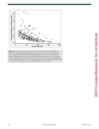
Cesarean section (Fig. 83).54
It is remarkable that for more
than 100 y investigators have been
reporting an inverse association with
latitude and many chronic illnesses
including common cancers,85
sev-
eral autoimmune diseases including
type 1 diabetes and multiple sclero-
sis73,134-139
as well as hypertension.159
In addition the revelation that expo-
sure to sunlight or UV radiation
could cure and prevent rickets12,13
led
to the widespread recommendation by health regulators and gov-
ernment agencies to encourage sensible sun exposure, i.e., amount
of sun that would be beneficial for producing vitamin D and
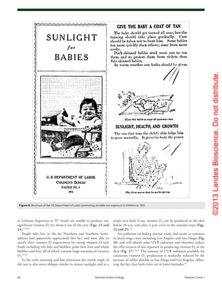
reducing risk for rickets while preventing sunburning (Fig. 8).
The global appreciation of the beneficial effects of vitamin D
for health lead to widespread vitamin D fortification throughout
Europe and the United States in the 1930s-1940s. Not only milk
but hot dogs, soda, custard, bread, cereals and even beer was forti-
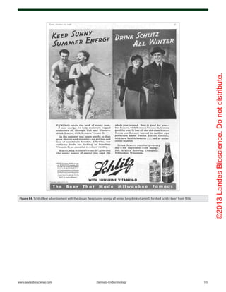
fied with vitamin D (Fig. 9). Schlitz even promoted their vitamin
D fortified beer in the winter with the slogan “if you want to keep
sunnyenergyallwinterlongdrinkvitaminDfortifiedSchlitzbeer”
(Fig. 84). They may have been correct now with the revelation
that vitamin D deficiency was associated with depression, seasonal
affective disorder and neurocognitive dysfunction.198-200,202,203,210
Unfortunately in the early 1950s the outbreak of hypercalce-
mia in British infants, who also had birth defects which included
altered facial features, mental retardation, and heart problems, was
incorrectly attributed to be over fortification of milk with vitamin
D since it was believed that these were signs of vitamin D intoxi-
cation.20,22
The more likely explanation is that these children had
a syndrome which is associated with a hypersensitivity to vitamin
D causing hypercalcemia and also with an elfin appearance and
heart problems.20
However because this “outbreak” was associated
with infants who had birth defects and mental retardation laws
were quickly passed forbidding the fortification of not only foods
but any consumer product including skin cream with vitamin D.
This legislation was quickly adopted in most European countries
and was used as a reason by other countries not to fortify milk
with vitamin D.
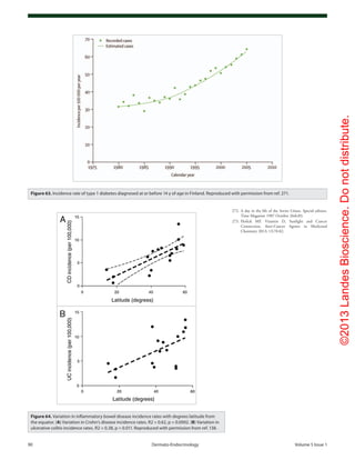
Figure 44. Geometric mean (95% CI) monthly variation in serum 25-hydroxyvitamin D [25(OH)D]
concentrations in men (■; n = 3725) and women (□; n = 3712) in the 1958 British birth cohort at age 45
y. The interaction between sex and month was significant [p = 0.02, linear regression analyses on log
25(OH)D]. n per sex and month ranged from 17 to 340: 98 in December 2003 for women and < 100 for
both sexes in December 2002 (n = 40 M, 37 F), January 2004 (n = 95 M, 75 F), February 2004 (n = 58 M, 70
F), and March 2004 (n = 22 M, 17 F). Reproduced with permission from ref. 61.](https://image.slidesharecdn.com/holickd-200529140344/85/Holick-vit-D-Health-25-320.jpg)
![©2013LandesBioscience.Donotdistribute.
www.landesbioscience.com Dermato-Endocrinology 87
224. Iakovlev VI. Prevention of ultraviolet deficiency in
children and adolescents. Gig Sanit 1981; 81-2;
PMID:7203052.
225. Sperti G. United States Patent Office, Patent Number
1,956,599. In: Google Patents; 1934.
226. Sperti G. Electric light source. In: Google Patents;
1936.
221. Houghton LA, Vieth R. The case against ergocalciferol
(vitamin D2
) as a vitamin supplement. Am J Clin Nutr
2006; 84:694-7; PMID:17023693.
222. Rollier A. Heliotherapy: Its Therapeutic, prophylactic
and social value. Am J Nurs 1927; 27:815-23.
223. Huldschinsky K. Heilung von Rachitis durch kün-
stliche Höhensonne. Dtsch Med Wochenschr 1919;
45:712-3; http://dx.doi.org/10.1055/s-0028-1137830.
219. Armas LAG, Hollis BW, Heaney RP. Vitamin D2
is much less effective than vitamin D3
in humans.
J Clin Endocrinol Metab 2004; 89:5387-91;
PMID:15531486; http://dx.doi.org/10.1210/jc.2004-
0360.
220. Trang HM, Cole DE, Rubin LA, Pierratos A, Siu
S, Vieth R. Evidence that vitamin D3
increases
serum 25-hydroxyvitamin D more efficiently than
does vitamin D2
. Am J Clin Nutr 1998; 68:854-8;
PMID:9771862.
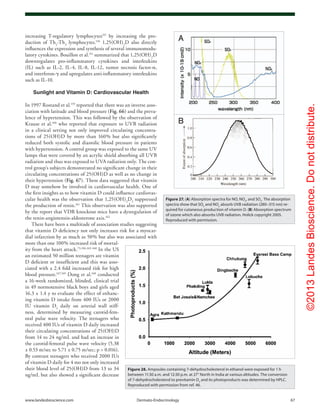
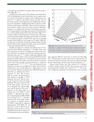
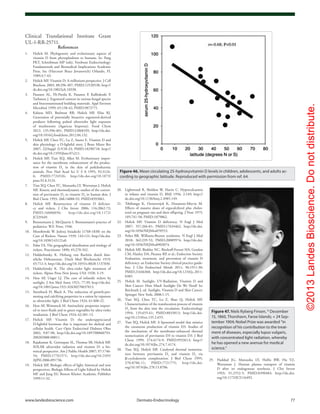
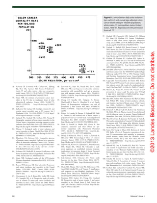
Figure 60. Metabolism of 25-hydroxyvitamin D [25(OH)D] to 1,25-dihydroxyvitamin D 1,25(OH)2
D for non-skeletal functions. When a monocyte/mac-
rophage is stimulated through its toll-like receptor 2/1 (TLR2/1) by an infective agent such as Mycobacterium tuberculosis (TB), or its lipopolysaccha-
ride (LPS) the signal upregulates the expression of vitamin D receptor (VDR) and the 25-hydroxyvitamin D-1-hydroxylase (1-OHase). 25(OH)D levels >
30 ng/mL provides adequate substrate for the 1-OHase to convert it to 1,25(OH)2
D. 1,25(OH)2
D returns to the nucleus where it increases the expression
of cathelicidin which is a peptide capable of promoting innate immunity and inducing the destruction of infective agents such as TB. It is also likely
that the 1,25(OH)2
D produced in the monocytes/macrophage is released to act locally on activated T (AT) and activated B (AB) lymphocytes which
regulate cytokine and immunoglobulin synthesis respectively. When 25(OH)D levels are ~30 ng/mL, it reduces risk of many common cancers.22-32
It is
believed that the local production of 1,25(OH)2
D in the breast, colon, prostate, and other cells regulates a variety of genes that control proliferation.
Once 1,25(OH)2
D completes the task of maintaining normal cellular proliferation and differentiation, it induces the 25-hydroxyvitamin D-24-hydrox-
ylase (24-OHase). The 24-OHase enhances the metabolism of 1,25(OH)2
D to calcitroic acid which is biologically inert. Thus, the local production of
1,25(OH)2
D does not enter the circulation and has no influence on calcium metabolism. The parathyroid glands have 1-OHase activity and the local
production of 1,25(OH)2
D inhibits the expression and synthesis of PTH. The production of 1,25(OH)2
D in the kidney enters the circulation and is able to
downregulate renin production in the kidney and to stimulate insulin secretion in the β-islet cells of the pancreas. Holick, copyright 2007. Reproduced
with permission.](https://image.slidesharecdn.com/holickd-200529140344/85/Holick-vit-D-Health-37-320.jpg)
![©2013LandesBioscience.Donotdistribute.
88 Dermato-Endocrinology Volume 5 Issue 1
242. Warren R, Gartstein V, Kligman AM, Montagna W,
Allendorf RA, Ridder GM. Age, sunlight, and facial
skin: a histologic and quantitative study. J Am Acad
Dermatol 1991; 25:751-60; PMID:1802896; http://
dx.doi.org/10.1016/S0190-9622(08)80964-4.
243. Cleaver JE, Crowley E. UV damage, DNA repair and
skin carcinogenesis. Front Biosci 2002; 7:d1024-43;
PMID:11897551.
244. Hollis BW. Circulating 25-hydroxyvitamin D levels
indicative of vitamin D sufficiency: implications for
establishing a new effective dietary intake recom-
mendation for vitamin D. J Nutr 2005; 135:317-22;
PMID:15671234.
245. Moore C, Murphy MM, Keast DR, Holick MF.
Vitamin D intake in the United States. J Am Diet
Assoc 2004; 104:980-3; PMID:15175600; http://
dx.doi.org/10.1016/j.jada.2004.03.028.
246. Käkelä R, Hyvärinen H, Vainiotalo P. Fatty acid
composition in liver and blubber of the Saimaa ringed
seal (Phoca hispida saimensis) compared with that of
the ringed seal (Phoca hispida botnica) and grey seal
(Halichoerus grypus from the Baltic. Comp Biochem
Physiol B 1993; 105:553-65; PMID:8365111; http://
dx.doi.org/10.1016/0305-0491(93)90088-M.
247. Wagner CL, Greer FR. Breastfeeding atSo, Nutrition
Co. Prevention of Rickets and Vitamin D Deficiency
in Infants, Children, and Adolescents. Pediatr 2008;
122:1142-52; http://dx.doi.org/10.1542/peds.2008-
1862.
248. Shah BR, Finberg L. Single-day therapy for nutritional
vitamin D-deficiency rickets: a preferred method. J
Pediatr 1994; 125:487-90; PMID:8071764; http://
dx.doi.org/10.1016/S0022-3476(05)83303-7.
249. El-Hajj Fuleihan G, Nabulsi M, Tamim H, Maalouf
J, Salamoun M, Khalife H, et al. Effect of vita-
min D replacement on musculoskeletal parameters in
school children: a randomized controlled trial. J Clin
Endocrinol Metab 2006; 91:405-12; PMID:16278262;
http://dx.doi.org/10.1210/jc.2005-1436.
250. Heaney RP, Davies KM, Chen TC, Holick MF, Barger-
Lux MJ. Human serum 25-hydroxycholecalciferol
response to extended oral dosing with cholecalciferol.
Am J Clin Nutr 2003; 77:204-10; PMID:12499343.
251. Malabanan A, Veronikis IE, Holick MF. Redefining
vitamin D insufficiency. Lancet 1998; 351:805-6;
PMID:9519960; http://dx.doi.org/10.1016/S0140-
6736(05)78933-9.
252. Pietras SM, Obayan BK, Cai MH, Holick MF. Vitamin
D2
treatment for vitamin D deficiency and insufficiency
for up to 6 years. Arch Intern Med 2009; 169:1806-8;
PMID:19858440; http://dx.doi.org/10.1001/archin-
ternmed.2009.361.
253. Holick MF. Vitamin D status: measurement, inter-
pretation, and clinical application. Ann Epidemiol
2009; 19:73-8; PMID:18329892; http://dx.doi.
org/10.1016/j.annepidem.2007.12.001.
254. Koutkia P, Chen TC, Holick MF. Vitamin D intoxica-
tion associated with an over-the-counter supplement.
N Engl J Med 2001; 345:66-7; PMID:11439958;
http://dx.doi.org/10.1056/NEJM200107053450115.
255. Adams JS, Lee G. Gains in bone mineral density with
resolution of vitamin D intoxication. Ann Intern
Med 1997; 127:203-6; PMID:9245225; http://dx.doi.
org/10.7326/0003-4819-127-3-199708010-00004.
256. Webb AR, DeCosta BR, Holick MF. Sunlight regulates
the cutaneous production of vitamin D3
by causing
its photodegradation. J Clin Endocrinol Metab 1989;
68:882-7; PMID:2541158; http://dx.doi.org/10.1210/
jcem-68-5-882.
257. Vieth R. Vitamin D toxicity, policy, and science. J Bone
Miner Res 2007; 22(Suppl 2):V64-8; PMID:18290725;
http://dx.doi.org/10.1359/jbmr.07s221.
258. Rajakumar K, Reis EC, Holick MF. Dosing error
with over-the-counter vitamin D supplement: a risk
for vitamin D toxicity in infants. Clin Pediatr (Phila)
2013; 52:82-5; PMID:22492833; http://dx.doi.
org/10.1177/0009922812439245.
235. Brash DE, Ziegler A, Jonason AS, Simon JA, Kunala
S, Leffell DJ. Sunlight and sunburn in human skin
cancer: p53, apoptosis, and tumor promotion. The
journal of investigative dermatology Symposium pro-
ceedings/the Society for Investigative Dermatology, Inc
[and] European Society for Dermatological Research
1996;1:136-42.
236. Kricker A, Armstrong BK, English DR, Heenan PJ.
Does intermittent sun exposure cause basal cell carci-
noma? a case-control study in Western Australia. Int
J Cancer 1995; 60:489-94; PMID:7829262; http://
dx.doi.org/10.1002/ijc.2910600411.
237. Kennedy C, Bajdik CD, Willemze R, De Gruijl FR,
Bouwes Bavinck JN; Leiden Skin Cancer Study. The
influence of painful sunburns and lifetime sun exposure
on the risk of actinic keratoses, seborrheic warts, mela-
nocytic nevi, atypical nevi, and skin cancer. J Invest
Dermatol 2003; 120:1087-93; PMID:12787139;
http://dx.doi.org/10.1046/j.1523-1747.2003.12246.x.
238. Chen TC. The Photobiology of Vitamin D. In: Holick
MF, ed. Vitamin D – Physiology, Molecular Biology
and Clinical Applications. Totowa, NJ: Humana Press;
1998:17-37.
239. Knaysi GA, Crikelair GF, Cosman B. The role of
nines: its history and accuracy. Plast Reconstr Surg
1968; 41:560-3; PMID:5654897; http://dx.doi.
org/10.1097/00006534-196806000-00008.
240. Holick MF. Vitamin D and sunlight: strategies for can-
cer prevention and other health benefits. Clin J Am Soc
Nephrol 2008; 3:1548-54; PMID:18550652; http://
dx.doi.org/10.2215/CJN.01350308.
241. Buettner PG, Raasch BA. Incidence rates of skin cancer
in Townsville, Australia. Int J Cancer 1998; 78:587-
93; PMID:9808527; http://dx.doi.org/10.1002/
(SICI)1097-0215(19981123)78:5<587::AID-
IJC10>3.0.CO;2-E.
227. Gronowitz E, Larkö O, Gilljam M, Hollsing A, Lindblad
A, Mellström D, et al. Ultraviolet B radiation improves
serum levels of vitamin D in patients with cystic fibro-
sis. Acta Paediatr 2005; 94:547-52; PMID:16188742;
http://dx.doi.org/10.1080/08035250410025276.
228. Chandra P, Wolfenden LL, Ziegler TR, Tian J, Luo M,
Stecenko AA, et al. Treatment of vitamin D deficiency
with UV light in patients with malabsorption syn-
dromes: a case series. Photodermatol Photoimmunol
Photomed 2007; 23:179-85; PMID:17803596; http://
dx.doi.org/10.1111/j.1600-0781.2007.00302.x.
229. Dabai NS, Pramyothin P, Holick MF. The effect
of ultraviolet radiation from a novel portable fluo-
rescent lamp on serum 25-hydroxyvitamin D3
lev-
els in healthy adults with Fitzpatrick skin types II
and III. Photodermatol Photoimmunol Photomed
2012; 28:307-11; PMID:23126292; http://dx.doi.
org/10.1111/phpp.12000.
230. Koutkia P, Lu Z, Chen TC, Holick MF. Treatment
of vitamin D deficiency due to Crohn’s disease with
tanning bed ultraviolet B radiation. Gastroenterology
2001; 121:1485-8; PMID:11729127; http://dx.doi.
org/10.1053/gast.2001.29686.
231. Tangpricha V, Turner A, Spina C, Decastro S, Chen
TC, Holick MF. Tanning is associated with optimal
vitamin D status (serum 25-hydroxyvitamin D concen-
tration) and higher bone mineral density. Am J Clin
Nutr 2004; 80:1645-9; PMID:15585781.
232. Holick MF. Sunlight and vitamin D for bone health
and prevention of autoimmune diseases, cancers,
and cardiovascular disease. Am J Clin Nutr 2004;
80(Suppl):1678S-88S; PMID:15585788.
233. Holick MF. The vitamin D epidemic and its health
consequences. J Nutr 2005; 135:2739S-48S;
PMID:16251641.
234. MacKie RM. Incidence, risk factors and prevention
of melanoma. Eur J Cancer 1998; 34(Suppl 3):S3-6;
PMID:9849401; http://dx.doi.org/10.1016/S0959-
8049(98)00003-3.
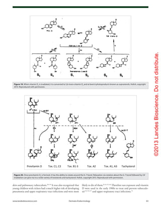
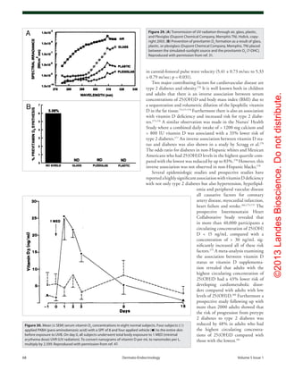
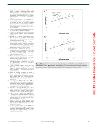
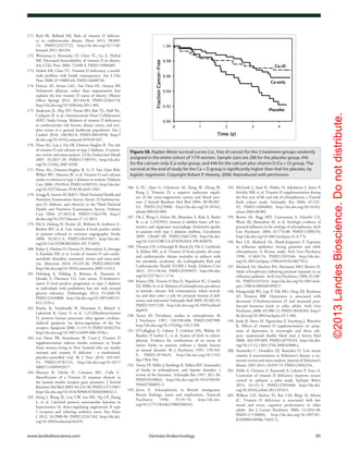
Figure 61. Prevalence of multiple sclerosis (MS) by latitude in the United States according to data
from Noonan et al.140
(×) and Wallin et al.141
(○). The dashed line is a quadratic fit to the data from
Noonan et al.,140
and the solid line is a fit to the data from Wallin et al.141
Reproduced with permis-
sion from ref. 134.](https://image.slidesharecdn.com/holickd-200529140344/85/Holick-vit-D-Health-38-320.jpg)
![©2013LandesBioscience.Donotdistribute.
www.landesbioscience.com Dermato-Endocrinology 89
268. Daly RM, Gagnon C, Lu ZX, Magliano DJ, Dunstan
DW, Sikaris KA, et al. Prevalence of vitamin D
deficiency and its determinants in Australian adults
aged 25 years and older: a national, population-
based study. Clin Endocrinol (Oxf) 2012; 77:26-35;
PMID:22168576; http://dx.doi.org/10.1111/j.1365-
2265.2011.04320.x.
269. Czarnecki D, Meehan CJ, Bruce F. The vitamin D
status of Australian dermatologists. Clin Exp Dermatol
2009; 34:621-38; PMID:19236422; http://dx.doi.
org/10.1111/j.1365-2230.2008.03002.x.
270. Nesby-O’Dell S, Scanlon KS, Cogswell ME, Gillespie
C, Hollis BW, Looker AC, et al. Hypovitaminosis D
prevalence and determinants among African American
and white women of reproductive age: third National
Health and Nutrition Examination Survey, 1988-1994.
Am J Clin Nutr 2002; 76:187-92; PMID:12081833.
271. Harjutsalo V, Sjöberg L, Tuomilehto J. Time trends
in the incidence of type 1 diabetes in Finnish chil-
dren: a cohort study. Lancet 2008; 371:1777-82;
PMID:18502302; http://dx.doi.org/10.1016/S0140-
6736(08)60765-5.
264. Holick MF. Phylogenetic and evolutionary aspects of
vitamin D from phytoplankton to humans. In: Pang
PKT, Schreibman MP (eds), Verebrate Endocrinology:
Fundamentals and Biomedical Implications. Academic
Press, Inc. (Harcourt Brace Jovanovich) Orlando, FL.
1989. 3:7-43.
265. Looker AC, Johnson CL, Lachner DA, Pfeiffer CM,
Schleicher RL, Sempos CT. Vitamin D Status: United
States, 2001-2006. NCHS Data Brief 2011; 56.
266. Hossein-nezhad A, Holick MF. Optimize dietary intake
of vitamin D: an epigenetic perspective. Curr Opin Clin
Nutr Metab Care 2012; 15:567-79; PMID:23075936;
http://dx.doi.org/10.1097/MCO.0b013e3283594978.
267. Holick MF, Binkley NC, Bischoff-Ferrari HA, Gordon
CM, Hanley DA, Heaney RP, et al. Guidelines for
preventing and treating vitamin D deficiency and
insufficiency revisited. J Clin Endocrinol Metab
2012; 97:1153-8; PMID:22442274; http://dx.doi.
org/10.1210/jc.2011-2601.
259. Hollis BW, Johnson D, Hulsey TC, Ebeling M,
Wagner CL. Vitamin D supplementation during preg-
nancy: double-blind, randomized clinical trial of safety
and effectiveness. J Bone Miner Res 2011; 26:2341-
57; PMID:21706518; http://dx.doi.org/10.1002/
jbmr.463.
260. Vieth R. Vitamin D supplementation, 25-hydroxyvi-
tamin D concentrations, and safety. Am J Clin Nutr
1999; 69:842-56; PMID:10232622.
261. Taylor JG. Yahweh and the sun: biblical and archaeo-
logical evidence for sun worship in ancient Israel: T&T
Clark; 1993.
262. Srivastava VC, Basham AL, Sharma G. Sun-worship in
ancient India: Indological Publications; 1972.
263. Holick MF. Evolution and function of vitamin
D. Recent Results Cancer Res 2003; 164:3-28;
PMID:12899511; http://dx.doi.org/10.1007/978-3-
642-55580-0_1.
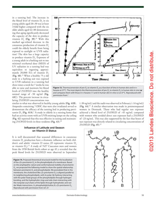
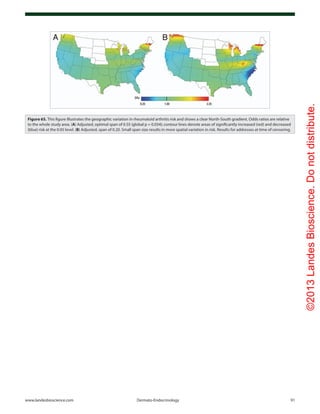
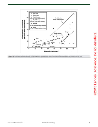
Figure 62. Age-standardized incidence rates of type 1 diabetes per 100,000 boys <14 years of age, by latitude, in 51 regions worldwide, 2002. Data
points are shown by dots; names shown adjacent to the dots denote location, where space allows. Where space was limited, numerical codes (below)
designate location. Source: data from WHO DiaMond [3]. Lux., Luxembourg. Numerical codes for areas: 2. Beja, Tunisia; 3. Gafsa, Tunisia; 4. Kairoan,
Tunisia; 5. Monastir, Tunisia; 7. Mauritius; 8. Wuhan, China; 9. Sichuan, China; 10. Huhehot, China; 16. Nanjing, China; 17. Jinan, China; 21. Harbin, China;
23. Changsha, China; 25. Hainan, China; 29. Hong Kong, China; 31. Israel; 32. Chiba, Japan; 33. Hokkaido, Japan; 34. Okinawa, Japan; 36. Novosibirsk,
Russia; 38. Antwerp, Belgium; 39. Varna, Bulgaria; 40. Denmark; 43. France; 44. Baden, Germany; 45. Attica, Greece; 48. Sicily, Italy; 49. Pavia, Italy; 50.
Marche, Italy; 52. Lazio, Italy; 59. Krakow, Poland; 61. Algarve, Portugal; 62. Coimbra, Portugal; 63. Madeira Island, Portugal; 64. Portalegre, Portugal; 65.
Bucharest, Romania; 67. Slovakia; 68. Catalonia, Spain; 71. Leicestershire, UK; 72. Northern Ireland, UK; 77. Allegheny, PA, USA; 80. Avellaneda, Argen-
tina; 82. Corrientes, Argentina; 87. Paraguay; 88. Lima, Peru; 90. Caracas, Venezuela; 97. Auckland, New Zealand. Data points not labelled because of
space constraints (latitude in degrees, rate per 100,000): 11. Dalian, China (39, 1.1); 12. Guilin, China (24, 0.6); 13. Beijing, China (40, 0.7); 14. Shanghai,
China (32. 0.7); 15. Chang Chun, China (44, 0.6); 18. Jilin, China (43, 0.4); 19. Shenyang, China (42, 0.4); 20. Lanzhou, China (36, 0.5); 22. Nanning, China (23,
0.3); 24. Zhengzhou, China (35, 0.2); 26. Tie Ling, China (42, 0.2); 27. Zunyi, China (28, 0.1); 28. Wulumuqi, China (44, 0.9); 35. Karachi, Pakistan (25, 0.5); 37.
Austria (48, 9.8); 46. Hungary (47, 8.7); 51. Turin, Italy (45, 11.9); 53. Lombardia, Italy (46, 7.6); 66. Slovenia (46, 6.8); 79. Chicago, IL, USA (42, 10.2). R2 = 0.25,
p < 0.001.](https://image.slidesharecdn.com/holickd-200529140344/85/Holick-vit-D-Health-39-320.jpg)

![©2013LandesBioscience.Donotdistribute.
98 Dermato-Endocrinology Volume 5 Issue 1
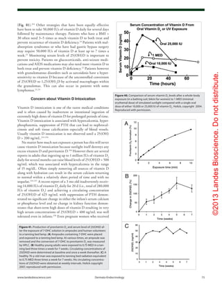
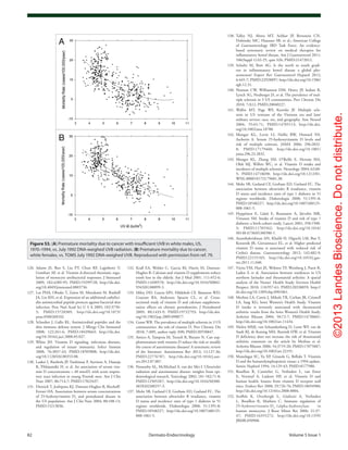
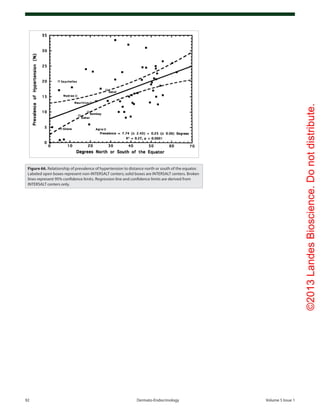
Figure 73. (A) Mean (SEM) serum levels of 25(OH)vitamin D in patients with cystic fibrosis, treated
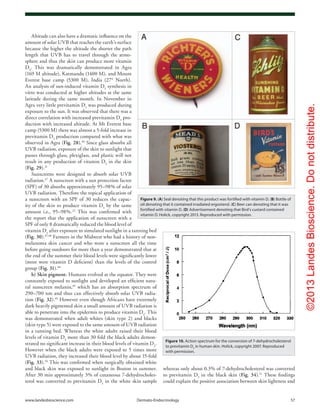
with UVB (▲) (n = 9), and non-treated CF patients as controls (●) (n = 14) at baseline and after 8,
16 and 24 weeks. There were significant differences between the groups at all time points except
at baseline (ANOVA, p < 0.0001). (B) Mean (± SEM) serum 25-hydroxyvitamin D concentration (ng/
mL) before and after 8 weeks of UV light to cystic fibrosis (CF) subjects. Serum 25-hydroxyvitamin
D [25(OH)D] levels in the five CF subjects at baseline were 21 ± 3 ng/ml, which increased to 27 ± 4
ng/ml at the end of 8 weeks (p = 0.05). Reproduced with permission from ref. 227.](https://image.slidesharecdn.com/holickd-200529140344/85/Holick-vit-D-Health-48-320.jpg)
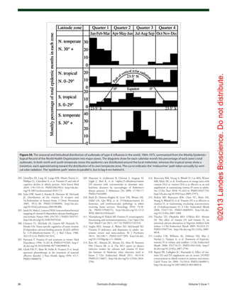
![©2013LandesBioscience.Donotdistribute.
104 Dermato-Endocrinology Volume 5 Issue 1
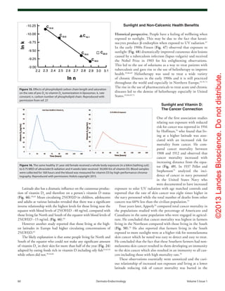
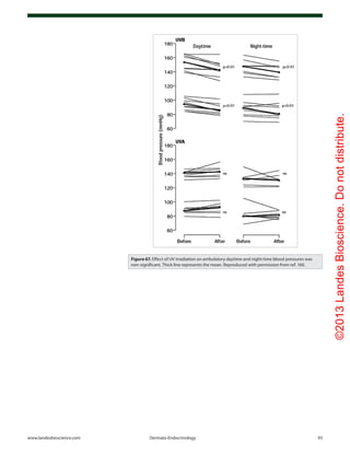
Figure 81. Mean serum 25-hydroxyvitamin D (25[OH]D) and calcium levels. Results are given
as mean (SEM) values averaged over 6-mo intervals. Time 0 is initiation of treatment. (A) Mean
25(OH)D levels in all patients treated with 50,000 IU of ergocalciferol (vitamin D2
) every 2 weeks
(maintenance therapy, n = 86). Forty-one of the patients were vitamin D insufficient or deficient
and first received 50,000-IU ergocalciferol weekly for 8 weeks before being placed on mainte-
nance therapy of 50,000 IU of ergocalciferol every 2 weeks. The mean 25(OH)D level of each 6-mo
interval was compared with initial mean 25(OH)D level and showed a significant difference of p <
0.001 for all time points. To convert 25(OH)D to nanomoles per liter, multiply by 2.496. (B) Mean
serum 25(OH)D levels in patients receiving maintenance therapy only. There were 38 patients who
were vitamin D insufficient (25[OH]D levels < 21–29 ng/mL and 7 patients who were vitamin D suf-
ficient (25[OH]D levels ≥ 30 ng/mL) who were treated only with maintenance therapy of 50,000 IU
of ergocalciferol (vitamin D2
) every 2 weeks. The mean 25(OH)D levels in each 6-mo interval were
compared with mean initial 25(OH)D levels and showed a significant difference of p < 0.001 for
all time points up to 48 mo. The data for interval months 60 and 72 were pooled, and there was
a significant difference of p < 0.01 compared with the baseline value. (C) Serum calcium levels.
Results for all 86 patients who were treated with 50,000 IU of ergocalciferol (vitamin D2
). The refer-
ence range for serum calcium level is 8.5 to 10.2 mg/dL (to convert to millimoles per liter, multiply
by 0.25). Reproduced with permission from ref. 252.](https://image.slidesharecdn.com/holickd-200529140344/85/Holick-vit-D-Health-54-320.jpg)